Abstract
Although the buccal fat pad (BFP) was originally used as an alternative method for the closure of small to medium-sized oroantral and oronasal communications, its use has now been extended to use after excision of oral pre malignant lesions. This report describes experience with this technique
Keywords: Buccal fat pad, Oral premalignant lesion, Pedicled flap, Oro-antral fistula
Introduction
A pedicled buccal fat pad (BFP) flap was first described in 1977 by Egyedi [1] for the closure of oroantral communications after oncological resections. In 1983, Neder [2] utilized the buccal fat pad as a free graft in the oral cavity. In 1986, Tideman et al. [3], showed that the pedicled buccal fat pad flap is epithelialized within 3–4 weeks and therefore, cover with a skin graft is not required. Since then, there have been several studies on the use of this flap for closure of oroantral and oronasal communications secondary to exodontia. It was only after the onset of this century that Rapidis et al. [4], Hao [5], and Dean et al. [6], used pedicled BFP flaps for reconstruction of medium sized post-surgical oral defects for malignant lesions. In 2005, Amin [7] showed how effectively a buccal fat pad can be utilized in post partial maxillectomy defects for neoplastic diseases. The procedural simplicity, with very low complication rates and excellent functional outcome, encouraged us to use a pedicled BFP as a reconstruction means for selective intraoral cancers.
Anatomy
The buccal fat pad lies in the masticatory space between the buccinator muscle medially and the masseter muscle laterally, and it is wrapped within a thin fascial envelope. The BFP is divided into 3 lobes (anterior, intermediate, and posterior). The posterior lobe has four extensions (buccal, pterygoid, pterygopalatine,and temporal).Several nutritional vessels exist in each lobe and together form a subcapsular plexus [8] (Figs. 1, 2, 3, 4, 5, 6).
Fig. 1.
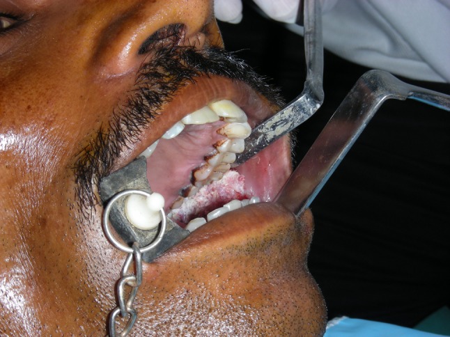
Verrcous leukoplakia
Fig. 2.
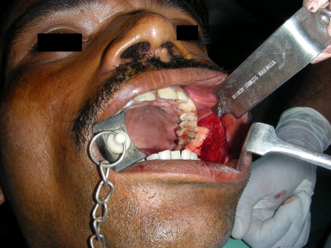
Lesion excised
Fig. 3.
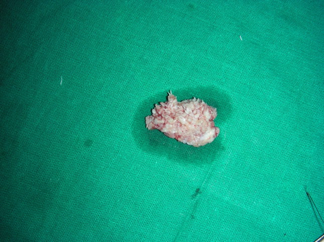
Excised specimen
Fig. 4.
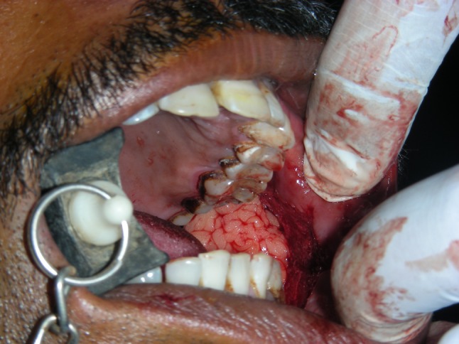
Buccal pad of fat brought to surgical site
Fig. 5.
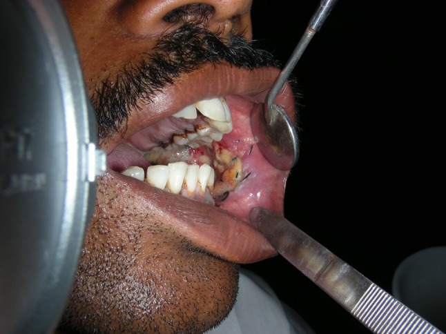
1 week post op
Fig. 6.
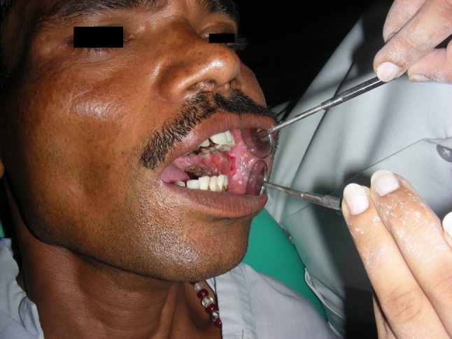
2nd week post op
The principal arteries supplying the BFP are derived from the buccal and deep temporal branches of the maxillary artery, from the transverse facial branch of the superficial temporal artery and from a few branches of the facial artery [3]. Morphologically, the buccal fat pad is quite different from subcutaneous fat and is similar to orbital fat [9].
The mean volume of BFP is 10 ml, with a mean thickness of 6 mm [10] and an approximate weight of 9.3 gm. It is capable of covering small to medium defects about 4 cm in diameter.
The physiological functions of BFP are (1) to fill the masticatory space, acting as cushion for the masticatory muscles, (2) to counteract negative pressure during suction in a newborn, and (3) as a rich venous net, with valve like structures, possibly involved in the exo-endocranial blood flow through the pterygoid plexus [11].
Patients and Methods
The BFP was used as a pedicled graft to reconstruct medium-sized surgical defects of the oral soft and hard tissues in 11 patients. 4 of the defects were in the maxilla, 3 in the retromandibular area, and 3 in the cheek and oral commissure. The BFP was left uncovered to epithelialize. The buccal fat pad was assessed daily and, oral rinsing was probihited for the first 4–5 days. The surgical field was cleaned twice daily with saline soaked gauze.
The patients were evaluated on the 7th day. All the patients were put on oral physiotherapy after 7th post op day. They were then evaluated after 2 weeks, 1 month and at every 3 months for 2 years.
Results
In the immediate postoperative period, most of the patients fared well except for 2 patients. One of them had a partial loss of graft with oro-antral fistula which can be attributed to continued smoking in post operative period which was sutured again. In the other case hematoma in the buccal fat pad was observed which healed with moderate fibrosis. All other patients had significantly improved mouth opening. In all the patients who had an uneventful immediate postoperative period, signs of buccal fat pad epithelialization had started by the end of the first week and the BFP was completely epithelialized at the end of the first month. Three months later, at the second follow-up visit, most of the patients had the buccal fat pad replaced by a thin whitish streak covered by normal mucosa, with very minimal fibrosis. One patient was lost in follow up & in one case there were signs of recurrence of lesion in adjacent site
Patient characteristics, lesion description and postreconstruction outcome of patients
| P | F/M | Age (years) | Indications | Anatomic location (all of in maxilla) | Follow-up (month) | Complications |
|---|---|---|---|---|---|---|
| 1 | M | 60 | OAF | MAXILLA | ||
| 2 | M | 30 | OSMF | BUCCAL MUCOSA | 6 | Hematoma |
| 3 | M | 23 | VERRUCOUS LEUKOPLAKIA | BUCCAL MUCOSA | 6 | |
| 4 | F | 25 | SPECKLED LEUKOPLAKIA | BUCCAL MUCOSA | 6 | |
| 5 | M | 37 | OAF | MAXILLA | 1 | |
| 6 | M | 37 | PLEOMORPHIC ADENOMA | PALATE | 3 | |
| 7 | F | 17 | OSMF | BUCCAL MUCOSA | ||
| 8 | M | 38 | LEUKOPLAKIA | BUCCAL MUCOSA | 18 | |
| 9 | M | 35 | OSMF | BUCCAL MUCOSA | 6 | |
| 10 | M | 44 | OAF | MAXILLA | 6 | Partial loss of graft |
| 11 | F | 36 | LEUKOPLAKIA | BUCCAL MUCOSA | 8 |
Conclusions
The buccal fat pad becomes an ideal choice for medium to even larger intraoral defects, because local flaps such as the buccal advancement flap, palatal pedicled flap, double layered closure flaps using buccal and palatal tissues, and other such procedures disappointingly produce large denuded areas and are unsuitable for large defects [12, 13]. The body and buccal extension (accounts for almost half of the total volume of buccal fat pad) are accessible through the oral cavity. A fact has to be borne in mind that excessive stretching in the flap invariably impairs the vascularity, so closure of larger defects cannot be guaranteed without producing flap necrosis or creating a new fistula. The findings support the view that the BFP is a useful, easy, and uncomplicated alternative method for the reconstruction of small to medium-sized surgical defects of the oral hard and soft tissues
Contributor Information
Shishir Mohan, Email: drshishirdent@yahoo.co.in.
Hasti Kankariya, Email: drhasti81@gmail.com.
Bhupendra Harjani, Email: bhupendraharjani@rediffmail.com.
References
- 1.Egyedi P (1977) Utilization of the buccal fat pad for closure of oroantral communication. J Maxillofac Surg 5:241–244 [DOI] [PubMed]
- 2.Neder A. Use of buccal fat pad for grafts. Oral Surg Oral Med Oral Pathol. 1983;55:349–350. doi: 10.1016/0030-4220(83)90187-1. [DOI] [PubMed] [Google Scholar]
- 3.Tideman H, Bosanquet A. Scott use of the buccal fat pad as pedicled graft. J Oral Maxillofac Surg. 1986;44:435–440. doi: 10.1016/S0278-2391(86)80007-6. [DOI] [PubMed] [Google Scholar]
- 4.Rapidis AD, Alexandridis CA, Eleftheriadis E. Angelopoulos: the use of the buccal fat pad for reconstruction of oral defects: review of the literature and report of 15 cases. Oral Maxillofac Surg. 2000;58:158–163. doi: 10.1016/s0278-2391(00)90330-6. [DOI] [PubMed] [Google Scholar]
- 5.Hao SP. Reconstruction of oral defects with the pedicled buccal fat pad ap. Otolaryngo Head Neck Surg. 2000;122:863–867. doi: 10.1016/S0194-5998(00)70015-5. [DOI] [PubMed] [Google Scholar]
- 6.Dean A, Alamillos F, Garcia-Lopez A, Sanchez J, Penalba M. The buccal fat pad in oral reconstruction. Head Neck. 2001;23:383–388. doi: 10.1002/hed.1048. [DOI] [PubMed] [Google Scholar]
- 7.Amin MA, Bailey BM, Swinson B, Witherow H. Use of the buccal fat pad in the reconstruction and prosthetic rehabilitation of oncological maxillary defects. Br J Oral Maxillofac Surg. 2005;43:148–154. doi: 10.1016/j.bjoms.2004.10.014. [DOI] [PubMed] [Google Scholar]
- 8.Zhang HM, Yan YP, Qi KM, Wang JQ, Liu ZF. Anatomical structure of the buccal fat pad and its clinical adaptations. Plast Reconstr Surg. 2002;109:2509–2518. doi: 10.1097/00006534-200206000-00052. [DOI] [PubMed] [Google Scholar]
- 9.Ilankovan V, Soames JV. Morphometric analysis of orbital buccal & subcutaneous fat in treatment of enopthalamos. Br J Maxillofac Surg. 1995;33:40–42. doi: 10.1016/0266-4356(95)90085-3. [DOI] [PubMed] [Google Scholar]
- 10.Loh FC, Loh HS. Use of the buccal fat pad for correction of intraoral defects: report of cases. J Oral Maxillofac Surg. 1991;49:413–416. doi: 10.1016/0278-2391(91)90382-V. [DOI] [PubMed] [Google Scholar]
- 11.Racz L, Maros TN, Seres-Sturm L (1989) Structural characteristics and functional significance of the buccal fat pad (corpus adiposum buccae). Morphol Embryol (Bucur) 35:73–77 [PubMed]
- 12.Guven O. A clinical study on oroantral › stulae. J Craniomaxillofac Surg. 1998;26:267–271. doi: 10.1016/S1010-5182(98)80024-3. [DOI] [PubMed] [Google Scholar]
- 13.El-Hakim IE, el-Fakharany AM. The use of the pedicled buccal fat pad (BFP) and palatal rotating aps in closure of oroantral communication and palatal defects. J Laryngol Otol. 1999;118:134–138. doi: 10.1017/s0022215100145335. [DOI] [PubMed] [Google Scholar]


