Abstract
Introduction
Statins reportedly have anti-inflammatory and anti-thrombotic effects aside from cholesterol-lowering. This study aimed to evaluate the effect of pre-existing statin use on platelet activation markers and clinical outcome in acute ischemic stroke patients.
Methods
This prospective study evaluated 172 patients with acute ischemic stroke divided in two groups: patients with pre-existing statin (n = 43) and without pre-existing statin (66 cases with statins initiated post-stroke and 63 without statin treatment). Platelet activation markers (CD62P and CD63) were measured by flow cytometry at different time points after stroke and analyzed with clinical outcome.
Results
The CD62P and CD63 expressions on platelets were significantly lower in the patients with pre-existing statin use compared to the patients without pre-existing statin use on Day 1 post-stroke (p < 0.05). The CD62P expression was significantly lower in the patients with pre-existing statin use on 90 days after the acute stroke (p < 0.05). Patients with pre-existing statin use had lower incidences of early neurologic deterioration (END) than those without treatment (p < 0.05). Among several baseline clinical variables, admission NIHSS score, history of coronary artery disease, and pre-existing statin use were independent predictions of good clinical outcome at three months.
Conclusions
Pre-existing statin use is associated with decreased platelet activity as well as improved clinical outcome and reduced END in patients with acute ischemic stroke.
Keywords: flow cytometry, ischemic stroke, outcome, platelet activation
Introduction
Stroke is a major cause of morbidity and one of the leading causes of death worldwide [1]. Atherothrombosis and inflammation play important roles in the pathogenesis of acute ischemic stroke [2-4]. A previous study demonstrates that platelet activity, measured by CD62P and CD63 expressions on platelets, are increased after acute ischemic stroke and reduced in patients who receive anti-platelet therapy [5-7]. Anti-platelet drugs are the most commonly used drugs for secondary prevention after ischemic stroke of non-cardio-embolic origin [8], but their efficacy is not completely satisfactory [9,10].
Statins, the 3-hydroxy 3-methyl-glutaryl coenzyme-A (HMG-CoA) reductase inhibitors, are medications originally used for the control of hypercholesterolemia [11]. However, there is increasing evidence that statins have anti-inflammatory and anti-thrombotic effects aside from their cholesterol-lowering effect [12,13]. Statin therapy has been shown to reduce cardiovascular events, including myocardial infarction, stroke, and death [14-16]. Moreover, early statin treatment may reduce the severity and improve the outcome of myocardial infarction, ischemic stroke, and intra-cerebral hemorrhage [17-20].
Although statin therapy is widely used in patients at high-risk for major vascular events, the benefits of pre-existing statin therapy in patients with acute ischemic stroke remain controversial. Multiple studies have demonstrated improved clinical outcomes in patients taking statins at stroke onset [18,19]; however, mechanisms conferring this protection have not been well studied. Thus, this prospective cohort study aimed to test the difference in platelet activity between patients taking statins before and after acute ischemic stroke by assessing CD62P and CD63 expression. This study also analyzed if prior statin treatment could reduce the neurologic deterioration and improve the functional outcome of patients with ischemic stroke.
Materials and methods
Study participants
Consecutive patients with acute ischemic stroke admitted to the Department of Neurology of Chang Gung Memorial Hospital-Kaohsiung, Taiwan, from August 2009 to July 2010 were evaluated. Acute ischemic stroke was defined as acute-onset loss of focal cerebral function persisting for at least 24 hours. The diagnosis of stroke was made based on clinical presentation, neurologic examination, and results of brain magnetic resonance imaging (MRI) with magnetic resonance angiography (MRA). Patients with cardio-embolic stroke were excluded, as well as those with underlying neoplasm, vasculitis, hematologic disorders that affect platelet count or function, end-stage renal disease, liver cirrhosis, and congestive heart failure. As pathogenesis and treatment could be different between patients with cardio-embolic and non-cardio-embolic ischemic stroke, those with cardio-embolism were excluded by clinical presentation, ECG, and cardiac ultrasound, while those who received intravenous thrombolytic therapy were also excluded.
To avoid the confounding factor of anti-platelet therapies or dosage effects on measured platelet activity, all patients taking one or more anti-platelet medication (e.g. aspirin, dipyradimole, or clopidogrel) prior to stroke onset were excluded from enrollment. All enrolled patients were treated with aspirin (100 mg/day) therapy post-stroke. The Institutional Review Committee on Human Research approved the study protocol and the participating subjects provided informed consent.
Demographic data, history of risk factors (i.e., hypertension, diabetes mellitus, dyslipidemia, cigarette smoking, and cardiovascular disease), and history of previous vascular events (i.e., myocardial infarction, angina, old stroke history) were obtained at baseline. Vascular risk factors included hypertension, blood pressure above 140/90 mmHg at two readings or use of anti-hypertensives; diabetes mellitus (DM), elevated blood glucose at two recordings, elevated hemoglobin A1c (HbA1c) or use of anti-diabetics; and dyslipidemia, total cholesterol above 200 mg/dL, triglycerides above 180 mg/dL or use of lipid-lowering medication [10].
Of the 220 patients with acute non-cardio-embolic ischemic stroke, 30 were excluded due to previous anti-platelet therapy before the stroke, six due to cardio-embolic stroke (e.g. paroxysmal atrial fibrillation on EKG), four due to hemorrhagic transformations of ischemic strokes on the first brain imaging, four with end-stage renal disease, and four with gastro-intestinal bleeding in the acute stage. The remaining 172 patients were classified into two groups: patients with pre-existing statin use (patients taking statins prior to stroke onset) and patients without pre-existing statin use (patients did not take statins prior to stroke onset). For further comparison, the patients without pre-existing statin use were divided to two sub-groups: the statin-initiated group (patients placed on statins after stroke onset) and the non-statin treatment group (patients not placed on statins before and after stroke onset).
The lipid-lowering regimens used for preventing ischemic stroke were according to the American Heart Association/American Stroke Association guidelines for diabetic or high-risk patients and included statins if the low density lipoprotein (LDL) was above 70 and in non-diabetics if the LDL was above 100 [21]. The etiologic sub-types of i schemic stroke were classified according to the TOAST (Trial of Org 10172 in Acute Stroke Treatment) criteria [22].
Clinical assessments
Detailed medical history was obtained from patients and their families whenever possible, with specific standardized questioning regarding prior use of drugs. All of the patients underwent complete neurologic examination upon enrollment and on follow up. Brain MRI with MRA, extra-cranial carotid sonography, and trans-cranial color-coded sonography were performed on ischemic stroke patients during hospitalization. Early neurologic deterioration (END) was defined as an increase of four or more points in National Institutes of Health Stroke Scale (NIHSS) during hospitalization [23]. Follow-up brain CT scans were performed if END was noted, including hemorrhagic transformation (hyperdense signal over the infarction area), enlarged cerebral infarction (increased area of original lower attenuation or new infarction lesions), and cerebral edema (abnormal hypodense signal surrounding the infarction area with sulcal effacement or mass effect) [24].
Neurologic deficits due to stroke were assessed using the NIHSS. Physical disability and handicap were evaluated using the Barthel index (BI) and modified Rankin Scale (mRS). Therapeutic outcomes were evaluated three months post-stroke and good outcome was defined as a three-month mRS of 0 to 2, while poor outcome was mRS of 3 to 6.
Blood sampling and assessment of platelet activity
Blood samples were collected by venipuncture of the forearm veins in acute stroke patients within 48 hours and on days 7 and 30 post-stroke. Flow cytometry for platelet activity markers was performed as previously described [7]. Platelet activity was assessed using platelet activation markers (CD62P and CD63). Briefly, sodium citrate containing blood was centrifuged for 15 minutes at 1,500 rpm at room temperature. The samples were incubated with saturating concentrations of phycoerythrin (PE)-labeled antibodies (Becton Dickinson Biosciences, CA, USA.) against CD62P (clone AK-4) and CD63 (Clone H5C6) with fluorescein isothiocyanate-labeled antibodies against CD61 (clone VI-PL2) for 30 minutes at room temperature in the dark. For control experiments, platelets were incubated with PE-coupled unspecific mouse IgG1 (Becton Dickinson Biosciences, CA, USA.) with the same ratio and concentration of fluorochrome-to-protein as specific IgG.
After immuno-labeling, the samples were analyzed by Coulter Epics XL flow cytometry (Beckman Coulter, Miami, USA.). Forward light scatter and CD61 expression were used for platelet identification. Platelet-bound anti-CD62P and anti-CD63 antibodies were determined by analyzing 10,000 platelets for PE-positive fluorescence.
Results were presented as percentages of antibody-positive platelets. Intra-assay variability based on repeated measurements of the same blood sample was low, with mean coefficients of variance of 7.5% (< 48 hours), 7.3% (day 7), and 6.9% (day 30) for stroke patients.
Statistical analysis
Data were presented as mean ± standard error of the mean and as comparisons between groups. Continuous variables, including age, cell counts, lipid profile, HbA1c, blood pressure, and CD62P and CD63 levels were analyzed by independent t-test between groups. The NIHSS score between the two groups were analyzed by the Mann-Whitney U test. Chi-square test or Fisher's exact test were used to compare proportions between the two patient groups. Repeated measures of ANOVA were used to compare platelet activity at different time points (< 48 hours and on Days 7 and 30 post-stroke), while Scheffe's multiple comparison analyzed the intra-individual course of parameters over time and compared the parameters of two different groups.
The independent t-test was used to compare the good and poor outcome groups. Multiple logistic regression analyses were used to determine the independent influence of different predictive variables on clinical outcome. The variables considered were age, gender, lipid profiles, coronary artery diseases, NIHSS score, pre-existing statin use, the levels of CD62P and CD63. A p < 0.05 was considered statistically significant. All statistical calculations were performed using the SAS software package, version 9.1 (2002, SAS Statistical Institute, Cary, North Carolina).
Ethics approval
The study was approved by Chang Gung Memorial Hospital's Institutional Review Committee on Human Research.
Results
Baseline characteristics between the patient groups
The group with pre-existing statin use had 43 patients whereas the group without pre-existing statin use had 129. Of these 129 patients, 66 were given statins after the stroke onset (statin-initiated group) and 63 were not given statins at all (non-statin treatment group). In patients with pre-existing statin use, 17 used atorvastatin (10 to 20 mg/d), 11 used fluvastatin (80 mg/d), 12 used rosuvastatin (5 to 10 mg/d), and 3 used simvastatin (10 to 40 mg/d). All of the patients with pre-existing statin use took statins for more than seven days and the final dose of statins was 24 hours before the onset of symptoms. In the statin-initiated group, 18 used atorvastatin (10 to 20 mg/d), 15 used fluvastatin (80 mg/d), 25 used rosuvastatin (5 to 10 mg/d), and 8 used simvastatin (10 to 40 mg/d). They all took the first dose of statin within 72 hours after stroke onset.
The demographic data of the study patients are shown in Table 1. The history of DM was significantly different between the two groups but there were no significant differences in age, sex, other vascular risk factors, and stroke sub-types. The median (inter-quartile range) NIHSS score on admission was significantly lower in patients with pre-existing statin use than those without pre-existing statin use (p < 0.05).
Table 1.
Baseline characteristics of stroke patients in the two groups
| With pre-existing statin use (n = 43) | Without pre-existing statin use (n = 129) | P value | |
|---|---|---|---|
| Age (years) (mean ± SEM) | 67.5 ± 1.4 | 64.0 ± 1.2 | 0.43 |
| Male (%) | 69.8 | 60.6 | 0.41 |
| Hypertension (%) | 83.7 | 74.2 | 0.39 |
| Diabetes mellitus (%) | 58.1 | 31.8 | 0.01 |
| Coronary artery diseases (%) | 7.0 | 3.0 | 0.37 |
| Intracranial atherosclerosis (%) | 30.2 | 24.2 | 0.64 |
| Stroke subtype | |||
| Small-vessel disease | 51.3 | 60.0 | 0.42 |
| Large-vessel disease | 48.7 | 40.0 | |
| Median NIHSS (IQR) scores on admission | 4 (2.0-7.0) | 5 (2.5-7.75) | 0.03 |
IQR, inter-quartile range; NIHSS, National Institutes of Health Stroke Scale; SEM, standard error of the mean.
Laboratory data were presented in Table 2. Serum total cholesterol, LDL-cholesterol, and triglyceride were significantly lower in patients with pre-existing statin use than those without pre-existing statin use. There were no significant differences between the two groups in terms of white blood cell (WBC), red blood cell (RBC) and platelet counts, HbA1c, prothrombin time (PT), activated partial thromboplastin time (APTT), high density lipoprotein (HDL)-cholesterol, and systolic and diastolic blood pressure. The CD62P and CD63 expressions on platelets were significantly lower in patients with pre-existing statin use than those without pre-existing statin use (p < 0.05).
Table 2.
Baseline laboratory data of stroke patients in the two groups
| With pre-existing statin use (n = 43) | Without pre-existing statin use (n = 129) | P value | |
|---|---|---|---|
| White blood cells (X103/ml) | 7.5 ± 0.4 | 7.5 ± 0.3 | 0.86 |
| Red blood cells (X106/ml) | 4.6 ± 0.1 | 4.7 ± 0.1 | 0.71 |
| Platelet counts (X104/ml) | 20.2 ± 1.1 | 21.9 ± 0.7 | 0.43 |
| Total cholesterol (mg/dl)† | 148.3 ± 4.3 | 218.8 ± 6.5 | < 0.0001 |
| LDL-cholesterol (mg/dl)† | 83.0 ± 3.7 | 142.9 ± 5.1 | < 0.0001 |
| HDL-cholesterol (mg/dl)† | 40.3 ± 1.6 | 42.8 ± 1.5 | 0.27 |
| Triglyceride (mg/dl) | 125.3 ± 8.3 | 176.3 ± 14.7 | 0.04 |
| HbA1c (%) | 7.3 ± 0.3 | 7.1 ± 0.3 | 0.43 |
| APTT (second) | 28.0 ± 0.5 | 27.9 ± 0.4 | 0.91 |
| PT (second) | 10.2 ± 0.1 | 10.1 ± 0.1 | 0.37 |
| Systolic BP (mmHg) | 138.7 ± 2.4 | 146.6 ± 2.4 | 0.13 |
| Diastolic BP (mmHg) | 80.1 ± 1.3 | 85.2 ± 1.6 | 0.10 |
| CD62P (%)† | 2.2 ± 0.6 | 3.9 ± 0.5 | 0.01 |
| CD63 (%)† | 1.2 ± 0.2 | 1.8 ± 0.5 | 0.03 |
APTT, activated partial thromboplastin time; BP, blood pressure; HbA1c, hemoglobin A1c; HDL, high density lipoprotein; LDL, low density lipoprotein; PT, prothrombin time.
† The data was expressed as primitive data, and the P value was obtained from those primitive data that were logarithmically transformed to improved normality and compared by independent -t test.
Serial changes in platelet activation markers between groups
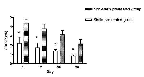
Expressions of CD62P between the patients with pre-existing statin use and without pre-existing statin use are shown in Figure 1. The percentage of platelets expressing CD62P was significantly lower in the patients with pre-existing statin used group on day 1 compared with those without pre-existing statin use. Although CD62P levels declined gradually in two groups, platelet CD62P levels were still significantly lower in patients with pre-existing statin use 90 days after the acute stroke (p < 0.05). Repeated analysis of variance (ANOVA) with Scheffe's multiple comparison showed that CD62P expressions between the two groups at four different time points (< 48 hours and on days 7, 30, and 90 post-stroke) were significantly different (p < 0.05).
Figure 1.
Serial changes of the percentages of CD62P expression in platelets between patients with pre-existing statin use and those without pre-existing statin use after acute ischemic stroke. *p < 0.05 patients with pre-existing statin use compared with patients without pre-existing statin use.
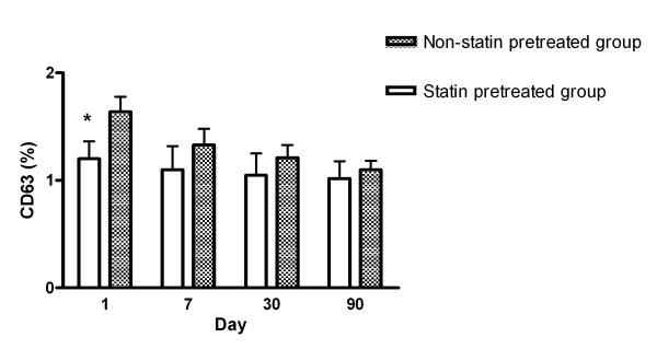
Expressions of CD63 between the patients with pre-existing statin use and those without pre-existing statin use are shown in Figure 2. The percentage of platelets expressing CD63 was significantly lower in patients with pre-existing statin use on day 1 than those without pre-existing statin use. However, there was no significant difference after day 7 post-stroke. Repeated measures comparison showed that CD63 expressions between the two groups at different time points were not different.
Figure 2.
Serial changes of the percentages of CD63 expression in platelets between patients with pre-existing statin use and those without pre-existing statin use after acute ischemic stroke. *p < 0.05 patients with pre-existing statin use compared with patients without pre-existing statin use.
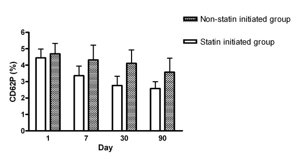
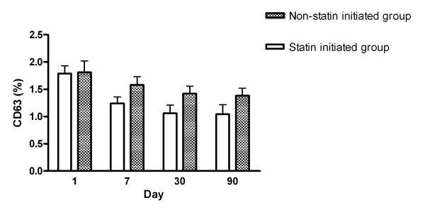
Expression of CD62P and CD63 between the statin-initiated and non-statin treatment groups are shown in Figures 3 and 4, respectively. The percentage of platelets expressing CD62P or CD63 was not different between the two sub-groups from days 1 to 90. Repeated measures comparison of CD62P or CD63 expressions at different time points were also not different between the two sub-groups.
Figure 3.
Serial changes of the percentages of CD62P expression in platelets between statin initiated group and non-statin treated group after acute ischemic stroke.
Figure 4.
Serial changes of the percentages of CD63 expression in platelets between statin initiated group and non-statin treated group after acute ischemic stroke.
Predictive factors for three-month outcome
Potential prognostic factors of the 172 acute stroke patients for three-month outcome are listed in Table 3. Although no patient died during the follow-up period, 133 had good outcomes and 39 had poor outcomes. Statistical analysis revealed that history of coronary artery diseases, NIHSS score on admission, pre-existing statin use, and platelet activation markers (CD62P and CD63) were significantly different between the good and poor outcome groups. Variables used in the stepwise logistic regression model included underlying age, gender, lipid profiles, coronary artery diseases, NIHSS score, pre-existing statin use, and CD62P and CD63 levels. After analysis, only NIHSS score (odds ratio (OR) = 1.10, 95% confidence interval (CI) = 1.03-1.17; P = 0.007), underlying coronary artery diseases (OR = 0.05, 95% CI = 0.01-0.38; P = 0.003), and pre-existing statin use (OR = 4.82, 95% CI = 1.22-19.03; P = 0.025) were independently associated with three-month outcome.
Table 3.
Prognostic factors of patients with acute ischemic stroke
| Good outcome (n = 133) | Poor outcome (n = 39) | P value | |
|---|---|---|---|
| Age (year) | |||
| Sex (female) (%) | 32.3 | 46.2 | 0.11 |
| Hypertension (%) | 77.4 | 84.6 | 0.33 |
| Diabetes mellitus (%) | 38.3 | 48.7 | 0.25 |
| Hyperlipidemia (%) | 50.4 | 38.5 | 0.19 |
| Coronary artery disease (%) | 1.5 | 12.8 | 0.002 |
| Intra-cranial atherosclerosis (%) | 23.3 | 30.8 | 0.34 |
| Stroke sub-type | |||
| Small vessel disease (%) | 57.1 | 48.7 | 0.24 |
| Large vessel disease (%) | 39.1 | 51.3 | |
| Statin therapy groups | |||
| Pre-existing statin use (%) | 29.3 | 10.3 | 0.016 |
| Median NIHSS (IQR) scores on admission | 3 (1-5) | 4 (3-8) | 0.009 |
| Laboratory data on admission | |||
| White blood cells (X103/ml) | 7.5 ± 0.2 | 7.6 ± 0.4 | 0.69 |
| Red blood cells (X106/ml) | 4.6 ± 0.1 | 4.6 ± 0.1 | 0.73 |
| Platelet counts (X104/ml) | 21.2 ± 0.6 | 19.5 ± 0.7 | 0.06 |
| Total cholesterol (mg/dl) | 183.0 ± 4.2 | 187.8 ± 8.5 | 0.61 |
| LDL-cholesterol (mg/dl) | 113.7 ± 3.7 | 114.3 ± 6.7 | 0.93 |
| Triglyceride (mg/dl) | 143.1 ± 7.8 | 147.2 ± 8.5 | 0.82 |
| HbA1c (%) | 7.0 ± 0.2 | 7.4 ± 0.4 | 0.33 |
| Systolic BP (mmHg) | 143.4 ± 1.8 | 140.6 ± 3.5 | 0.46 |
| Diastolic BP (mmHg) | 83.8 ± 1.1 | 82.8 ± 2.1 | 0.67 |
| Platelet markers on admission | |||
| CD62P (%)† | 3.45 ± 0.38 | 5.39 ± 0.89 | 0.024 |
| CD63 (%)† | 1.41 ± 0.12 | 1.99 ± 0.31 | 0.035 |
Good outcome, three-month mRS (0 to 2); poor outcome, three-month modified Rankin Scale (3 to 6).
BP, blood pressure; HbA1c, hemoglobin A1c; IQR, inter-quartile range; LDL, low density lipoprotein; NIHSS, National Institutes of Health Stroke Scale;
† The data was expressed as primitive data, however, the p value was obtained from logarithmically transformed of primitive data to improved normality.
Early neurologic deterioration and statin therapy
Of 172 acute ischemic patients, 14 suffered from END while hospitalized during the study period. Of the 14 END patients, four had hemorrhagic transformation, five had larger cerebral infarction, and five had increased of cerebral edema by follow-up brain CT. Patients with statin treatment after acute stroke (e.g. pre-existing statin use and statin-initiated groups) had significantly lower incidence of END (5 in 109 cases) compared with those without statin treatment (9 in 63 cases; p < 0.05). Furthermore, statin initiation after acute stroke also had significantly lower incidence of END (1 in 66 cases) compared with those without statin treatment (9 in 63 cases; P = 0.008).
Discussion
The present study examined the expression of serial platelet activation markers (CD62P and CD63) and the three-month outcome after acute non-cardio-embolic ischemic stroke and produced four major findings. First, the severity of the initial stroke (NIHSS score on admission) is relatively low in patients with statin treatment before stroke onset. Second, CD62P and CD63 expressions in platelets on admission are significantly lower in patients with pre-existing statin use than those without pre-existing statin use. Furthermore, CD62P expressions between groups at four different time points (< 48 hours and on days 7, 30, and 90) are significantly different (p < 0.05). Third, underlying coronary artery diseases, NIHSS score on admission, and pre-existing statin use are independently associated with three-month outcome. Finally, patients with statin treatment in the acute phase of stroke have significantly lower incidences of END compared with those without statin treatment.
In the current study, platelet activation levels differed between the statin groups. However, they did not significantly predict good outcome. In contrast, statin use or non-use did predict outcome. This suggests that either platelet activity detected in vitro by flow cytometry may not actually reflect the in vivo platelet effects of statins, or that statins improve outcome via other mechanisms. The mechanisms by which statins benefit patients with acute ischemic stroke remain unclear and are likely to be multi-factorial. Increasing evidence shows that statins have pleiotropic effects beyond their lipid-lowering effects [25]. Statins interfere with platelet aggregation and have anti-inflammatory, anti-oxidative, and anti-apoptotic properties [26-29]. Previous studies have demonstrated that platelet aggregation and leukocyte activity are significantly increased after ischemic stroke and contribute to the severity of brain damage [4,30]. The present study is the first to demonstrate that platelet activation markers (CD62P and CD63) are significantly inhibited in patients taking statins prior to an acute non-cardio-embolic ischemic stroke. These results are consistent with those of previous studies and further corroborate the anti-platelet effect of statins [31]. This anti-platelet effect may be via improved endothelial function and increased systemic nitric oxide bioavailability that inhibits exaggerated platelet activation [26].
There is mounting evidence that statin therapy may alter vascular atherosclerosis and reduce cardiovascular events, including myocardial infarction, stroke, and death [13,14,32]. The JUPITER study further demonstrates that rosuvastatin significantly reduces the incidence of major cardiovascular events in apparently healthy persons without hyperlipidemia but with elevated high-sensitivity C-reactive protein levels [33]. Furthermore, the abrupt withdrawal of statin therapy may increase the risk of cardiovascular events and death because of rebound of inflammatory response [23,34,35].
Recent studies also show that prior or early use of statins may reduce the severity and improve the outcome of myocardial infarction, ischemic stroke, and intra-cerebral hemorrhage [17-20]. Consistent with previous research, the current study reveals that pre-treatment with statins may be associated with reduced clinical severity and favorable three-month outcome in patients with acute ischemic stroke. Recent clinical research demonstrates that high CD62P and CD40L expressions on admission are strongly associated with poor clinical outcomes after acute ischemic stroke [36]. Therefore, stronger inhibition of platelet activity in the acute phase of ischemic stroke may be beneficial in improving clinical outcome.
The causes of END after acute ischemic stroke remain unknown but may be related to hemorrhagic transformation, progression of cerebral infarction, or cerebral edema. Previous researchers hypothesize that early leukocyte-endothelium interaction and substantial inflammatory reaction may play important roles in patients with stroke in progression [4,19]. In the current study, although the causes of END are heterogeneous between groups, statin treatment in the acute phase of stroke reduces the incidence of END compared with those without statin treatment after stroke. Because of the limitations of case number and study design, the role of statin therapy in END cannot be explained.
This study has several limitations. First, this is an observational study and it is possible that unmeasured or unknown confounders may have influenced the results. Second, although patients with prior anti-platelet therapy before acute stroke have been excluded, the expression of the platelet activation markers may be influenced by other drugs (e.g. beta-blocker, ACE inhibitors, and calcium channel blockers) that may cause potential bias in the statistical analysis [37]. Third, whether any particular statin or dosage has superior effects has not been tested because of the relatively small number of patients. Fourth, the baseline NIHSS is lower in the pre-existing statin use group. This may be based on very early effects of statins (such as increased perfusion and tissue salvage/collateral flow, which would be plausible if NIHSS measurement was performed at 12 hours after stroke onset) or may represent an imbalance in stroke severity between the two groups suggesting a bias of study design because it is not randomized. Fifth, patients with cardio-embolic stroke were excluded from this study according to medical history, ECG, and/or echocardiography. Thus, the effect of statin pre-treatment in patients with cardio-embolic stroke was unclear. Finally, this is a cohort study in a single-center with a relatively small patient number. Thus, it is possible that there is a smaller-than-expected magnitude of the protective effects exerted by statins. Randomized, larger-scale trials are warranted to establish whether early statin therapy has important beneficial effects in patients with acute ischemic stroke.
Conclusions
Statin therapy in acute non-cardio-embolic ischemic stroke can improve the three-month outcome and prevent END through potential anti-platelet effects. More prospective, longitudinal observational studies are warranted to evaluate the relation between dosage and choice of different statins in treating non-cardio-embolic stroke patients, to determine how to prevent END, and to improve neurologic outcome.
Key messages
1. Prior statin therapy is associated with reduced platelet activity in patients with acute ischemic stroke.
2. Pre-treatment with statins is associated with the severity of acute ischemic stroke.
3. Our study confirms previous studies demonstrating that pre-existing statin use is associated with improved clinical outcomes in acute ischemic stroke.
Abbreviations
ANOVA: analysis of variance; APTT: activated partial thromboplastin time; BI: Barthel index; CI: confidence interval; DM: diabetes mellitus; END: early neurologic deterioration; HbA1c: hemoglobin A1c; HDL: high density lipoprotein; HMG-CoA: 3-hydroxy 3-methyl-glutaryl coenzyme-A; LDL: low density lipoprotein; MRA: magnetic resonance angiography; MRI: magnetic resonance imaging; mRS: modified Rankin Scale; NIHSS: National Institutes of Health Stroke Scale; OR: odds ratio; PE: phycoerythrin; PT: prothrombin time; RBC: red blood cell; WBC: white blood cell.
Competing interests
The authors declare that they have no competing interests.
Contributor Information
Nai-Wen Tsai, Email: tsainw@yahoo.com.tw.
Tsu-Kung Lin, Email: tklin@adm.cgmh.org.tw.
Wen-Neng Chang, Email: cwenneng@ms19.hinet.net.
Chung-Ren Jan, Email: crjan@isca.vghks.gov.tw.
Chi-Ren Huang, Email: suika68@adm.cgmh.org.tw.
Shang-Der Chen, Email: chensd@adm.cgmh.org.tw.
Kuei-Yueh Cheng, Email: naiwen@adm.cgmh.org.tw.
Yi-Fang Chiang, Email: if_maymay@yahoo.com.tw.
Hung-Chen Wang, Email: m82whc@yahoo.com.tw.
Tzu-Ming Yang, Email: ma6046@gmail.com.
Yu-Jun Lin, Email: lin-yu-jun@yahoo.com.tw.
Wei-Che Lin, Email: u64lin@yahoo.com.tw.
Hsueh-Wen Chang, Email: tsainw@hotmail.com.
Lian-Hui Lee, Email: napaj@adm.cgmh.org.tw.
Cheng-Hsien Lu, Email: chlu99@ms44.url.com.tw.
Acknowledgements
This study was supported by grants from Chang Gung Memorial Hospital (Research Project CMRPG881011) and the National Science Council (Research Project NSC98-2314-B-182A-069).
Authors' contributions
NWT and HCW participated in the design of the study and drafted the manuscript. CRJ, KYC, and YFC carried out the flow cytometry study. TKL, CRH, and SDC participated in the sequence alignment and clinical evaluation of patients. WCL interpreted the neuro-imaging studies. HWC, TMY, and YJL performed the statistical analysis. CHL, LHL and WNC conceived the study, and participated in its design and coordination, and helped draft the manuscript. All authors read and approved the final manuscript.
References
- Suwanwela N, Koroshetz WJ. Acute ischemic stroke: overview of recent therapeutic developments. Annu Rev Med. 2007;58:89–106. doi: 10.1146/annurev.med.58.070605.115306. [DOI] [PubMed] [Google Scholar]
- Ruggeri ZM. Platelets in atherothrombosis. Nat Med. 2002;8:1227–1234. doi: 10.1038/nm1102-1227. [DOI] [PubMed] [Google Scholar]
- Huang J, Upadhyay UM, Tamargo RJ. Inflammation in stroke and focal cerebral ischemia. Surg Neurol. 2006;66:232–245. doi: 10.1016/j.surneu.2005.12.028. [DOI] [PubMed] [Google Scholar]
- Tsai NW, Chang WN, Shaw CF, Jan CR, Huang CR, Chen SD, Chuang YC, Lee LH, Lu CH. The value of leukocyte adhesion molecules in patients after ischemic stroke. J Neurol. 2009;256:1296–1302. doi: 10.1007/s00415-009-5117-3. [DOI] [PubMed] [Google Scholar]
- Marquardt L, Ruf A, Mansmann U, Winter R, Schuler M, Buggle F, Mayer H, Grau AJ. Course of platelet activation markers after ischemic stroke. Stroke. 2002;33:2570–2574. doi: 10.1161/01.STR.0000034398.34938.20. [DOI] [PubMed] [Google Scholar]
- Cha JK, Jeong MH, Jang JY, Bae HR, Lim YJ, Kim JS, Kim SH, Kim JW. Serial measurement of surface expressions of CD63, P-selectin and CD40 ligand on platelets in atherosclerotic ischemic stroke. A possible role of CD40 ligand on platelets in atherosclerotic ischemic stroke. Cerebrovasc Dis. 2003;16:376–382. doi: 10.1159/000072560. [DOI] [PubMed] [Google Scholar]
- Tsai NW, Chang WN, Shaw CF, Jan CR, Chang HW, Huang CR, Chen SD, Chuang YC, Lee LH, Lu CH. Serial change in platelet activation markers with aspirin and clopidogrel after acute ischemic stroke. Clin Neuropharmacol. 2010;33:40–45. doi: 10.1097/WNF.0b013e3181b8abc6. [DOI] [PubMed] [Google Scholar]
- Hankey GJ. Secondary prevention of recurrent stroke. Stroke. 2005;36:218–221. doi: 10.1161/01.STR.0000153048.87248.3b. [DOI] [PubMed] [Google Scholar]
- van der Worp HB, van Gijn J. Clinical practice. Acute ischemic stroke. N Engl J Med. 2007;357:572–579. doi: 10.1056/NEJMcp072057. [DOI] [PubMed] [Google Scholar]
- Goldstein LB, Adams R, Alberts MJ, Appel LJ, Brass LM, Bushnell CD, Culebras A, Degraba TJ, Gorelick PB, Guyton JR, Hart RG, Howard G, Kelly-Hayes M, Nixon JV, Sacco RL. Primary prevention of ischemic stroke: a guideline from the American Heart Association/American Stroke Association Stroke Council: cosponsored by the Atherosclerotic Peripheral Vascular Disease Interdisciplinary Working Group; Cardiovascular Nursing Council; Clinical Cardiology Council; Nutrition, Physical Activity, and Metabolism Council; and the Quality of Care and Outcomes Research Interdisciplinary Working Group: the American Academy of Neurology affirms the value of this guideline. Stroke. 2006;37:1583–1633. doi: 10.1161/01.STR.0000223048.70103.F1. [DOI] [PubMed] [Google Scholar]
- Alberts AW. Discovery, biochemistry and biology of lovastatin. Am J Cardiol. 1988;62:10J–15J. doi: 10.1016/0002-9149(88)90002-1. [DOI] [PubMed] [Google Scholar]
- Kwak BR, Mach F. Statins inhibit leukocyte recruitment: new evidence for their anti-inflammatory properties. Arterioscler Thromb Vasc Biol. 2001;21:1256–1258. [PubMed] [Google Scholar]
- Veillard NR, Mach F. Statins: the new aspirin? Cell Mol Life Sci. 2002;59:1771–1786. doi: 10.1007/PL00012505. [DOI] [PMC free article] [PubMed] [Google Scholar]
- Tonkin A, John R. Prevention of cardiovascular events and death with pravastatin in patients with coronary heart disease and a broad range of initial cholesterol levels. The Long-Term Intervention with Pravastatin in Ischaemic Disease (LIPID) Study Group. N Engl J Med. 1998;339:1349–1357. doi: 10.1056/NEJM199811053391902. [DOI] [PubMed] [Google Scholar]
- Moonis M, Kane K, Schwiderski U, Sandage BW, Fisher M. HMG-CoA reductase inhibitors improve acute ischemic stroke outcome. Stroke. 2005;36:1298–1300. doi: 10.1161/01.STR.0000165920.67784.58. [DOI] [PubMed] [Google Scholar]
- Amarenco P, Goldstein LB, Szarek M, Sillesen H, Rudolph AE, Callahan A, Hennerici M, Simunovic L, Zivin JA, Welch KM. Effects of intense low-density lipoprotein cholesterol reduction in patients with stroke or transient ischemic attack: the Stroke Prevention by Aggressive Reduction in Cholesterol Levels (SPARCL) trial. Stroke. 2007;38:3198–3204. doi: 10.1161/STROKEAHA.107.493106. [DOI] [PubMed] [Google Scholar]
- Fonarow GC, Wright RS, Spencer FA, Fredrick PD, Dong W, Every N, French WJ. Effect of statin use within the first 24 hours of admission for acute myocardial infarction on early morbidity and mortality. Am J Cardiol. 2005;96:611–616. doi: 10.1016/j.amjcard.2005.04.029. [DOI] [PubMed] [Google Scholar]
- Marti-Fabregas J, Gomis M, Arboix A, Aleu A, Pagonabarraga J, Belvis R, Cocho D, Roquer J, Rodriguez A, Garcia M, Molina-Porcel L, Díaz-Manera J, Martí-Vilalta J-L. Favorable outcome of ischemic stroke in patients pretreated with statins. Stroke. 2004;35:1117–1121. doi: 10.1161/01.STR.0000125863.93921.3f. [DOI] [PubMed] [Google Scholar]
- Greisenegger S, Mullner M, Tentschert S, Lang W, Lalouschek W. Effect of pretreatment with statins on the severity of acute ischemic cerebrovascular events. J Neurol Sci. 2004;221:5–10. doi: 10.1016/j.jns.2004.01.015. [DOI] [PubMed] [Google Scholar]
- Leker RR, Khoury ST, Rafaeli G, Shwartz R, Eichel R, Tanne D. Prior use of statins improves outcome in patients with intracerebral hemorrhage: prospective data from the National Acute Stroke Israeli Surveys (NASIS) Stroke. 2009;40:2581–2584. doi: 10.1161/STROKEAHA.108.546259. [DOI] [PubMed] [Google Scholar]
- Sacco RL, Adams R, Albers G, Alberts MJ, Benavente O, Furie K, Goldstein LB, Gorelick P, Halperin J, Harbaugh R, Johnston SC, Katzan I, Kelly-Hayes M, Kenton EJ, Marks M, Schwamm LH, Tomsick T. Guidelines for prevention of stroke in patients with ischemic stroke or transient ischemic attack: a statement for healthcare professionals from the American Heart Association/American Stroke Association Council on Stroke: co-sponsored by the Council on Cardiovascular Radiology and Intervention: the American Academy of Neurology affirms the value of this guideline. Stroke. 2006;37:577–617. doi: 10.1161/01.STR.0000199147.30016.74. [DOI] [PubMed] [Google Scholar]
- Adams HP Jr, Bendixen BH, Kappelle LJ, Biller J, Love BB, Gordon DL, Marsh EE. Classification of subtype of acute ischemic stroke. Definitions for use in a multicenter clinical trial. TOAST. Trial of Org 10172 in Acute Stroke Treatment. Stroke. 1993;24:35–41. doi: 10.1161/01.STR.24.1.35. [DOI] [PubMed] [Google Scholar]
- Blanco M, Nombela F, Castellanos M, Rodriguez-Yanez M, Garcia-Gil M, Leira R, Lizasoain I, Serena J, Vivancos J, Moro MA, Dávalos A, Castillo J. Statin treatment withdrawal in ischemic stroke: a controlled randomized study. Neurology. 2007;69:904–910. doi: 10.1212/01.wnl.0000269789.09277.47. [DOI] [PubMed] [Google Scholar]
- Lovblad KO, Baird AE. Computed tomography in acute ischemic stroke. Neuroradiology. 2010;52:175–187. doi: 10.1007/s00234-009-0633-5. [DOI] [PubMed] [Google Scholar]
- Koh KK. Effects of statins on vascular wall: vasomotor function, inflammation, and plaque stability. Cardiovasc Res. 2000;47:648–657. doi: 10.1016/S0008-6363(00)00146-2. [DOI] [PubMed] [Google Scholar]
- Schafer A, Fraccarollo D, Eigenthaler M, Tas P, Firnschild A, Frantz S, Ertl G, Bauersachs J. Rosuvastatin reduces platelet activation in heart failure: role of NO bioavailability. Arterioscler Thromb Vasc Biol. 2005;25:1071–1077. doi: 10.1161/01.ATV.0000161926.43967.df. [DOI] [PubMed] [Google Scholar]
- Puccetti L, Pasqui AL, Pastorelli M, Bova G, Cercignani M, Palazzuoli A, Angori P, Auteri A, Bruni F. Time-dependent effect of statins on platelet function in hypercholesterolaemia. Eur J Clin Invest. 2002;32:901–908. doi: 10.1046/j.1365-2362.2002.01086.x. [DOI] [PubMed] [Google Scholar]
- Lefer DJ. Statins as potent antiinflammatory drugs. Circulation. 2002;106:2041–2042. doi: 10.1161/01.CIR.0000033635.42612.88. [DOI] [PubMed] [Google Scholar]
- Laufs U, Gertz K, Dirnagl U, Bohm M, Nickenig G, Endres M. Rosuvastatin, a new HMG-CoA reductase inhibitor, upregulates endothelial nitric oxide synthase and protects from ischemic stroke in mice. Brain Res. 2002;942:23–30. doi: 10.1016/S0006-8993(02)02649-5. [DOI] [PubMed] [Google Scholar]
- Htun P, Fateh-Moghadam S, Tomandl B, Handschu R, Klinger K, Stellos K, Garlichs C, Daniel W, Gawaz M. Course of platelet activation and platelet-leukocyte interaction in cerebrovascular ischemia. Stroke. 2006;37:2283–2287. doi: 10.1161/01.STR.0000236638.75591.61. [DOI] [PubMed] [Google Scholar]
- Cipollone F, Mezzetti A, Porreca E, Di Febbo C, Nutini M, Fazia M, Falco A, Cuccurullo F, Davi G. Association between enhanced soluble CD40L and prothrombotic state in hypercholesterolemia: effects of statin therapy. Circulation. 2002;106:399–402. doi: 10.1161/01.CIR.0000025419.95769.F0. [DOI] [PubMed] [Google Scholar]
- Crouse JR, Byington RP, Bond MG, Espeland MA, Craven TE, Sprinkle JW, McGovern ME, Furberg CD. Pravastatin, Lipids, and Atherosclerosis in the Carotid Arteries (PLAC-II) Am J Cardiol. 1995;75:455–459. doi: 10.1016/S0002-9149(99)80580-3. [DOI] [PubMed] [Google Scholar]
- Ridker PM, Danielson E, Fonseca FA, Genest J, Gotto AM Jr, Kastelein JJ, Koenig W, Libby P, Lorenzatti AJ, MacFadyen JG, Nordestgaard BG, Shepherd J, Willerson JT, Glynn RJ. Rosuvastatin to prevent vascular events in men and women with elevated C-reactive protein. N Engl J Med. 2008;359:2195–2207. doi: 10.1056/NEJMoa0807646. [DOI] [PubMed] [Google Scholar]
- Gertz K, Laufs U, Lindauer U, Nickenig G, Bohm M, Dirnagl U, Endres M. Withdrawal of statin treatment abrogates stroke protection in mice. Stroke. 2003;34:551–557. doi: 10.1161/01.STR.0000054055.28435.BF. [DOI] [PubMed] [Google Scholar]
- Li JJ, Li YS, Chen J, Yang JQ. Rebound phenomenon of inflammatory response may be a major mechanism responsible for increased cardiovascular events after abrupt cessation of statin therapy. Med Hypotheses. 2006;66:1199–1204. doi: 10.1016/j.mehy.2005.06.035. [DOI] [PubMed] [Google Scholar]
- Yip HK, Liou CW, Chang HW, Lan MY, Liu JS, Chen MC. Link between platelet activity and outcomes after an ischemic stroke. Cerebrovasc Dis. 2005;20:120–128. doi: 10.1159/000086802. [DOI] [PubMed] [Google Scholar]
- Spencer CG, Gurney D, Blann AD, Beevers DG, Lip GY. Von Willebrand factor, soluble P-selectin, and target organ damage in hypertension: a substudy of the Anglo-Scandinavian Cardiac Outcomes Trial (ASCOT) Hypertension. 2002;40:61–66. doi: 10.1161/01.HYP.0000022061.12297.2E. [DOI] [PubMed] [Google Scholar]


