Abstract
The use of vital blue dyes in sentinel lymph node mapping and biopsy is gaining popularity in the surgical management of cancer. However, intraoperative use of these dyes has associated risks for the patient. This case report and review of the literature present current medical knowledge about one of the vital blue dyes, isosulfan blue, and the associated clinical risks of this dye when used in the perioperative management of patients who undergo sentinel lymph node mapping and biopsy.
Keywords: Anaphylaxis, cancer, isosulfan blue, methemoglobinemia, pulse oximetry, vital dyes
INTRODUCTION
The vital blue dye isosulfan blue (CAS No. 68238-36-8; Lymphazurin 1%) is a Food and Drug Administration–approved dye for sentinel lymph node mapping.1-5 When subcutaneously administered, the dye travels within the afferent lymphatics to assist the surgeon in the identification of the primary or sentinel lymph node.5 Although the use of vital dyes such as isosulfan blue allows surgeons to avoid nontherapeutic lymph node dissection, adverse reactions ranging from mild allergic reactions to anaphylaxis have been reported in the medical literature.6-9
Isosulfan blue is a member of a family of triphenylmethane-based dyes. These dyes are chemically similar to commonly used industrial dyes in household items such as paper, textiles, detergents, and cosmetics.10 Prior sensitization through widespread exposure from common household items containing these dyes is the explanation for an immunoglobulin E–mediated hypersensitivity reaction, with a reported incidence of 0.1%-2%.7,10-18 Other known complications of isosulfan blue are transient discoloration of the skin (blue urticaria), interference with pulse oximetry saturation readings (factitious hypoxia), or development of methemoglobinemia.8,19-24 The following report describes 2 clinical events involving isosulfan blue at our institution.
CASE 1
An 83-year-old woman was scheduled for vulvectomy with sentinel node biopsy for invasive squamous cell carcinoma of the vulva. Her past medical history was significant for anxiety, essential hypertension, and hypothyroidism. A positron emission tomography scan to determine the spread and distribution of the vulvar carcinoma also identified a left adrenal nodule and bilateral thyroid nodules. During the preoperative assessment, elevated norepinephrine and normetanephrine values were also present. However, the patient displayed no active signs or symptoms consistent with a diagnosis of pheochromocytoma, such as headache, palpitations, sweating, or pallor. She had no other significant past medical or surgical history, and her family history was negative for the presence of adrenal tumors, thyroid cancer, or genetically inherited syndromes. Her home medications included metoprolol, levothyroxine, and rabeprazole. Although the patient reported gastrointestinal irritation following aspirin ingestion, she denied any medication allergies or food sensitivities.
Computed tomography of the chest, electrocardiogram, and noninvasive renal and carotid studies were all within normal limits. Nevertheless, she was admitted 4 days prior to her scheduled surgical procedure for inpatient management of suspected pheochromocytoma. Treatment included hydration and administration of the long-acting alpha-antagonist, doxazosin.
On the morning of surgery, the patient was sedated and underwent central venous and radial artery cannulations. Following placement of these invasive monitors, the patient underwent a titrated general anesthetic and orotracheal intubation without incident. After the patient was placed in the lithotomy position, surgery commenced with an intradermal injection of 4 mL of isosulfan blue in the vulva lateral to the squamous cell carcinoma. For the duration of the case, the patient remained hemodynamically stable with intermittent intravenous nitroglycerin titrated for blood pressure control. Pulse oximetry saturation readings were stable (98% to 100%) during vulvectomy with sentinel node biopsy.
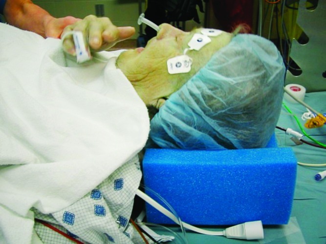
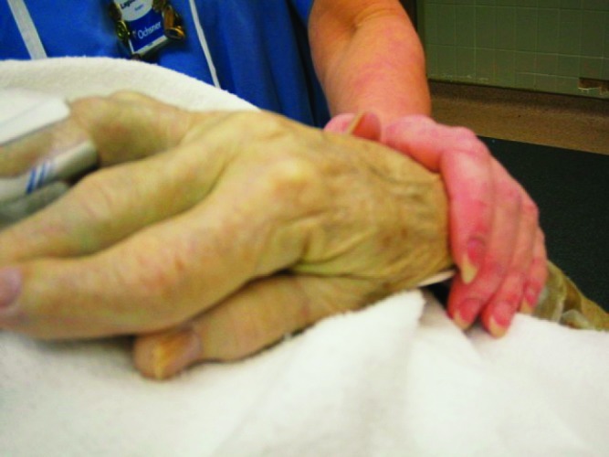
Upon completion of surgery and while the patient was returned to the supine position, an abrupt decrease in pulse oximetry saturation (94%) occurred with the development of a marked blue-green hue on the skin of the upper extremities, chest, and face (blue urticaria) (Figure 1). As 100% oxygen was administered to the patient, as confirmed by the flow-meter bobbins and the oxygen analyzer, attention turned to the anesthetic circuit, which was determined to be patent, and bilateral breath sounds were auscultated from the chest. Following emergence from general anesthesia, the patient was following appropriate verbal commands with extubation of the trachea. Arterial blood gas (ABG) analysis by the point-of-care i-STAT 1 (Abbott Point of Care, Princeton, NJ) demonstrated acceptable partial pressure gas measurements without evidence of acidosis or hypoxia (factitious hypoxia) (Table). Upon transfer to the postanesthesia care unit (PACU), a detailed report about the clinical assessment of the blue urticaria and associated factitious hypoxia was reported to PACU staff, the patient, and the patient's family (Figure 2).
Figure 1.

Discoloration of skin following successful emergence from general anesthesia with the patient awake and following commands.
Figure 2.

A comparison of skin color prior to transport to the postanesthesia care unit.
Because the i-STAT 1 point-of-care ABG analyzer does not measure, but rather calculates hemoglobin saturation, respiratory therapy analyzed an arterial blood sample with a co-oximeter blood gas analyzer (NPT7, Radiometer America, Westlake, OH) and recorded a normal oxyhemoglobin saturation without abnormal elevations in methemoglobin or carboxyhemoglobin levels, which confirmed the clinical diagnosis of factitious hypoxia (Table).
Given these normal laboratory values and the absence of any clinical indicators of cardiovascular or respiratory compromise in the PACU, the decrease in the intraoperative pulse oximetry saturation readings (factitious hypoxia) and the presence of the blue urticaria were attributed to systemic absorption of the isosulfan blue dye. However, because of the intensity of the blue urticaria (Figures 1 and 2), the patient was further monitored in the PACU for 24 hours and then transferred to a regular postsurgical floor. Over the course of the next 2 days, skin color returned to normal and the patient was discharged on postoperative day 3 with no clinical signs of blue urticaria.
CASE 2
A 62-year-old female was diagnosed with left breast cancer after a screening mammogram found a focal asymmetric density. Core biopsy revealed a well-differentiated invasive infiltrating ductal carcinoma that measured approximately 6 mm on diagnostic mammogram. The patient decided after extensive counseling to proceed with breast-conserving therapy and was scheduled for a left breast wire localization lumpectomy with sentinel lymph node biopsy and possible axillary lymph node dissection. The patient had a medical history of asthma, obesity, and a sulfur allergy. Her surgical history included a childhood appendectomy, cesarean section, and dilation and curettage. She denied any adverse events with anesthesia in her past procedures and had no family history of anesthetic complications.
Following the induction of general anesthesia, a laryngeal mask airway was inserted to maintain a patent upper airway. Approximately 15 minutes following anesthetic induction, the surgical team injected 0.45 mCi of technetium-labeled sulfur colloid into the left breast along with 5 mL of isosulfan blue dye for further sentinel lymph node identification. Within minutes, the patient developed marked hypotension and bradycardia that responded to intravenous injections of ephedrine, diphenhydramine, dexamethasone, and fluids. The hemodynamic instability corrected within 5 minutes, and surgeons proceeded with the case. After 23 hours of observation in the PACU, the patient was discharged without further complications.
DISCUSSION
Intradermal and parenchymal injections of vital blue dyes, such as isosulfan blue, can lead to systemic absorption through lymphatic channels and vascular beds near tumor sites.5,25,26 The numerous adverse events reported from isosulfan blue and the structurally related patent blue are thought to arise from circulating complexes of protein-bound dye molecules.21,27,28 The consequences of systemic circulation include reports of interference with pulse oximetry readings, the development of blue urticaria, and anaphylaxis.6,9,11,18,19,29-32
In patients such as described in Case 1, the first priority should be to rule out causes of real arterial hypoxemia by checking oxygen delivery to the patient. Next, confirm the integrity of the anesthetic breathing circuit by switching to manual ventilation and observing the movements of the chest and the circuit system flow valves.21 Inadvertent endobronchial intubation should be ruled out by checking for the presence of bilateral breath sounds to confirm that the endotracheal tube is still appropriately positioned following changes in surgical positioning of the patient. Although vital blue dyes have been reported to generate erroneous pulse oximetry readings (factitious hypoxia), an arterial blood sample for co-oximetry analysis should be obtained to determine if the declines in pulse oximetry are clinically relevant or factitious.22,24,30,33
Although case reports and large retrospective studies have reported decreases in pulse oximetry readings following injections of the vital dye isosulfan blue,22,23,29,32,34,35 other studies have evaluated the magnitude and duration of the effect of isosulfan blue on pulse oximetry readings.24,31 In a study of 33 patients undergoing isosulfan blue administration for sentinel lymph node biopsy, all patients experienced peak decreases in pulse oximetry saturations within 30 minutes, but no adverse events were reported.31
A second study compared the effects of 2 vital blue dyes, patent blue and isosulfan blue, on pulse oximetry saturation readings, oxygen saturation, and partial oxygen pressure values in 16 patients undergoing sentinel lymph node biopsy. Although the 2 dyes interfered with pulse oximetry saturation readings, only isosulfan blue demonstrated significant differences in pulse oximetry saturation values when compared to control values.24 However, these pulse oximetry desaturations were not accompanied by real hemoglobin desaturations when determined by blood-gas analysis. In this study, peak interference of the dye was observed 15 minutes after injection.24
Pulse Oximetry
The sensor used in pulse oximetry emits both red and infrared light at a wavelength between 660 and 910 nm.36,37 The blood partially absorbs the transmitted light and the sensor absorbs the remainder, during which time the transmitted signal is analyzed to calculate peripheral arterial saturation based on the difference.36,37 Because isosulfan blue dye has a peak absorption wavelength of 638 nm, which is similar to reduced hemoglobin, the dye interferes with the sensor, leading to overestimation of hemoglobin desaturation.24,31
Multiple investigations comparing ABG analysis with pulse oximetry saturations have shown that declines in pulse oximetry saturation following vital dye administrations are factitious and not clinically relevant.24,31,34 However, blood gas analysis with co-oximetry should be strongly considered in cases of hemodynamic instability to confirm adequate oxyhemoglobin content and to rule out elevated levels of methemoglobin that can occur following co-administration of ester-local anesthetics.22,38-41 In contrast to the many commercial blood gas analyzers that calculate oxygen content, a co-oximeter accurately measures all hemoglobin moieties.42-45 Although pulse oximetry desaturation in the presence of isosulfan blue can be profound (SpO2 89%-90%),32,34 in the majority of cases, the pulse oximetry desaturation would not be interpreted as clinically significant (SpO2 > 90%).24,31,34
Anaphylactic reaction to the vital dye, or possibly the sulfur colloid, occurred in Case 2. Anaphylactic-like reactions to the vital dye have been described in the literature and carry a reported incidence ranging from 0.1% to 2%.7,10-18 The largest available series using isosulfan blue in 4,975 patients reported an incidence of anaphylaxis of 0.1%.16 Although anaphylactic reactions to the vital blue dyes have been known, the first case of an anaphylactic reaction to isosulfan blue was reported in 1985.9 Since that time, a number of allergic reactions, including shock, have been reported.7,8,11-16,46-48 The hemodynamic instability in Case 2 quickly resolved following administration of antihistamines, steroids, vasopressor support, and fluids, and this therapy is similar to what has been reported in other similar case studies.12,13,15,20
However in some patients, persistent systemic hypotension can occur despite administration of large doses of vasopressors.7,49 For those patients, it may be prudent to obtain oxyhemoglobin and methemoglobin values with a co-oximeter and serum tryptase assays and/or skin testing.50,51 Finally, in a study of 639 patients who underwent sentinel lymph node biopsy for breast cancer using isosulfan blue, 7 anaphylactic reactions were reported, of which 2 reactions were biphasic in nature.47 In those 2 patients, recurrences of anaphylaxis appeared during postoperative monitoring (6 and 8 hours after surgery), and both responded well to antiallergic treatments.47
In an attempt to reduce the incidence and severity of anaphylactic reactions to isosulfan blue, the benefits of prophylactic therapy were studied in 1,013 consecutive patients who underwent sentinel lymphadenectomy for breast carcinoma.15 A total of 667 patients (65.8%) received prophylactic therapy involving the administration of steroids (100 mg of hydrocortisone, or 20 mg of methylprednisolone, or 4 mg of dexamethasone), 50 mg of diphenhydramine, and 20 mg of famotidine before or at the induction of general anesthesia and prior to isosulfan blue dye injection. Thirty-three patients (3.3%) received prophylaxis but no dye injections. Twelve patients (1.2%) received dye injections but no prophylactic therapy, and the remaining 301 patients (29.7%) received no prophylactic therapy or dye. Blue urticaria and facial edema were observed in 3 of 667 patients (0.4%) receiving the prophylaxis regimen and in 1 of 12 patients (8.3%) receiving the dye but no prophylaxis. No episodes of hypotension occurred, and no patients required vasopressors, ventilatory support, or intensive care observation.
Other interesting findings in this study were the incidence of adverse reactions to agents other than the blue dye and the presence of increased wound complications.15 Two patients developed anaphylaxis out of 667 patients (0.3%) who received prophylaxis and dye, and 3 patients developed anaphylaxis out of 301 patients (1.0%) who did not receive prophylactic therapy or the dye. Although the findings are clinically relevant, they were not statistically significant. Finally, wound healing complication rates doubled but did not reach statistical significance. The findings that prophylactic therapy did not decrease the incidence of anaphylaxis to isosulfan blue, but did decrease the morbidity associated with the dye, suggest that routine prophylaxis should be considered in patients receiving isosulfan blue for lymphatic mapping and sentinel lymph node biopsy.15
CONCLUSION
The use of vital blue dyes such as isosulfan blue for sentinel lymph node mapping and biopsy carries the risks of skin pigmentation changes, interference in pulse oximetry saturation readings, and anaphylaxis in a small number of cases. One should expect a decrease in pulse oximetry saturation readings with isosulfan blue, but in the majority of these cases, the changes in pulse oximetry saturation readings will not be clinically significant. However, when conducting ABG analysis, one should use a co-oximeter to accurately measure the oxyhemoglobin content of the blood and minimize the diagnosis of factitious hypoxia. A high index of suspicion and appropriate clinical management will minimize the potential morbidity when using these vital blue dyes in the perioperative management of patients undergoing sentinel lymph node mapping and biopsy.
Table.
Serial Arterial Blood Gas Assessments in a Patient Following Lymphatic Injection of the Vital Blue Dye, Isosulfan Blue (CAS No. 68238-36-8; Lymphazurin 1%)
Footnotes
1Dr Haque is now with Southeast Anesthesiology Consultants, Charlotte, NC.
The authors have no financial or proprietary interest in the subject matter of this article.
This article meets the Accreditation Council for Graduate Medical Education and the American Board of Medical Specialties Maintenance of Certification competencies for Patient Care and Medical Knowledge.
REFERENCES
- 1.Echt ML, Finan MA, Hoffman MS, Kline RC, Roberts WS, Fiorica JV. Detection of sentinel lymph nodes with lymphazurin in cervical, uterine, and vulvar malignancies. South Med J. 1999 Feb;92(2):204–208. doi: 10.1097/00007611-199902000-00008. [DOI] [PubMed] [Google Scholar]
- 2.O'Boyle JD, Coleman RL, Bernstein SG, Lifshitz S, Muller CY, Miller DS. Intraoperative lymphatic mapping in cervix cancer patients undergoing radical hysterectomy: A pilot study. Gynecol Oncol. 2000 Nov;79(2):238–243. doi: 10.1006/gyno.2000.5930. [DOI] [PubMed] [Google Scholar]
- 3.Plante M, Renaud MC, Roy M. Sentinel node evaluation in gynecologic cancer. Oncology (Williston Park) 2004 Jan;1888-90(1):75–87. 95–96. discussion. [PubMed] [Google Scholar]
- 4.Levenback C, Burke TW, Gershenson DM, Morris M, Malpica A, Ross MI. Intraoperative lymphatic mapping for vulvar cancer. Obstet Gynecol. 1994 Aug;84(2):163–167. [PubMed] [Google Scholar]
- 5.Hirsch JI, Tisnado J, Cho SR, Beachley MC. Use of isosulfan blue for identification of lymphatic vessels: experimental and clinical evaluation. AJR Am J Roentgenol. 1982 Dec;139(6):1061–1064. doi: 10.2214/ajr.139.6.1061. [DOI] [PubMed] [Google Scholar]
- 6.Laurie SA, Khan DA, Gruchalla RS, Peters G. Anaphylaxis to isosulfan blue. Ann Allergy Asthma Immunol. 2002 Jan;88(1):64–66. doi: 10.1016/S1081-1206(10)63595-8. [DOI] [PubMed] [Google Scholar]
- 7.Cimmino VM, Brown AC, Szocik JF, et al. Allergic reactions to isosulfan blue during sentinel node biopsy—a common event. Surgery. 2001 Sep;130(3):439–442. doi: 10.1067/msy.2001.116407. [DOI] [PubMed] [Google Scholar]
- 8.Leong SP, Donegan E, Heffernon W, Dean S, Katz JA. Adverse reactions to isosulfan blue during selective sentinel lymph node dissection in melanoma. Ann Surg Oncol. 2000 Jun;7(5):361–366. doi: 10.1007/s10434-000-0361-x. [DOI] [PubMed] [Google Scholar]
- 9.Longnecker SM, Guzzardo MM, Van Voris LP. Life-threatening anaphylaxis following subcutaneous administration of isosulfan blue 1% Clin Pharm. 1985 Mar-Apr;4(2):219–221. [PubMed] [Google Scholar]
- 10.Thevarajah S, Huston TL, Simmons RM. A comparison of the adverse reactions associated with isosulfan blue versus methylene blue dye in sentinel lymph node biopsy for breast cancer. Am J Surg. 2005 Feb;189(2):236–239. doi: 10.1016/j.amjsurg.2004.06.042. [DOI] [PubMed] [Google Scholar]
- 11.Kaufman G, Guth AA, Pachter HL, Roses DF. A cautionary tale: anaphylaxis to isosulfan blue dye after 12 years and 3339 cases of lymphatic mapping. Am Surg. 2008 Feb;74(2):152–155. [PubMed] [Google Scholar]
- 12.Komenaka IK, Bauer VP, Schnabel FR, et al. Allergic reactions to isosulfan blue in sentinel lymph node mapping. Breast J. 2005 Jan-Feb;11(1):70–72. doi: 10.1111/j.1075-122X.2005.21574.x. [DOI] [PubMed] [Google Scholar]
- 13.Montgomery LL, Thorne AC, Van Zee KJ, et al. Isosulfan blue dye reactions during sentinel lymph node mapping for breast cancer. Anesth Analg. 2002 Aug;95(2):385–388. doi: 10.1097/00000539-200208000-00026. [DOI] [PubMed] [Google Scholar]
- 14.Raut CP, Daley MD, Hunt KK, et al. Anaphylactoid reactions to isosulfan blue dye during breast cancer lymphatic mapping in patients given preoperative prophylaxis. J Clin Oncol. 2004 Feb 1;22(3):567–568. doi: 10.1200/JCO.2004.99.276. [DOI] [PubMed] [Google Scholar]
- 15.Raut CP, Hunt KK, Akins JS, et al. Incidence of anaphylactoid reactions to isosulfan blue dye during breast carcinoma lymphatic mapping in patients treated with preoperative prophylaxis: results of a surgical prospective clinical practice protocol. Cancer. 2005 Aug 15;104(4):692–699. doi: 10.1002/cncr.21226. [DOI] [PubMed] [Google Scholar]
- 16.Golshan M, Nakhlis F. Can methylene blue only be used in sentinel lymph node biopsy for breast cancer? Breast J. 2006 Sep-Oct;12(5):428–430. doi: 10.1111/j.1075-122X.2006.00299.x. [DOI] [PubMed] [Google Scholar]
- 17.Scherer K, Studer W, Figueiredo V, Bircher AJ. Anaphylaxis to isosulfan blue and cross-reactivity to patent blue V: case report and review of the nomenclature of vital blue dyes. Ann Allergy Asthma Immunol. 2006 Mar;96(3):497–500. doi: 10.1016/S1081-1206(10)60921-0. [DOI] [PubMed] [Google Scholar]
- 18.Sandhu S, Farag E, Argalious M. Anaphylaxis to isosulfan blue dye during sentinel lymph node biopsy. J Clin Anesth. 2005 Dec;17(8):633–635. doi: 10.1016/j.jclinane.2005.03.006. [DOI] [PubMed] [Google Scholar]
- 19.Momeni R, Ariyan S. Pulse oximetry declines due to intradermal isosulfan blue dye: a controlled prospective study. Ann Surg Oncol. 2004 Apr;11(4):434–437. doi: 10.1245/ASO.2004.05.015. [DOI] [PubMed] [Google Scholar]
- 20.Daley MD, Norman PH, Leak JA, et al. Adverse events associated with the intraoperative injection of isosulfan blue. J Clin Anesth. 2004 Aug;16(5):332–341. doi: 10.1016/j.jclinane.2003.09.013. [DOI] [PubMed] [Google Scholar]
- 21.Hoskin RW, Granger R. Intraoperative decrease in pulse oximeter readings following injection of isosulfan blue. Can J Anaesth. 2001 Jan;48(1):38–40. doi: 10.1007/BF03019812. [DOI] [PubMed] [Google Scholar]
- 22.Burgoyne LL, Jay DW, Bikhazi GB, De Armendi AJ. Isosulfan blue causes factitious methemoglobinemia in an infant. Paediatr Anaesth. 2005 Dec;15(12):1116–1119. doi: 10.1111/j.1460-9592.2005.01578.x. [DOI] [PubMed] [Google Scholar]
- 23.Wear KD, Karsif K, Turner J. Staining of endotracheal tube with isosulfan blue dye after sentinel node mapping: a case report. Breast J. 2003 Jan-Feb;9(1):47–48. doi: 10.1046/j.1524-4741.2003.09116.x. [DOI] [PubMed] [Google Scholar]
- 24.Piñero A, Illana J, García-Palenciano C, et al. Effect on oximetry of dyes used for sentinel lymph node biopsy. Arch Surg. 2004 Nov;139(11):1204–1207. doi: 10.1001/archsurg.139.11.1204. [DOI] [PubMed] [Google Scholar]
- 25.Steele SR, Martin MJ, Mullenix PS, Olsen SB, Andersen CA. Intraoperative use of isosulfan blue in the treatment of persistent lymphatic leaks. Am J Surg. 2003 Jul;186(1):9–12. doi: 10.1016/s0002-9610(03)00113-2. [DOI] [PubMed] [Google Scholar]
- 26.Hill AD, Tran KN, Akhurst T, et al. Lessons learned from 500 cases of lymphatic mapping for breast cancer. Ann Surg. 1999 Apr;229(4):528–535. doi: 10.1097/00000658-199904000-00012. [DOI] [PMC free article] [PubMed] [Google Scholar]
- 27.Kieckbusch H, Coldewey SM, Hollenhorst J, Haeseler G, Hillemanns P, Hertel H. Patent blue sentinel node mapping in cervical cancer patients may lead to decreased pulse oximeter readings and positive methaemoglobin results. Eur J Anaesthesiol. 2008 May;25(5):365–368. doi: 10.1017/S0265021508003578. Epub 2008 Feb 13. [DOI] [PubMed] [Google Scholar]
- 28.Chia YY, Liu K, Kao PF, Sun GC, Wang KY. Prolonged interference of patent blue on pulse oximetry readings. Acta Anaesthesiol Sin. 2001 Mar;39(1):27–32. [PubMed] [Google Scholar]
- 29.Wisely NA, Zeiton Z. Use of isosulfan blue in breast surgery interferes with pulse oximetry. Anaesthesia. 2005 Jun;60(6):625–626. doi: 10.1111/j.1365-2044.2005.04247.x. [DOI] [PubMed] [Google Scholar]
- 30.Heinle E, Burdumy T, Recabaren J. Factitious oxygen desaturation after isosulfan blue injection. Am Surg. 2003 Oct;69(10):899–901. [PubMed] [Google Scholar]
- 31.Vokach-Brodsky L, Jeffrey SS, Lemmens HJ, Brock-Utne JG. Isosulfan blue affects pulse oximetry. Anesthesiology. 2000 Oct;93(4):1002–1003. doi: 10.1097/00000542-200010000-00022. [DOI] [PubMed] [Google Scholar]
- 32.Coleman RL, Whitten CW, O'Boyle J, Sidhu B. Unexplained decrease in measured oxygen saturation by pulse oximetry following injection of Lymphazurin 1% (isosulfan blue) during a lymphatic mapping procedure. J Surg Oncol. 1999 Feb;70(2):126–129. doi: 10.1002/(sici)1096-9098(199902)70:2<126::aid-jso12>3.0.co;2-p. [DOI] [PubMed] [Google Scholar]
- 33.Rizzi RR, Thomas K, Pilnik S. Factious desaturation due to isosulfan dye injection. Anesthesiology. 2000 Oct;93(4):1146–1147. doi: 10.1097/00000542-200010000-00042. [DOI] [PubMed] [Google Scholar]
- 34.El-Tamer M, Komenaka IK, Curry S, Troxel AB, Ditkoff BA, Schnabel FR. Pulse oximeter changes with sentinel lymph node biopsy in breast cancer. Arch Surg. 2003 Nov;138(11):1257–1260. doi: 10.1001/archsurg.138.11.1257. [DOI] [PubMed] [Google Scholar]
- 35.Mateo D, Suescun MC, Cahisa M, Ruiz P, García M, Miranda L. [Decreasing oxygen saturation detected by pulse oximetry after the administration of isosulfan blue] Rev Esp Anestesiol Reanim. 2002 Feb;49(2):114–115. Spanish. [PubMed] [Google Scholar]
- 36.Ralston AC, Webb RK, Runciman WB. Potential errors in pulse oximetry. III: effects of interferences, dyes, dyshaemoglobins and other pigments. Anaesthesia. 1991 Apr;46(4):291–295. doi: 10.1111/j.1365-2044.1991.tb11501.x. [DOI] [PubMed] [Google Scholar]
- 37.Schnapp LM, Cohen NH. Pulse oximetry. Uses and abuses. Chest. 1990 Nov;98(5):1244–1250. doi: 10.1378/chest.98.5.1244. [DOI] [PubMed] [Google Scholar]
- 38.Guay J. Methemoglobinemia related to local anesthetics: a summary of 242 episodes. Anesth Analg. 2009 Mar;108(3):837–845. doi: 10.1213/ane.0b013e318187c4b1. [DOI] [PubMed] [Google Scholar]
- 39.Young B. Intraoperative detection of methemoglobinemia in a patient given benzocaine spray to relieve discomfort from a nasogastric tube: a case report. AANA J. 2008 Apr;76(2):99–102. [PubMed] [Google Scholar]
- 40.Kwok S, Fischer JL, Rogers JD. Benzocaine and lidocaine induced methemoglobinemia after bronchoscopy: a case report. J Med Case Reports. 2008 Jan 23;2:16. doi: 10.1186/1752-1947-2-16. [DOI] [PMC free article] [PubMed] [Google Scholar]
- 41.Wurdeman RL, Mohiuddin SM, Holmberg MJ, Shalaby A. Benzocaine-induced methemoglobinemia during an outpatient procedure. Pharmacotherapy. 2000 Jun;20(6):735–738. doi: 10.1592/phco.20.7.735.35175. [DOI] [PubMed] [Google Scholar]
- 42.Mack E. Focus on diagnosis: co-oximetry. Pediatr Rev. 2007 Feb;28(2):73–74. doi: 10.1542/pir.28-2-73. [DOI] [PubMed] [Google Scholar]
- 43.Haymond S, Cariappa R, Eby CS, Scott MG. Laboratory assessment of oxygenation in methemoglobinemia. Clin Chem. 2005 Feb;51(2):434–444. doi: 10.1373/clinchem.2004.035154. Epub 2004 Oct 28. [DOI] [PubMed] [Google Scholar]
- 44.Mathews PJ., Jr Co-oximetry. Respir Care Clin N Am. 1995 Sep;1(1):47–68. [PubMed] [Google Scholar]
- 45.García Carmona T, Domíneguez De Villota E, Mosquera Gonzalez M, Muedra Lopez A, Ruíz De Andrés S, Estada Girauta J. [P 50: Comparison between the values based on calculated saturations from oxygen contents (Van Slyke) and on optically measured saturations (Co-Oximetry)] Rev Clin Esp. 1975 Jul 31;138(2):149–153. Spanish. [PubMed] [Google Scholar]
- 46.Soni M, Saha S, Korant A, et al. A prospective trial comparing 1% lymphazurin vs 1% methylene blue in sentinel lymph node mapping of gastrointestinal tumors. Ann Surg Oncol. 2009 Aug;16(8):2224–2230. doi: 10.1245/s10434-009-0529-y. Epub 2009 May 30. [DOI] [PubMed] [Google Scholar]
- 47.Albo D, Wayne JD, Hunt KK, et al. Anaphylactic reactions to isosulfan blue dye during sentinel lymph node biopsy for breast cancer. Am J Surg. 2001 Oct;182(4):393–398. doi: 10.1016/s0002-9610(01)00734-6. [DOI] [PubMed] [Google Scholar]
- 48.Lyew MA, Gamblin TC, Ayoub M. Systemic anaphylaxis associated with intramammary isosulfan blue injection used for sentinel node detection under general anesthesia. Anesthesiology. 2000 Oct;93(4):1145–1146. doi: 10.1097/00000542-200010000-00041. [DOI] [PubMed] [Google Scholar]
- 49.Sprung J, Tully MJ, Ziser A. Anaphylactic reactions to isosulfan blue dye during sentinel node lymphadenectomy for breast cancer. Anesth Analg. 2003 Apr;96(4):1051–1053. doi: 10.1213/01.ANE.0000048709.61118.52. [DOI] [PubMed] [Google Scholar]
- 50.Schwartz LB, Metcalfe DD, Miller JS, Earl H, Sullivan T. Tryptase levels as an indicator of mast-cell activation in systemic anaphylaxis and mastocytosis. N Engl J Med. 1987 Jun 25;316(26):1622–1626. doi: 10.1056/NEJM198706253162603. [DOI] [PubMed] [Google Scholar]
- 51.Schwartz LB. Tryptase, a mediator of human mast cells. J Allergy Clin Immunol. 1990 Oct;86((4 Pt 2)):594–598. doi: 10.1016/s0091-6749(05)80222-2. [DOI] [PubMed] [Google Scholar]


