Summary
In the safety stenting, it is important to get to know the characteristics of a plaque. In petrous carotid artery stenosis, it is difficult to know the characteristics of the plaque. We paid our attention to the MPRAGE (Magnetization Prepared Rapid Acquisition with Gradient Echo) method on high resolving power MRI.
By the MPRAGE method, low intensity was observed in these lesions of all cases. This result suggested that the plaque in petrous portion was a fibrous plaque. This method is useful to get to know the characteristics of a plaque in petrous portion before endovascular treatment.
Key words: Petrous carotid stenosis, plaque characterization, MPRAGE
Introduction
The stenting to ICS or the origin of vertebral-artery stenosis have recently been performed much number as magnifying of indication, improvement of an instrument and so on1,2.
The stenting to ICS in bifurcation became quite possible in detailed to plaque characterization with various modalities, such as an echo check and helical CT, progresses, and examination3-7.
On the other hand, in petrous portion, it was not well known about its plaque characterization as the anatomical characteristics or the difficulties of diagnostic by an ultrasonography or computed tomography.
In some references, the high resolving power MRI was picturized, therefore it was possible to evaluate the plaque characteristics such as soft plaque containing cholesterol, necrosis, haemorrhage, etc. and hard plaque containing fibrous tissue and calcification8,9. In this study, we evaluated plaque characterization using the MPRAGE method on MRI in ICS on petrous portion.
Methods
Four patients among treated cases with DSA from November 2003 to August, 2004 are evaluated ICS in petrous portion with MRIMPRAGE methods. The detail of the high resolving power MRI has been reported as the procedure, and progress of the image pick-up procedure.We paid our attention to the MPRAGE method in the various image pick-up procedures. This procedure is the method to emphasize the tissue contrast. In ICS of bifurcation, this procedure was also used as control and evaluated effectiveness in plaque characterization.
In these four cases, the signal intensity in the MRI-MPRAGE method was estimated as plaque figures and perioperative complication of these patients was investigated.
Results
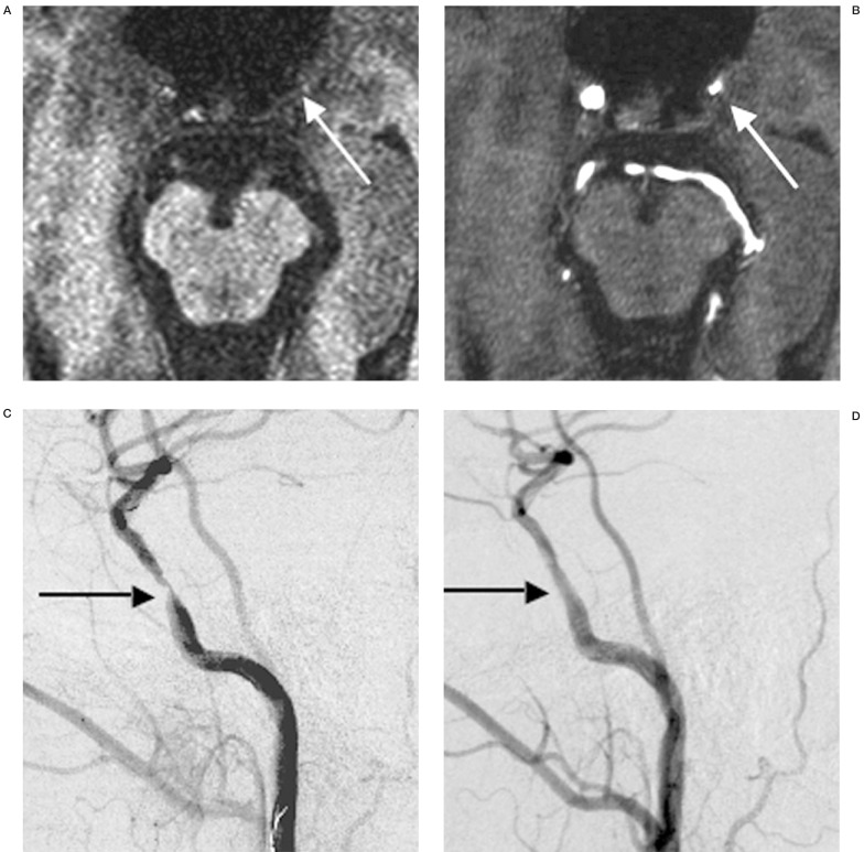
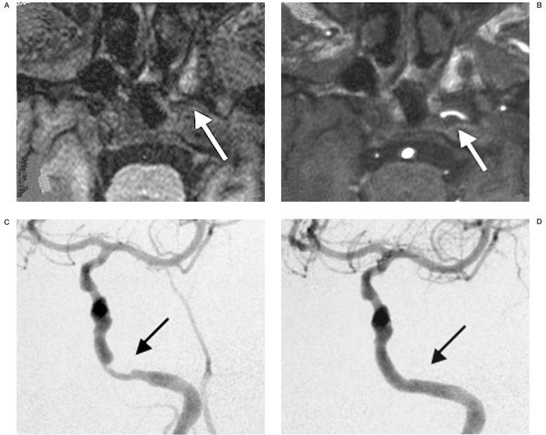
In the MPRAGE method, low intensity was observed in these lesions of all cases (table 1). The first case was performed PTA (figure 1), the second case was performed the stenting in pertous portion (figure 2), and the others were considered as follow-up. A clinical complication was not observed in those treated cases and distal embolism image was not accepted in DWI study on post-operative MRI.
Table 1.
Summary of all cases.
| Case | age/sex | rate of stenosis |
MPRAGE | treatment |
|---|---|---|---|---|
| 1 | 59/male | 80% | low | PTA |
| 2 | 74/male | 80% | low | Stenting |
| 3 | 60/male | 87% | low | Medical Tx. |
| 4 | 56/male | 66% | low | Medical Tx. |
Figure 1.
Case1. A) MPRAGE image demonstrating low intensity plaque of petrous carotid artery stenosis (arrow). B) Source image of 3D-TOF demonstrating petrous carotid artery stenosis (arrow). C) preoperative angiogram demonstrating severe petrous carotid artery stenosis(arrow). D) postoperative angiogram demonstrating good result on PTA(arrow).
Figure 2.
Case2. A) MPRAGE image demonstrating low intensity plaque of petrous carotid artery stenosis (arrow). B) Source image of 3D-TOF demonstrating petrous carotid artery stenosis (arrow). C) preoperative angiogram demonstrating severe petrous carotid artery stenosis (arrow). D) postoperative angiogram demonstrating good result on stenting (arrow).
Discussion
Carotid stenting (CAS) is beginning to spread quickly as a cure over carotid artery stenosis. One of the serious complications of this CAS is distal embolism. Although various protection device as the preventive measures is developed, it is thought that the prevention effect of distal embolism is not perfect and CAS to the carotid stenosis containing the component of soft plaque is high-risk at present. Therefore, when performing safe stent placement, preoperative plaque characteristic evaluation is very important.
The stenting to ICS in petrous portion is beginning to be performed by the development with the thin diameter of divice. Until now, it has been thought that the characterization of the plaque in this portion is hard plaque consisted of fibrous tissue. However, the characterization of the plaque of this part is actually unknown. As a plaque characterization appraisal method, the cervical echo check has been widely used from the former. This procedure cannot be used in the internal carotid artery surrounded by the petrous bone. Although the procedure of an intravascular echo check is possible also by this part, there are problems, such as invasiveness and the possibility of the embolic complication in an advanced stenosis lesion.
Recently, the usefulness of the high resolving power MRI has been reported as the procedure of the plaque evaluation in ICS with development of an instrument and progress of the image pick-up procedure. Therefore we investigate on these patients with these lesions by the MPRAGE method with the various image pick-up procedures. Since a pathology specimen was not obtained, the decision was not completed, but as for this, plaque of the petrous carotid artery stenosis resulted in supporting that it is fibrous plaque as was said from the former.
Conclusions
In the petrous carotid artery stenosis, only endovascular treatment is possible for recovering an antegrade blood flow. In a case uncontrollable by medication, endovascular treatment is an important therapeutic procedure and it is thought that the role will become large from now on. It is required to grasp the characterization of a plaque beforehand in order to treat more safety CAS. Characteristic diagnosis of the plaque using this MPRAGE method did not have an attack either, it is the procedure of doing simple, it was thought that qualitative diagnosis whether a plaque is hard at least or it is soft was attained, and it was thought that it was a useful procedure.
The MRI-MPRAGE method can expect the useful method for the investigation the plaque characterization of the petrous carotid artery stenosis.
References
- 1.Tsutsumi M, Kazekawa K, et al. Improved cerebral perfusion after simultaneous stenting for tandem stenoses of the internal carotid artery-two case reports. Neurol Med Chir (Tokyo). 2003;43:386–390. doi: 10.2176/nmc.43.386. [DOI] [PubMed] [Google Scholar]
- 2.Terada T, Tsuura M, et al. Endovascular therapy for stenosis of the petrous or cavernous portion of the internal carotid artery: percutaneous transluminal angioplasty compared with stent placement. J Neurosurg. 2003;98:491–497. doi: 10.3171/jns.2003.98.3.0491. [DOI] [PubMed] [Google Scholar]
- 3.Nagai Y, Kitagawa K, et al. Significance of earlier carotid atherosclerosis for stroke subtypes. Stroke. 2001;32:1780–1785. doi: 10.1161/01.str.32.8.1780. [DOI] [PubMed] [Google Scholar]
- 4.Hattori S, Hattori Y, Kasai K. Plaque score in the diagnosis of arteriosclerosis. Nippon Rinsho 62 Sup. 2004;3:269–271. [PubMed] [Google Scholar]
- 5.O'brien SP, Siggel B, et al. Carotid plaque spaces relate to symptoms and ultrasound scattering. Ultrasound Med Biol. 2004;32:381–385. doi: 10.1016/j.ultrasmedbio.2004.02.002. [DOI] [PubMed] [Google Scholar]
- 6.Niwa Y, Katano H, Yamada K. Calcification in carotid atheromatous plaque: delineation by 3D-CT angiography, compared with pathological findings. Neurol Res. 2004;26:778–784. doi: 10.1179/016164104225014120. [DOI] [PubMed] [Google Scholar]
- 7.Lovett JK, Gallagher PJ, et al. Histological correlates of carotid plaque surface morphology on lumen contrast imaging. Circulation. 2004;12:2190–2197. doi: 10.1161/01.CIR.0000144307.82502.32. [DOI] [PubMed] [Google Scholar]
- 8.Murphy RE, Moody AR, et al. Prevalence of complicated carotid atheroma as detected by magnetic resonance direct thrombus imaging in patients with suspected carotid artery stenosis and previous acute cerebral ischemia. Circulation. 2003;24:3053–3058. doi: 10.1161/01.CIR.0000074204.92443.37. [DOI] [PubMed] [Google Scholar]
- 9.Moody AR, Murphy RE, et al. Characterization of complicated carotid plaque with magnetic resonance direct thrombus imaging in patients with cerebral ischemia. Circulation. 2003;24:3047–3052. doi: 10.1161/01.CIR.0000074222.61572.44. [DOI] [PubMed] [Google Scholar]


