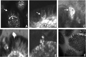
Figure 1.
Confocal endomicroscopic imaging of epithelial cell shedding in the terminal ileum. (A) Fluorescein images capillaries beneath epithelial cells (block arrow) and the lateral intracellular space between epithelial cells (line arrow). (B) Epithelial cells become permeable to fluorescein prior to shedding (line arrow). (C–E) Fluorescein fluorescence signal is intense as shedding cells move out of the epithelial monolayer. (F) Intensely fluorescent shedding epithelial cells seen en face.

