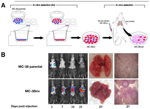
Figure 1.
Cell culture and mouse model: murine model of metastasis, in vivo monitoring, and ex vivo proof of metastases. (A) MC-38 parental cells (heterogeneous, blue and red) were subjected to 6 sequential passages through matrigel-coated transwells (enrichment of invasive subpopulations of MC-38 cells [red]) called “MC-38inv.” After in vivo passage, a stabilized cell line (pink cells) called “MC-38met” was established. (B) MC-38inv cells were tested alongside MC-38 parental cells for the ability to form lung metastasis in a tail vein assay. The figure shows representative tumor progression in live mice by bioluminescent imaging (days 1–21) and at the time of autopsy (day 21).

