Abstract
We evaluated sentence comprehension of variety of sentence constructions and components of short term memory in 53 individuals with acute ischemic stroke, to test some current hypotheses about the role of Broca's area in these tasks. We found that some patients show structure-specific, task-independent deficits in sentence comprehension, with chance level of accuracy on passive reversible sentences, more impaired comprehension of object-cleft than subject-cleft sentences, and more impaired comprehension of reversible than irreversible sentences in both sentence-picture matching and enactment tasks. In a dichotomous analysis, this pattern of “asyntactic comprehension” was associated with dysfunctional tissue in left angular gyrus, rather than dysfunctional tissue in Broca's area as previously proposed. Tissue dysfunction in left Brodmann area (BA) 44, part of Broca's area, was associated with phonological short term memory (STM) impairment defined by forward digit span ≤ 4. Verbal working memory defined by backward digit span ≤ 2 was associated with tissue dysfunction left premotor cortex (BA 6). In a continuous analysis, patients with acute ischemia in left BA 44 were impaired in phonological STM. Patients with ischemia in left BA 45 and BA 6 were impaired in passive, reversible sentences, STM, and verbal working memory. Patients with ischemia in left BA 39 were impaired in passive reversible sentences, object cleft sentences, STM, and verbal working memory. Therefore, various components of working memory seem to depend on a network of brain regions that include left angular gyrus and posterior frontal cortex (BA 6, 44, 45); left BA 45 and angular gyrus (BA 39) may have additional roles in comprehension of syntax such as thematic role checking.
Keywords: Asyntactic Comprehension, Working Memory, Broca's Area, Angular Gyrus
Introduction
It is widely agreed that patients with Broca's aphasia or damage to Broca's area often have impaired syntactic processing (Caplan, 1987). Functional imaging studies also show activation of Broca's area during syntax processing (Just et al., 1996; Friederici et al., 2003, Hashimoto and Sakai, 2002; Marcus et al., 2003; Musso et al., 2003), although these studies also show activation in other areas (Caplan, 2002). However, the type of deficit in syntactic comprehension observed in these patients is widely debated. Grodzinsky (2000) has made the strong claim that Broca's area is the “neural home to mechanisms involved in the computation of transformational relations between moved phrasal constituents and their extraction sites.” On the basis of the “trace deletion hypothesis” (TDH), according to which Broca's aphasic patients do not maintain traces of movement in sentence constructions derived by transformational movement, such as verbal passive and object relative clause constructions, he predicted chance or below chance performance on comprehension of these sentences in all patients with lesions of Broca's area (but see Berndt et al., 1996; Hickok and Avrutin, 1995; Caplan et al., 1996 for arguments against this prediction).
The influence of working memory deficits on sentence comprehension has also been controversial. Some investigators have viewed verbal working memory (VWM) as a single resource that supports a variety of language processes (Just and Carpenter, 1992). Others view working memory as a set of resources that each support different language comprehension functions, and distinguish VWM (which enables manipulation during storage) from short-term memory (STM) or articulatory loop (Caplan and Waters, 1999). Functional imaging studies show that articulatory loop tasks and comprehension of movement-derived sentences (Meltzer et al,, 2010; Santi and Grodzinsky, 2007) are both associated with activation in Broca's area (Paulesu et al., 1993), and many studies have found an association between VWM or STM and sentence comprehension (Vallar and Baddeley, 1984). These associations might be observed because the two tasks both initially depend on Broca's area. It is important to determine if there are deficits specific to particular tasks or sentence structures (or an interaction between tasks and structures) to distinguish general deficits in processing semantically reversible sentences (e.g. due to impaired working memory) from specific syntactic deficits (e.g. trace deletion). Caplan et al. (2006) studied patients with chronic stroke and found none with structure-specific, task-independent deficits. However, the failure to find such patients in the chronic stage could be due to differential practice of one task in language therapy. For example, sentence-picture matching is often used in language therapy, while enactment of sentences is rarely used.
The study of patients acutely after stroke would eliminate the variable of differential practice of one task during therapy. Moreover, the study of acute aphasia allows investigation of structure-function relationships in the brain before the opportunity for extensive reorganization of these relationships through neuroplasticity. Thus, here we investigated the relationship between asyntactic comprehension, short-term storage/verbal working memory, and acute tissue dysfunction in Broca's area (operationally defined for this study as the area of the MNI atlas where any subjects had cytoarchitecture of Brodmann's area, hereafter BA, 44 or 45 in an autopsy study by Amunts et al., 1999). We initially tested the following hypotheses:
In acute stroke, some patients show structure-specific, task-independent deficits in sentence comprehension, with chance (or below) level of accuracy on passive reversible sentences, more impaired comprehension of object-cleft than subject-cleft sentences, and more impaired comprehension of reversible compared to irreversible sentences in both sentence-picture matching and enactment tasks (a pattern we call “asyntactic comprehension”);
In acute stroke, asyntactic comprehension is associated with impaired working memory, but not with dysfunctional tissue (infarct and/or hypoperfusion) in Broca's area.
However, literature published during our study and our own results led us to revise and refine our hypotheses in a post hoc analysis, and to evaluate separately the roles of BA 44 and 45 in sentence comprehension and verbal working memory, as well as to identify other areas of the brain involved in cortical networks underlying these functions. For example, Rogalsky and Hickok (2011) reviewed evidence from functional imaging studies and lesion studies regarding the role of Broca's area in sentence comprehension. They argued that there is little evidence in support of a specific syntax processing role. Rather, they presented evidence that BA 44 is likely to be involved in an articulatory rehearsal component of working memory necessary for sentence comprehension, and indicated that further support is needed from lesion studies. They argued that the role of BA 45 in sentence comprehension is less clear, but suggested that it appears to have a cognitive control function, such as semantic integration (e.g. as proposed by Hagoort, 2005) or thematic role checking (Caplan et al., 2008a; Caplan et al., 2008b). Therefore, we tested the additional hypothesis that in acute stroke, ischemia in BA 44, but not BA 45, is associated with impaired verbal STM or VWM. We also evaluated whether BA 44 or BA 45 is more strongly associated with tasks that require thematic role checking (e.g. comprehension of semantically reversible sentences), although we did not administer the ideal test to distinguish between deficits in thematic role checking, semantic integration, and other aspects of cognitive control.
Methods
Participants
We enrolled a consecutive series of 53 right-handed participants with first-ever acute left hemisphere symptomatic ischemic stroke who gave consent or who assented and indicated a family member who provided informed consent, and were admitted to the hospital within 24 hours of onset of stroke symptoms. Additional exclusion criteria were: non-native speakers of English, prior neurological disease, known uncorrected visual or hearing loss, acute stroke limited to the brainstem or cerebellum, diminished level of consciousness or requiring intubation, ongoing intravenous sedation, presence of any ferromagnetic implant (cardiac pacemakers, aneurysm clip) or other contraindication to MRI, pregnancy, allergy to Gadolinium contrast or renal failure (estimated glomerular filtration rate <60), or severe claustrophobia. Patients were enrolled the first day of admission to the hospital. They had MRI and language testing the same day.
Experimental Language Tasks
Patients underwent a battery of language tasks, including standardized and unstandardized tasks. However, the focus of this study is on the following experimental tasks.
Sentence Picture Matching (SPM)
Participants saw two pictures on a computer screen and simultaneously heard and saw a written sentence underneath the picture. The participant had to press a key to indicate which picture matched the sentence they heard and saw. There were a total of 80 experimental trials. There were 20 sentences of each category: (a) active, (b) passive, (c) subject-cleft, and (d) object-cleft (see examples below):
The father kicked the niece.
The niece was kicked by the father.
It was the father that kicked the niece.
It was the niece that the father kicked.
Within these categories each contained10 semantically reversible sentences (e.g. “It was the man that kicked the girl”) and 10 semantically irreversible sentences (e.g. “It was the man that kicked the tree”). For the reversible sentences, the foil represented the reversal of the object and the subject (e.g. “It was the girl that kicked the man”). For semantically irreversible sentences, foils contained a different object or agent (e.g. “It was the chicken that the mother held” vs “It was the fish that mother held.”)
Enactment
Participants heard and saw a sentence on the computer screen and were asked to act out the sentence using laminated paper figures/pictured objects. The figures/objects represented the subjects and objects in each sentence. The number of trials and sentence categories were the same as the SPM task. The vocabulary words were the same, but the exact sentences were not used twice. Responses were scored as correct if the participant selected the correct agent and showed that it was doing something to the correct object.
For both tasks participants were permitted to respond with either right or left hands, and the tasks were untimed, so motor difficulties did not appear to affect accuracy.
Short-term and Working Memory
STM and VWM were assessed with forward and backward digit span, respectively. Note that forward digit span was used to assess short-term storage (articulatory loop), while backward digit span was used to assess verbal working memory, because only the latter requires some manipulation during storage. Each span length was assessed by two trials, giving credit for the maximum span length for which the patient was successful on at least one trial.
Magnetic Resonance Imaging
Magnetic Resonance Imaging included T1, T2, Fluid Attenuated Inversion Recovery (to rule out old infarcts), Susceptibility Weighted Images (to rule out hemorrhage), diffusion-weighted images (DWI), Apparent Diffusion Coefficient (ADC) maps, and dynamic-susceptibility contrast echo-planar Perfusion Weighted Images (PWI) obtained parallel to the AC-PC line on Siemens 1.5 Tesla clinical scanner or Philips 3 Tesla research scanner. PWI was obtained with power-injection of 20 cc of Gadolinium at 5 cc/sec. Five mm thick slices, typically 17-20 slices to achieve whole brain coverage, were obtained for most sequences. A subset of participants also had high resolution (1 mm slice thickness) MPRAGE scans. Scans were initially analyzed for dysfunction in Broca's area on DWI, ADC, and PWI by the senior author (AH) and one technician without knowledge of results of the language assessment. Each sequence was registered to the MNI atlas, and we determined whether each patient's infarct and/or area of hypoperfusion covered (1) part, (2) all, or (0) none of the area corresponding to cytoarchitectural areas 44 and 45 in the probabilitistic map of Broca's area based on an autopsy study (Amunts et al., 1999). There was >90% point-to-point agreement between the 2 judges; discrepancies were resolved by joint review. Scans were re-analyzed to evaluate for ischemia in 12 other Brodmann's areas: 6, 10, 11,18, 19, 20, 21, 22, 37, 38, 39, 40 on a standard atlas (Damasio and Damasio, 1989) by technicians masked to the language scores. There was 100% point-to-point agreement between the 2 judges on 10 scans evaluated by both technicians. Dysfunctional tissue was defined as bright on DWI and dark on ADC and/or >4 sec delay in time to peak arrival of Gadolinium to the region compared to the homologous region in the right hemisphere (measured with ImageJ; http://rsb.info.nih.gov/ij/download.html). This degree of hypoperfusion corresponds to dysfunctional tissue defined by PET (Zaro-Weber, et al. 2010 ; Sobesky, et al. 2004).
Statistical Analysis
Dichotomous Tests
We first identified the pattern of asyntactic comprehension as a dichotomous variable (present or absent). The presence of asyntactic comprehension required: performance not significantly above chance level of accuracy on passive reversible sentences on at least one test (SPM or enactment); ≥ 10 percentage points lower accuracy on passive compared to active sentences and object-cleft compared to subject-cleft sentences, and ≥ 10 percentage points lower accuracy on reversible compared to irreversible sentences. These criteria (e.g. the 10 point difference) are arbitrary, but we chose them prospectively. Other tests of the same relationships that we carried out did not depend on these arbitrary criteria (see continuous analyses). We identified associations between ischemia (hypoperfusion/infarct) in all or part of Broca's area as defined above and (1) this pattern of asyntactic comprehension and (2) impaired STM (forward digit span ≤ 2) or VWM (backward digit span ≤ 4) using chi square tests. As a secondary analysis, we identified associations between ischemia (hypoperfusion/infarct) in all or part of 12 additional Brodmann's areas and (1) this pattern of asyntactic comprehension and (2) impaired STM (forward digit span ≤ 2) or VWM (backward digit span ≤ 4) using chi square tests. We applied a Bonferroni correction for 48 comparisons (12 Brodmann's areas × 4 tests), so that an alpha level of p<0.001 was considered above chance.
Comparisons across Continuous Variables
We used multivariate linear regression, with age and education as independent variables (entered together), and each of the sentence comprehension and digit span tests as dependent variables to determine whether these variables independently or together predicted performance on any of the behavioral tests.
We then identified patients with ischemia in anatomic regions of interest found to be associated with impairments of verbal STM, VWM, or asyntactic comprehension by chi square tests: BA 44, 45, 6 (premotor cortex), and 39 (angular gyrus). We compared their performance on sentence comprehension and working memory tasks to patients without ischemia in each of these areas using independent samples t-tests. We could not use ANOVA to compare performance across several lesion groups (e.g. those with ischemia in BA 44, 45, 39, etc.) at once, because many patients would fall into two or more groups.
Results
The age of participants ranged from 18 to 91 years (mean 59.3; sd 15.2). Education ranged from 7 to 24 years (mean 14.0; sd 3.4). Of the 53 patients, 30 were women.
Dichotomous Analysis
Very few patients showed ischemia in the entire area for any area; therefore we combined patients who showed ischemia in part of the area and all of the area as showing ischemia in the area. A total of 14 patients showed asyntactic comprehension on at least one test. Of the 14 patients with asyntactic comprehension, six patients showed this pattern on both sentence-picture matching and enactment, six patients showed the pattern only on one test (three in spm and three in enactment), and two failed to complete one or more tests. Many more patients showed impaired comprehension of syntactically complex sentences, but did not meet the criteria for asyntactic comprehension (e.g. because they also failed to comprehend active sentences or showed no difference between subject-cleft and object-cleft sentences).
Broca's area: Dichotomous Analysis
Asyntactic comprehension on at least one test was not significantly associated with tissue dysfunction in BA 44 (χ2=3.5; df=1; p=0.06), but there was a trend toward an association with tissue dysfunction in BA 45 that was not significant after the Bonferroni correction (χ2=5.1; df=1; p=0.02). These data are summarized in Figure 1.
Figure 1.
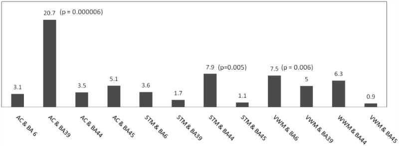
Results of dichotomous analyses: Chi square values for the associations between asyntactic comprehension, short term memory, verbal working memory and each of the Brodmann's areas of interest.
Phonological STM as defined by forward digit span ≤ 4 was strongly associated with with tissue dysfunction in BA 44 (χ2=7.9; df=1; p=0.005), but not associated with BA 45 (χ2=1.1; df=1; p=0.3). VWM as defined by backward digit span ≤ 2 was not significantly associated with tissue dysfunction in either area, but showed a trend toward an association with ischemia in BA 44 (χ2=6.3; df=1; p=0.01) only.
Although there is substantial overlap in patients who had ischemia in BA 44 and 45, some patients showed ischemia in part of one area but not the other, so that this dichotomous analysis revealed a disassociation between areas associated with asyntactic comprehension (BA 45) and areas associated with STM and VWM (BA 44). For example, the patient whose scans are shown in Figure 2 showed ischemia in part of BA 44 (pars opercularis), but not BA 45, and was 90% accurate in passive reversible sentences, and 95% accurate or above in comprehending all other sentence types, but had a forward digit span of only 3 (impaired).
Figure 2.
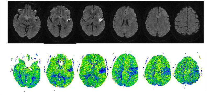
Top. DWI (left) and PWI (right) scans of a patient whose accuracy in sentence comprehension was 90% for passive reversible sentences, 95% for active reversible and 100% for object-cleft and subject-cleft sentences, but reduced forward digit span of 3 (and backward digit span of 3), at the time of acute dysfunctional tissue within part of BA 44 but not BA 45.
There was not a significant association between asyntactic comprehension and STM measured by forward digit span ≤ 4 (χ2=3.6; df=1; p=0.06) or between asyntactic comprehension and VWM measured by backward digit span ≤ 2 (χ2=2.5; df=1; p=0.12). Many patients who had limited spans did not have asyntactic comprehension.
Other areas associated with asyntactic comprehension
Asyntactic comprehension on at least one test was also strongly associated with tissue dysfunction in left angular gyrus (χ2=20.7; df=1; p=0.000006).
Thus, although there was only a trend for asyntactic comprehension to be associated with ischemia in left BA 45 in the dichotomous analysis, asyntactic comprehension was significantly associated with ischemia in left angular gyrus. For example, the patient in Figure 3, who had tissue dysfunction in BA 39 and 40 (supramarginal gyrus), but not BA 44 or 45, was only 40% correct for passive reversible sentences, but 85% correct for active reversible sentences. He was 85% correct for cleft-object sentences, and 100% correct for cleft-subject cleft sentences. This man had forward digit span of 3, and was unable to do backwards digits.
Figure 3.
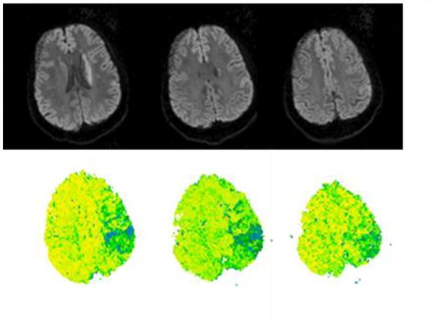
DWI (top panel) and PWI (lower panel) scans of a patient with asyntactic comprehension and impaired short-term memory associated with ischemia in left angular gyrus. He was 40% correct for passive reversible sentences, but 85% correct for active reversible sentences. He was 85% correct for cleft-object sentences, and 100% correct for cleft-subject cleft sentences. Forward digit span of 3, and was unable to do backwards digits. The acute infarct on DWI involves only the caudate. PWI shows significant hypoperfusion (>4 sec. delay in TTP relative to the homologous regions in the right hemisphere; see methods) in angular gyrus (BA 39) and supramarginal gyrus (BA 40), but not Broca's area (BA 44 or 45).
Other areas associated with impaired short-term/working memory
Reduced phonological STM defined by forward digit span ≤ 4 was not associated with tissue dysfunction in any of the areas after correction for multiple comparisons. Without correction, BA 6 (premotor cortex), 39 (angular gyrus), and 40 (supramarginal gyrus) showed a trend toward an association between ischemia and reduced verbal STM. After Bonferroni correction, reduced verbal working memory defined by backward digit span ≤ 2 was associated with tissue dysfunction left BA 6 (Fisher's exact: (p=0.0006) in posterior frontal cortex.
Analysis of Comprehension and Working/ Short-term Memory as Continuous Variables
There were no significant differences between men and women on any of the behavioral tests by independent t-tests. Also, neither education nor age were independently or together associated with accuracy of performance on any of the sentence types or digit spans using multivariate linear regression (r2=0.014; p=0.998 for active, reversible sentences to r2=0.115; p=0.112 for backward digit span).
Broca's Area: Continuous Analysis
We evaluated the effects of ischemia in left BA 44 and BA 45 on various tasks and demographics using independent samples t-tests to compare patients with ischemia in each area to patients without ischemia in that area. Results are summarized in Figures 4-6. Importantly, there were no significant differences between participants with ischemia and participants without ischemia in either BA 44 or 45 in terms of age or education.
Figure 4.
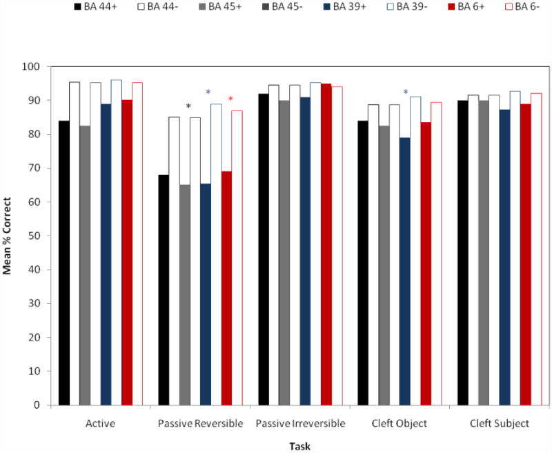
Results of continuous analysis: Mean scores for sentence-picture matching for various sentence types for patients with and without ischemia in each of the Brodmann areas significantly associated with asyntactic comprehension or STM/VWM in the dichotomous analysis. A significant difference by independent t-test is marked by *.
Figure 6.
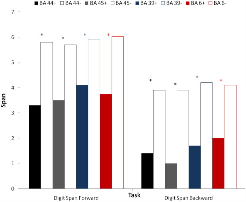
Results of continuous analysis: Mean scores for forward and backward digit span (VWM) for patients with and without ischemia in each of the Brodmann areas significantly associated with asyntactic comprehension, or with reduced forward digit span (STM), or backward digit span (VWM) in the dichotomous analysis. A significant difference by independent t-test is marked by *.
Those with ischemia in left BA 44 were significantly more impaired than those without in digit span forward (mean 3.3± SD 2.4 vs. 5.8±1.9; t=2.9; df=50; p=0.006); and digit span backward (mean 1.4±1.6 vs. 3.9±1.4; t=3.6; df=51; p=0.001). They were not significantly more impaired in comprehending any of the sentence types in sentence-picture matching or enactment than patients without ischemia in BA 44.
Patients with ischemia in left BA 45 were significantly more impaired than those without only in comprehension of passive reversible sentences in sentence picture matching (mean 65.0 ± 26.5% vs. 84.9 ± 7.2% correct; t=2.1; df=49; p=0.039); and digit span forward (mean 3.5 ± 2.6 vs. 5.7 ± 2.0; t=2.0; df=50; p=0.047); and digit span backward (mean 1.0 ± 1.4 vs. 3.9 ± 1.7; t=3.5; df=51; p=0.001.
Other Areas: Continuous Analysis
Having identified significant associations between infarct in left angular gyrus (BA 39) and asyntactic comprehension, as well as an association between premotor cortex (BA 6) and reduced digit span, as dichotomous variables, we evaluated the sentence comprehension and digit span performance in participants with and without ischemia in these regions of interest.
There was no significant difference between participants with or without ischemia in BA 39 or 6 in age or education by independent t-tests.
Acute ischemia in left BA 39 (angular gyrus) was associated with significant impairment in comprehension of passive reversible sentences in sentence-picture matching (65.4 ±19.9 vs. 88.9 ± 13.2; t=14.5; p<0.0001), cleft-object sentences in sentence-picture matching (79.0 ± 17.8 vs. 91.0 ±13.0; t=2.6; df=49; p=0.01) and in enactment (79.8 ± 13.6 vs. 95.6 ± 5.0; p=0.015), as well as digit span forward (mean 4.1 ± 2.0 vs 5.9 ± 2.0; t=2.8; df=50; p=0.007) and digit span backward (mean = 1.7 ±1.5 vs 4.2 ± 1.6; t= 5.0; df=51; p<0.0001).
Similarly, acute ischemia in left BA 6 (premotor area) was only associated with significant impairment in comprehension of passive reversible sentences in sentence picture matching (69.0 ± 18.5 vs. 86.9 ±17.0; df=49; t=2.9; p=0.005), as well as digit span forward (mean 3.8 ±1.8 vs 6.0 ±1.9; df=50; p<0.0001) and digit span backward (mean = 2.0 ± 1.7 vs 4.1 ± 1.7; t= 3.9; df=51; p=0.001).
Discussion
We confirmed our first hypothesis, that in patients with acute stroke, some patients show structure-specific, task-independent deficits in sentence comprehension, with particular impairment of movement-derived sentences. We can speculate that the difference between acute and chronic stroke findings might reflect differential practice in particular types of tasks after stroke that reduces the task demands for tasks like sentence-picture matching, and allows better performance on passive sentences, reversible sentences, and those with cleft-object clauses in the practiced task but not in unpracticed tasks. Nevertheless, some patients showed asyntactic comprehension in just one task – either enactment or sentence-picture matching, which could be due to random variability in performance or an interaction between the task demands and sentence structures that is not consistent across patients.
Our results concerning the relationship between ischemia in Broca's area and its effect on performance on measures of short term memory (forward digit span) or working memory (backward digit span) and syntax comprehension were complex, and consistent with an emerging story from the functional imaging literature (see Rogalsky and Hickok, 2011, for review).
First, we found an association between impaired verbal STM and ischemia in BA 44, or pars opercularis, consistent with the hypothesis that this region is essential for an articulatory loop component of phonological STM (Paulesu et al., 1993; Caplan et al., 2000; Rogalsky et al., 2008). Recent evidence from articulatory suppression combined with fMRI (Caplan, et al., 2000, Rogalsky et al., 2008) indicates that at least pars opercularis portion of Broca's area is involved in phonological STM at the level of articulatory rehearsal or integrating information maintained by articulatory rehearsal with higher level representations (Rogalsky and Hickok, 2011). This hypothesis is consistent with findings indicating that acute ischemia in BA 44 results in impairments of digit span (shown in this paper) as well as articulation (Hillis et al., 2002). However, ischemia in BA 44 alone was not associated with asyntactic comprehension or impaired comprehension of syntactically complex sentences (e.g. passive reversible sentences or cleft-object sentences) in the dichotomous analysis. These two results are consistent with previous studies from neurologically impaired individuals showing that patients with severely impaired ability to repeat verbatim (e.g. digit span of two) are sometimes able to comprehend syntactically complex sentences (Martin, 1993). There was also an association between impaired STM and ischemia in nearby BA 6 (premotor cortex), consistent with numerous functional imaging studies (Swartz et al., 1996; D'Esposito et al., 1999; D'Esposito and Postle, 1999). It is clear from both functional imaging studies and lesion studies that a network of regions underlies the complex cognitive processes of working memory, of which the phonological loop (which seems to depend on BA 44 and BA 6) is one component.
Secondly, we found a trend toward an association between asyntactic comprehension and ischemia in BA 45, or pars triangularis (p=0.02, which was not significant after correction for multiple comparisons), but no trend for pars opercularis (BA 44). Moreover, patients with ischemia in BA 45 were significantly impaired, by t-test, on comprehension of passive reversible sentences, compared to patients without lesions in this area. This result is consistent with the proposal that this region may be critical for thematic role checking or reanalysis. This finding is in keeping with a previous case report of acute ischemia in Broca's area associated with impairment in simple reversible questions (e.g. Is a horse larger than a dog?) and active and passive reversible sentences (The girl chased the man; the boy was chased by the dog) on a video-sentence matching task, which resolved when Broca's area was reperfused (Davis et al., 2009). Thus, more anterior parts of Broca's area, such as pars triangularis, may be involved in more “cognitive control” aspects of sentence comprehension, or more specifically, thematic role checking/reanalysis (Caplan et al., 2008a; Caplan et al., 2008b). Evidence for the latter hypothesis comes from distance effects in Broca's area for semantically unconstrained sentences (e.g. The lawyer that the banker irritated filed a lawsuit), but not semantically constrained sentences (e.g. The thief that that policeman arrested was known to carry a knife.). However, we found no significant impairment in comprehension of object cleft sentences associated with acute ischemia in left BA 45, which we might have expected on the account that these patients are impaired in thematic role checking or reanalysis. Patients with ischemia in BA 45 did have lower scores on object cleft sentences than patients without ischemia in this region, but the difference was not significant, possibly due to insufficient power or “lucky guessing” in some cases. Nevertheless, the possibility of a deficit in thematic role checking or cognitive control aspects of syntax processing associated with acute lesions of pars triangularis (BA 45), reflected in impaired comprehension of passive reversible sentences and reversible questions in at least in some patients, would not contradict previously published findings that many individuals with chronic lesions involving Broca's area have more selective deficits (or no deficits), as other areas of the brain may assume some normal roles of Broca's area during recovery.
In the continuous analysis ischemia in left BA 45 was associated with reductions in both forward and backward digit span, indicating that it may also have a role in working memory. Indeed, working memory may support thematic role checking. Alternatively, the association may have been significant because many patients with ischemia in BA 45 also have ischemia in BA 44. Furthermore, there is substantial overlap in the locations of these cytoarchetecteural fields across individual brains (Amunts et al., 1999).
Our original hypotheses were about the association between deficits and Broca's area (BA 44 and 45), because this area has been the focus of lesion studies and functional imaging studies. However, having failed to identify an association between asyntactic comprehension and acute ischemia in BA 44, nor an association between STM/VWM and BA 45 by chi square tests, we carried out additional analyses to determine if there are other regions of acute ischemia associated with asyntactic comprehension and/or impairments in STM or VWM. We identified in the dichotomous analysis a strong association between asyntactic comprehension and left angular gyrus, another region that is considered to be important in a neural network underlying STM (Swartz et al., 1996; Thomason et al., 2009; Metzak et al., 2011). Furthermore, in the continuous analysis, ischemia in the left angular gyrus was associated with impairments in comprehension of both passive reversible and object cleft sentences (but not other sentences), and associated with reduced forward and backward digit span by independent samples t-tests. This area is frequently activated in functional imaging studies of working memory and sentence comprehension, but receives far less attention in the literature than Broca's area. Although the connections between Broca's area and Wernicke's area are also emphasized in discussing anatomy underlying sentence comprehension, there are also white matter tracks connecting Broca's area and angular gyrus (Catani et al., 2005). Furthermore, there is strong evidence of functional connectivity between Broca's area and angular gyrus during both sentence comprehension (Prat and Just, 2010) and at rest (Greicius et al., 2003, Fox and Raichle, 2007). Therefore, the role of left angular gyrus in sentence comprehension deserves further exploration.
We also found in the continuous analysis that ischemia in left BA 6 (like BA 45) was associated with impaired comprehension of passive reversible sentences, and reduced digit span forward and backward. So, this area, too, may have a role in comprehension of syntactically complex sentences.
The mean scores for patients with ischemia across Brodmann areas 44, 45, and 6 did not differ very much for any sentence type or digit span task. This result is not very surprising, because there was substantial overlap in these groups; that is, several patients had ischemia in two or more of these areas. However, some patients had ischemia involving only one area, and had selective impairment or preservation of specific sentence types or tasks, such that some distinct lesion-deficit associations were revealed with the dichotomous analyses.
We were not able to obtain high resolution (1 mm slice thickness) T1 scans in all participants in this study for a voxel-based analysis, so we focused on 12 Brodmann areas in the frontal, temporal, and parietal cortex (which have been proposed as components of networks underlying language comprehension or working memory) as specific regions of interest. Future studies will evaluate specific voxels associated with syntactic comprehension deficits. Our preliminary results are consistent with the proposal that comprehension of certain types of syntactically complex sentences (e.g. passive reversible sentences and perhaps object cleft sentences) depend on at least posterior frontal regions (BA 6, 45) and angular gyrus, although these areas might have distinct roles in the task. We recently reported complementary evidence indicating that acute ischemia in most of the regions of interest within the left temporal cortex anterior to BA 37 disrupted comprehension of all sentence types, while ischemia in BA 45, 6, and 39 selectively disrupted comprehension of syntactically complex sentences (Trupe, et al., 2011 [abstract]).
In summary, we have provided evidence in favor of distinct anatomical and cognitive causes of the pattern of sentence comprehension impairment that we and others have referred to as “asyntactic comprehension”, as argued by many other authors (e.g. Berndt et al., 1996, Caplan et al., 2006). One cause seems to be disrupted thematic role checking, which may be a type of cognitive control that relies on left BA 45. Another cause may be some aspect of verbal working memory that supports manipulation of the information held in short-term storage, which seems to rely on at least left BA 6 and perhaps left angular gyrus. The important role of the left angular gyrus in comprehension of syntactically complex sentences requires further investigation. Phonological STM, specifically the articulatory loop component, seems to rely on left BA 44, but can be quite limited without disrupting sentence comprehension.
Figure 5.
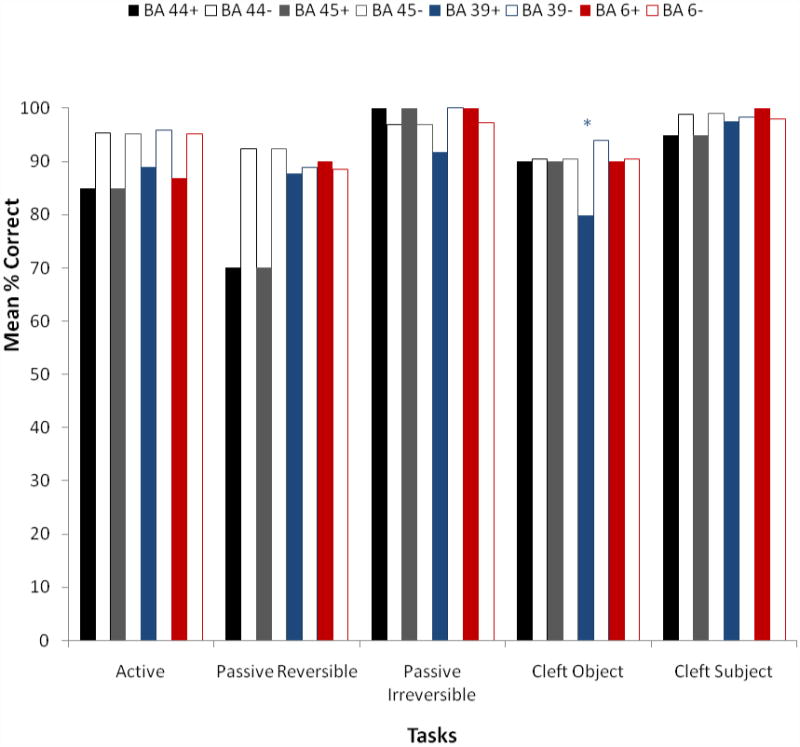
Results of continuous analysis: Mean scores for enactment for various sentence types for patients with and without ischemia in each of the Brodmann areas significantly associated with asyntactic comprehension or STM/VWM in the dichotomous analysis. A significant difference by independent t-test is marked by *.
Acknowledgments
This study was supported by NIH RO1 DC 05375. We gratefully acknowledge this support, the participation of the patients, and the very helpful comments of two anonymous reviewers on earlier versions of the paper.
Footnotes
Publisher's Disclaimer: This is a PDF file of an unedited manuscript that has been accepted for publication. As a service to our customers we are providing this early version of the manuscript. The manuscript will undergo copyediting, typesetting, and review of the resulting proof before it is published in its final citable form. Please note that during the production process errors may be discovered which could affect the content, and all legal disclaimers that apply to the journal pertain.
References
- Amunts K, Schleicher A, Burgel U, Mohlberg H, Uylings HBM, Zilles K. Broca's region revisited: Cytoarchitecture and intersubject variability. Journal of Comparative Neurology. 1999;412(2):319–941. doi: 10.1002/(sici)1096-9861(19990920)412:2<319::aid-cne10>3.0.co;2-7. [DOI] [PubMed] [Google Scholar]
- Berndt R, Mitchum CC, Haendiges AN. Comprehension of reversible sentences in “agrammatism”: A meta-analysis. Cognition. 1996;58(3):289–308. doi: 10.1016/0010-0277(95)00682-6. [DOI] [PubMed] [Google Scholar]
- Caplan D. Discrimination of normal and aphasic subjects on a test of syntactic comprehension. Neuropsychologia. 1987;25(1B):173–184. doi: 10.1016/0028-3932(87)90129-1. [DOI] [PubMed] [Google Scholar]
- Caplan D, Chen E, Waters G. Task-dependent and task-independent neurovascular responses to syntactic processing. Cortex. 2008a;44(3):257–275. doi: 10.1016/j.cortex.2006.06.005. [DOI] [PMC free article] [PubMed] [Google Scholar]
- Caplan D, Stanczak L, Waters G. Syntactic and thematic constraint effects on blood oxygenation level dependent signal correlates of comprehension of relative clauses. Journal of Cognitive Neuroscience. 2008b;20(4):643–656. doi: 10.1162/jocn.2008.20044. [DOI] [PMC free article] [PubMed] [Google Scholar]
- Caplan D, Hildebrandt N, Makris N. Location of lesions in stroke patients with deficits in syntactic processing in sentence comprehension. Brain. 1996;119(Pt 3):933–949. doi: 10.1093/brain/119.3.933. [DOI] [PubMed] [Google Scholar]
- Caplan D. The neural basis of syntactic processing: A critical look. New York, NY: Psychology Press; 2002. [Google Scholar]
- Caplan D, DeDe G, Michaud J. Task-Independent and task-specific syntactic deficits in aphasic comprehension. Aphasiology. 2006;9(11):893–920. [Google Scholar]
- Catani M, Jones DK, Ffytche DH. Perisylvian language networks of the human brain. Annals of Neurology. 2005;57(1):8–16. doi: 10.1002/ana.20319. [DOI] [PubMed] [Google Scholar]
- Davis C, Kleinman JT, Newhart M, Gingis L, Pawlak M, Hillis AE. Speech and language functions that require a functioning Broca's Area. Brain and Language. 2008;105(1):50–58. doi: 10.1016/j.bandl.2008.01.012. [DOI] [PubMed] [Google Scholar]
- D'Esposito M, Postle BR. The dependence of span and delayed-response performance on prefrontal cortex. Neuropsychologia. 1999;37(11):1303–1315. doi: 10.1016/s0028-3932(99)00021-4. [DOI] [PubMed] [Google Scholar]
- D'Esposito M, Postle BR, Ballard D, Lease J. Maintenance versus manipulation of information held in working memory: an event related fMRI study. Brain and Cognition. 1999;41(1):66–86. doi: 10.1006/brcg.1999.1096. [DOI] [PubMed] [Google Scholar]
- Fox MD, Raichle ME. Spontaneous fluctuations in brain activity observed with functional magnetic resonance imaging. Nature Reviews Neuroscience. 2007;8(9):700–711. doi: 10.1038/nrn2201. [DOI] [PubMed] [Google Scholar]
- Greicius MD, Krasnow B, Reiss AL, Menon V. Functional connectivity in the resting brain: A network analysis of the default mode hypothesis. Proceedings of the National Academy of Sciences of the United States of America. 2003;100(1):253–258. doi: 10.1073/pnas.0135058100. [DOI] [PMC free article] [PubMed] [Google Scholar]
- Friederici AD, Ruschemeyer SA, Hahne A, Fiebach CJ. The role of left inferior frontal and superior temporal cortex in sentence comprehension: localizing syntactic and semantic processes. Cerebral Cortex. 2003;13(2):170–177. doi: 10.1093/cercor/13.2.170. [DOI] [PubMed] [Google Scholar]
- Grodzinsky Y. The neurology of syntax: Language use without Broca's area. Behavioral Brain Science. 2000;23(1):1–21. doi: 10.1017/s0140525x00002399. [DOI] [PubMed] [Google Scholar]
- Hashimoto R, Sakai KL. Specialization in the left prefrontal cortex for sentence comprehension. Neuron. 2002;35(3):589–597. doi: 10.1016/s0896-6273(02)00788-2. [DOI] [PubMed] [Google Scholar]
- Hickok G, Avrutin S. Representation, referentiality, and processing in agrammatic comprehension: Two case studies. Brain and Language. 1995;50(1):10–26. doi: 10.1006/brln.1995.1038. [DOI] [PubMed] [Google Scholar]
- Just MA, Carpenter PA. A capacity theory of comprehension: Individual differences in working memory. Psychological Review. 1992;99(1):122–149. doi: 10.1037/0033-295x.99.1.122. [DOI] [PubMed] [Google Scholar]
- Just MA, Carpenter PA, Keller TA, Eddy WF, Thulborn KR. Brain activation modulated by sentence comprehension. Science. 1996;274(5284):114–116. doi: 10.1126/science.274.5284.114. [DOI] [PubMed] [Google Scholar]
- Marcus GF, Vouloumanos A, Sag IA. Does Broca's play by the rules? Nature Neuroscience. 2003;6(7):651–652. doi: 10.1038/nn0703-651. [DOI] [PubMed] [Google Scholar]
- Metzak P, Feredoes E, Takane Y, Wang L, Weinstein S, Cairo T, Ngan ET, Woodward TS. Constrained principal component analysis reveals functionally connected load-dependent networks involved in multiple stages of working memory. Human Brain Mapping. 2011;32(6):856–71. doi: 10.1002/hbm.21072. [DOI] [PMC free article] [PubMed] [Google Scholar]
- Meltzer JA, McArdle JJ, Schafer RJ, Braun AR. Neural aspects of sentence comprehension: syntactic complexity, reversibility, and reanalysis. Cerebral Cortex. 2010;20(8):1853–1864. doi: 10.1093/cercor/bhp249. [DOI] [PMC free article] [PubMed] [Google Scholar]
- Musso M, Moro A, Glauche V, Rijntjes M, Reichenbach J, Buchel C, Weiller C. Broca's area and the language instinct. Nature Neuroscience. 2003;6(7):774–781. doi: 10.1038/nn1077. [DOI] [PubMed] [Google Scholar]
- Prat CS, Just MA. Exploring the neural dynamics underpinning individual differences in sentence comprehension. Cerebral Cortex. 2010;21(8):1747–1760. doi: 10.1093/cercor/bhq241. [DOI] [PMC free article] [PubMed] [Google Scholar]
- Rogalsky C, Matchin W, Hickok G. Broca's area, sentence comprehension, and working memory: an fMRI Study. Frontiers of Human Neuroscience. 2008;2:14. doi: 10.3389/neuro.09.014.2008. [DOI] [PMC free article] [PubMed] [Google Scholar]
- Rogalsky C, Hickok G. The role of Broca's area in sentence comprehension. Journal of Cognitive Neuroscience. 2011;23(7):1664–80. doi: 10.1162/jocn.2010.21530. [DOI] [PubMed] [Google Scholar]
- Sobesky J, Zaro-Weber O, Lehnhardt FG, Hesselmann V, Thiel A, Dohmen C, Jacobs A, Neveling M, Heiss WD. Which time-to-peak threshold best identifies penumbral flow? A comparison of perfusion-weighted magnetic resonance imaging and positron emission tomography in acute ischemic stroke. Stroke. 2004;35(12):2843–2847. doi: 10.1161/01.STR.0000147043.29399.f6. [DOI] [PubMed] [Google Scholar]
- Swartz BE, Halgren E, Simpkins F, Mandelkern M. Studies of working memory using 18FDG-positron emission tomography in normal controls and subjects with epilepsy. Life Sciences. 1996;58(22):2057–2064. doi: 10.1016/0024-3205(96)00198-1. [DOI] [PubMed] [Google Scholar]
- Thomason ME, Race E, Burrows B, Whitfield-Gabrieli S, Glover GH, Gabrieli JD. Development of spatial and verbal working memory capacity in the human brain. Journal of Cognitive Neuroscience. 2009;21(2):316–332. doi: 10.1162/jocn.2008.21028. [DOI] [PMC free article] [PubMed] [Google Scholar]
- Trupe LA, Gottesman RF, Hillis AE. Acute ischemia in left angular gyrus impairs comprehension of syntax. Stroke. 2011;42(3):e162. [Google Scholar]
- Vallar G, Baddeley A. Fractionation of working memory: Neuropsychological evidence for a phonological short term store. Journal of Verbal Learning and Verbal Behavior. 1984;23:151–162. [Google Scholar]
- Zaro-Weber O, Moeller-Hartmann W, Heiss WD, Sobesky J. Maps of time to maximum and time to peak for mismatch definition in clinical stroke studies validated with positron emission tomography. Stroke. 2010;41(12):2817–2821. doi: 10.1161/STROKEAHA.110.594432. [DOI] [PubMed] [Google Scholar]


