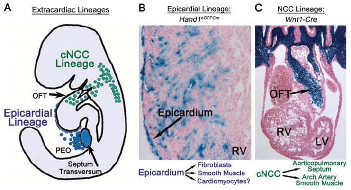
Figure 2. Extracardiac Lineages of the heart.

A, diagram of extracardiac lineages. B, Epicardial lineage shown by X-gal staining of Hand1eGFPCre R26R activation in right ventricle at E15.5 showing epicardial, cardiac fibroblast and coronary smooth muscle expression. C, Neural crest cell lineage, shown by X-gal staining of Wnt1-Cre R26R activation in OFT at E11.5. PEO, proepicardial organ; OFT, outflow tract; RV, right ventricle; LV, left ventricle; cNCC, cardiac neural crest cell. Left: diagram of epicardial and cNCC lineage contributions to heart development.
