

Summary
Background:
Metabolic syndrome is a result of multiple risk factors of atherosclerosis and diabetes. Obesity is an especially well recognized etiological factor. A rapidly increasing number of obese people constitutes a major social health problem in the developed, as well as developing countries. Bariatric surgeries are among methods of obesity treatment that gain on popularity. They include adjustable silicone gastric banding (ASGB), and adjustable laparoscopic gastric banding (ALGB).
Material/Methods:
The aim of our study was to analyze and present the most typical radiological images obtained during 130 upper gastrointestinal tract examinations in patients after ASGB or ALGB in the last three years.
Results/Conclusions:
ASGB and ALGB are effective and safe. However, they are connected with some postoperative complications. Application of these surgical procedures requires periodic, long-term radiological evaluations and cooperation between surgeons and radiologists. The radiologist must be familiar with bariatric surgical techniques, their complications and typical radiological presentations.
Keywords: bariatric radiology, adjustable silicone gastric banding, ASGB, vertical banded gastroplasty, VBG
Background
Obesity constitutes one of the greatest social challenges of the XXI century. Especially alarming are the tendencies in Europe and North America. In the last 20 years, the number of obese individuals in Europe rose threefold, and today, every second adult European citizen is obese. Obesity among children is nowadays 10 times more common than 30 years ago. For example in Poland, the incidence of overweight and obesity among children (individuals below 15 years of age) rose more than two times within the period of 6 years (from 8% in 1994 to 18% in 2000). In Europe, over one million deaths of adults per year result from weight-related diseases [1,2]. The most common cause of obesity is the imbalance between the caloric value of the consumed food and the energy expenditure. Change of nutritional patterns and lifestyle by increasing the physical activity, is the simplest therapeutic method and, as it turns out, not easy to implement and sometimes also not sufficient [3,4].
In many extremely obese patients, bariatric surgery constitutes the only effective but also increasingly common therapeutic method. A dynamic development of surgical techniques of obesity treatment, as well as increasing experiences of surgeons have substantially reduced the number of postoperative complications [5–7]. However, all the complications that still appear and the need of postoperative radiological follow-up in patients after bariatric surgeries (including the ones with banding) require from a radiologist precise knowledge of possible radiological images and the potential consequences of surgical procedures performed. This report aimed to present the above mentioned issues, important for the radiologist.
Material and Methods
As many as 130 examinations of the upper gastrointestinal tract in patients after surgery for obesity (ASGB or VBG) were subjected to a retrospective analysis. The examinations were performed in the last 3 years (December 2006 – December 2009) and used water contrast media: sodium amidotrizoate (Gastrografin) and iohexol (Omnipaque 350), as well as barium sulphate to visualise: the correctness of banding, the tightness and functioning of the implanted system, the capacity of the created upper gastric segment, the width of ASGB lumen, and occurring complications. The analysis of the material included the most characteristic radiological images for different diagnoses.
Discussion
Linnear was the pioneer of bariatric surgeries, called ‘excluding’ ones. In the years 1950–1960 he perfomed anastomosis the jejunum and ileum in order to obtain a shorter passage and an artificial ‘short bowel syndrome’. His procedures had many adverse effects. However, there was observed a significant weight loss. The adverse effects were mainly disturbing the bile acid circulation, causing an inhibited absorption of lipids and lipid-soluble vitamins, as well as vit. B12, presenting as osteoporosis, night blindness, and neuropathy. Patients operated on with the use of this method revealed gallbladder lithiasis, and diarrhoeal syndrome, leading to a severe dehydration. In the early 1960’s, theoretical basics of the second group of bariatric surgeries, i.e. gastric restriction surgeries, were developed. This significantly decreased the number of adverse effects.
Thinking about the methods of gastric restriction, Mason and Ito performed a total horizontal section of the stomach, anastomosing the created upper segment with the first intestinal loop. The results of the procedure which included the features of restriction and exclusion (in the context of weight loss) were good, but there appeared complications of anastomoses and bile reflux to the ‘upper stomach’ and esophagus [8].
In the beginning of the 1980’s, Mason, one of the pioneers of gastroplasty, changed the direction of the staples from a horizontal to a vertical one, and he placed the ‘pseudopylorus’ near the lesser curvature, and limited it with a teflon band in order to avoid its widening. Today, this procedure (called vertical banded gastroplasty, VBG) constitutes the basis of the classical bariatric surgery (Figure 1A–C). Taking into account the experiences of different centers, the number and types of complications are similar to the ones in ASGB. The efficacy of the mentioned procedures among the most commonly operated patients (with BMI ranging from 35 to 50 kg/m2) amounts to 30–60% of body mass reduction, and with a properly controlled diet, it pertains for many years [6,9,10].
Figure 1A.

VBG with a band – diagram.
Figure 1C.

VBG with a band – profile scopy.
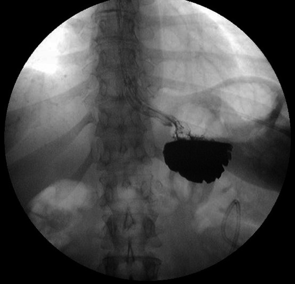
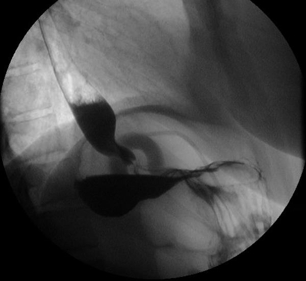
Nowadays, after many years of constant improvements and modifications, the most frequently used procedures of bariatric surgery are the methods of gastric restriction, i.e. decreasing the volume of the stomach. Their most popular example is the ASGB surgery [6,7]. The idea of this surgery is to place the band under the gastric cardia, around the whole stomach, dividing it into two parts (Figure 2A, B). This surgery was carried out for the first time by Wilkinson, Kolle and Molin. Kuzmak modified it by introducing a band in the form of a torus (tube-shaped) which can be filled with fluid (in practice this is mostly saline or contrast medium), allowing for a regulation of the lumen. As in gastroplasties, the upper segment is able to embrace up to a few dozens of millimeters of food (20–25 ml mostly). Further periodical follow-up requires a close cooperation of the radiologist and operating surgeon. Contrast-enhanced examination of the upper gastrointestinal tract, with fluoroscopy, remains the best method of lumen regulation. The first radiological evaluation is normally performed after 1–2 days following the procedure. It is aimed to control the components of the whole system, and especially the exact location of the band, and to exclude a possible perforation of the gastrointestinal tract. It is important to pay attention to the level of banding – it should be located a few cm below the diaphragm. The angle between the ASBG and the vertical plane should normally amount to 50–60°. The band regulates the width of the lumen and the time of food passage from the upper stomach segment by an individually adjusted degree of lumen contraction. Edema of the mucous membrane, caused by every procedure, does not allow for a final regulation of the lumen in the first radiological examination. Normally, this is done after 6–8 weeks from the moment of banding. The targeted lumen is 3.0–3.5 mm in width [11]. After the procedure, it is necessary to monitor the patient for some time because in the progressing process of weight loss, the gastric volume decreases as well, leading to band loosening – and thus to lumen widening which requires ‘tightening’ by introduction of approx. 0.5–1.5 ml of saline to the system. The procedure is normally performed under fluoroscopic guidance. A regulatory port is placed under the skin on the abdomen (Figure 3). Currently, ASGB is usually performed laparoscopically. Laparoscopy is also used in case of complications requiring band removal [6]. It is worth underscoring that during banding, the gastrointestinal tract is not dissected, which reduces the number of complications and shortens the time of postoperative care. Placing the band too high (mostly in the abdominal segment of the esophagus (Figure 4)) reduces body mass loss. At the same time, it increases the incidence of gastroesophageal reflux, inflammation of the gastric and esophageal mucous membrane, and disturbed motor activity of the esophagus (hyperkinesis in the form of tertiary contractions which increase the time of esophageal stasis of the refluxed material) (Figure 5). Gastroesophageal reflux disease (GERD) is the most frequently reported adverse effect, found in 12–22% of patients with a band. In an increased form, it often leads to ASGB removal or to repeated bariatric surgeries, by means of a different method, e.g. Roux-en-Y [6,9,12]. After performing one radiography and not knowing a detailed history of surgery, it is not always possible to evaluate whether the band was originally placed too high (in the abdominal segment of the esophagus) or whether it slipped out of place (Figure 6). In case of band displacement, which happens in 18–36% of postoperative cases, the lumen of the band may become wider. However, it is more often that the lumen tightens up and the upper gastric pouch gets enlarged and starts hanging over the band [6,7,9,12,13]. The lack of body mass loss or constant vomiting after the consumption of even the smallest amounts of food, are usually the causes of visit to the surgeon. The stomach may be hanging over the band also if the band is placed at a too big angle (horizontal placement), if the upper, surgically created gastric pouch is too large (over 20–25 cm3 in volume) or is expanded to as much as 100–150 cm3. The incidence of ‘upper stomach’ hanging over the band was reported in 12–15% of cases [6,7,12]. A plain scopic examination in such cases will show a fluid level in the upper gastric pouch after contrast medium administration – its visible retention in the part hanging over the band (Figure 7A, B). Such images should be differentiated with a cascade stomach (cascade in the fundus) which develops irrespective of ASGB. Characteristic for the stomach hanging over the band is that the upper gastric pouch is located at the level of or, more frequently, below the band, i.e. different than in the cascade stomach, with the upper part of the abdomen lying above the band (Figure 8A, B). One of the most serious complications of ASGB is the migration of the band through the abdominal wall, to the abdominal lumen. This situation is an absolute indication for band removal. It is believed that the most frequent cause of band migration is the weakening of the gastric wall, due to a local, long-term ischemia caused by the compression of the band. Such severe complications are found in up to 1–4% of all performed procedures [12,13]. Very often, patients do not experience any signs or symptoms but they may report stomach aches. Radiological examinations show a strange contrast passage, i.e. inside the GI tract but outside the lumen of the band (Figure 9A–C). If the migration is accompanied by abdominal wall perforation, one of the radiological symptoms is the presence of air under the phrenic domes [14,15]. Another severe complication requiring band removal is the gastric stenosis at the level of the band, which cannot be regulated by any functional loosening of ASGB. The incidence of this complication is evaluated for 10%. Relatively rare (1–8%) are the complications connected with the site of the port. These are mostly infections and abscesses (which however were not found in the studies presented by us). In 1% of cases, the ports get inverted [6,11]. The least frequent (0.3–0.5%) complications are when the band breaks or opens, which can occur many years after the procedure. The most common symptom reported by the patients is the body mass gain (Figure 10A–C).
Figure 2A.

Scopy of a correctly placed gastric band.
Figure 2B.

Diagram of a correctly placed gastric band, with α angle between the band and the vertical line of approx. 55°.
Figure 3.

Scopy of a needle in a port. Regulation of the degree of band constriction.
Figure 4.

High placement of the band – abdominal esophagus.
Figure 5.

Disturbed motor activity of the esophagus – tertiary contractions.
Figure 6.

ASGB displacement. A wide lumen of the band.
Figure 7A.
The upper gastric segment hanging over the band, AP scopy.
Figure 7B.

The upper gastric segment hanging over the band, profile scopy.
Figure 8A.

Stomach with a cascade in the fundus. AP scopy.
Figure 8B.
Stomach with a cascade in the fundus. Profile scopy.
Figure 9A.

A migrating band. AP radiography.
Figure 9C.

A migrating band. Profile scopy.
Figure 10A.

Band opening.AP scopy.
Figure 10C.

Band opening. AP scopy.
Conclusions
Publications of the most recent results of prospective randomized trials comparing different obesity surgeries contributed to the advancement in bariatric surgeries. It is important that a similar progress was made simultaneously in the field of radiological imaging of the performed procedures. ASGB (ALGB) and VBG are effective, safe, and increasingly more popular methods of surgical treatment in obesity. Their application requires a constant cooperation between surgeons and radiologists, and a thorough and comprehensive evaluation of the system by the radiologist: correctness of implantation (including port placement), tightness of the system, level and angle of the band, size of the upper gastric segment, width of the lumen of the band, and exclusion of potential adverse effects.
Figure 1B.

VBG with a band – AP scopy.
Figure 9B.

A migrating band. AP scopy.
Figure 10B.

Band opening. Profile scopy.
References:
- 1.The WHO European Ministerial Conference on Counteracting Obesity; Turkey. 15–17.11.2006; http://www.euro.who.int/obesity. [Google Scholar]
- 2.The challenge of obesity in the WHO European Region; Copenhagen, Bucharest. 12 September 2005; http://www.euro.who.int/obesity. [Google Scholar]
- 3.Piechota G, Karwat D, Małkiewicz J, et al. Sedentary Lifestyle between population of patients with overweight and obesity. Polish Journal of Environmental Studies. 2007;16(5A):394–98. [Google Scholar]
- 4.Piechota G, Karwat D, Małkiewicz J, et al. Nutritional habits of patients with overweight and obesity. Polish Journal of Environmental Studies. 2007;16(5A):390–93. [Google Scholar]
- 5.Flum DR, Belle SH, King WC, et al. The Longitudinal Assessment of Bariatric Surgery (LABS) Consortium Perioperative Safety in the Longitudinal Assessment of Bariatric Surgery. NEJM. 2009;361(5):445–54. doi: 10.1056/NEJMoa0901836. [DOI] [PMC free article] [PubMed] [Google Scholar]
- 6.Scozzari G, Farinella E, Bonnet G, et al. Laparoscopic Adjustable Silicone Gastric Banding vs Laparoscopic Vertical Banded Gastroplasty in Morbidly Obese Patients: Long-Term Results of a Prospective Randomized Controlled Clinical Trial. Obesity Surgery. 2009;19(8):1108–15. doi: 10.1007/s11695-009-9871-1. [DOI] [PubMed] [Google Scholar]
- 7.Parikh MS, Laker S, Weiner M, et al. Objective Comparison of Complications Resulting from Laparoscopic Bariatric Procedures. J Am Coll Surg. 2006;202(2):252–61. doi: 10.1016/j.jamcollsurg.2005.10.003. [DOI] [PubMed] [Google Scholar]
- 8.McGregor A. American Society for Metabolic and Bariatric Surgery; 1999. The Story for Surgery Obesity. www.ams.bsorg. [Google Scholar]
- 9.Morino M, Toppino M, Bonnet G, et al. Laparoscopic Adjustable Silicone Gastric Banding Versus Vertical Banded Gastroplasty in Morbidly Obese Patients: A Prospective Randomized Controlled Clinical Trial. Ann Surg. 2003;238:835–42. doi: 10.1097/01.sla.0000098627.18574.72. [DOI] [PMC free article] [PubMed] [Google Scholar]
- 10.Kwiatkowski A, Paśnik K, Stanowski E, et al. Regression of metabolic syndrome depending on type of bariatric surgery. Videosurgery and other Miniinvasive Techniques. 2009;4(2):53–58. [Google Scholar]
- 11.Wiesner W, Schoeb O, Hauser RS, et al. Adjustable Laparoscopic Gastric Banding in Patients with Morbid Obesity: Radiographic Management, Results and Postoperative Complications. Radiology. 2000;216:389–94. doi: 10.1148/radiology.216.2.r00au28389. [DOI] [PubMed] [Google Scholar]
- 12.Favretti F, Enzi G, Pizzirani E, et al. Adjustable Silicon Gastric Banding (ASGB): the Italian experience. Obes Surg. 1993;3(1):53–56. doi: 10.1381/096089293765559773. [DOI] [PubMed] [Google Scholar]
- 13.O’Brien PE, Dixon JB, Brown W, et al. The laparoscopic adjustable gastric band (Lap-Band): a prospective study of medium-term effect on weight, health and quality of life. Obes Surg. 2002;12(5):652–60. doi: 10.1381/096089202321019639. [DOI] [PubMed] [Google Scholar]
- 14.Pretolesi F, Camerini G, Gianetta E, et al. Intraluminal penetration of the band in patients with adjustable silicone gastric banding: radiological findings. EJR. 2001;11:412–16. doi: 10.1007/s003300000720. [DOI] [PubMed] [Google Scholar]
- 15.Hainaux B, Agneessens E, Rubesova E, et al. Intragastric Band Erosion After Laparoscopic Adjustable Gastric Banding for Morbid Obesity: Imaging Characteristics of an Underreported Complication. AJR. 2005;184:109–12. doi: 10.2214/ajr.184.1.01840109. [DOI] [PubMed] [Google Scholar]