Summary
Background:
At present, there is a number of diagnostic imaging procedures allowing for the evaluation of atherosclerosis. The earliest, subclinical stage of atherosclerosis can be visualized with the development of computed tomography (CT) and ultrasound (US) techniques. Therefore, the purpose of this study was to assess the degree of coronary artery calcification and carotid intima-media thickness in diabetic subjects divided into different age groups.
Material/Methods:
Fifty-six men, aged from 18 to 72 were included in the study. Participants were divided into 4 groups according to age (18–30, 31–45, 46–60 and more than 60 years). Two tests were performed: coronary calcium score (CS) determination and intima-media thickness (IMT) in ultrasound. CS was performed using a multi-slice scanner. Images were analyzed using the Agatson method. Ultrasound examinations were performed using a 9–12-MHz linear transducer.
Results:
The correlation coefficient between calcium score index (CSI) and age of patients was 0.52 (p<0.001). The correlation between duration of diabetes and CSI was significantly lower (r=0.3; p<0.05). The increase of IMT is associated with age to a much greater extent and the correlation coefficient was 0.63 (p<0.001). IMT depended on the duration of diabetes, but the correlation was also weak (r=0.35; p<0.01).
Conclusions:
Comparison of the findings obtained in the presented study and in the group of healthy subjects proves that influence of diabetes on vascular deterioration may be observed, even among young individuals. Obtained results allow to make the following conclusions: 1. Calcium score index remains low in the group of male patients with diabetes before the age of 45. 2. Intima-media thickness correlates well with age (r=0.6; p<0.05) and weaker with the duration of diabetes (r=0.35; p<0.05). 3. IMT assessment may be a useful tool to identify the increased predisposition to atherosclerosis, also before the age of 30.
Keywords: calcium score, IMT, computed tomography, ultrasound, diabetes
Background
It has been widely demonstrated that atherosclerotic process affects everyone but its intensity depends on genetic predispositions, environmental factors and co-morbidities [1]. It was demonstrated in post-mortem examinations that atherosclerosis may commence in childhood or adolescence while its symptoms occurring in the older population come as a consequence of gradually increasing morphological changes, their mineralization with calcium phosphate and regressive changes [2–4]. Currently, the diagnostics should be shifted toward identifying preclinical changes, as they are reversible, easier and less expensive to treat. Over the past few years there has been a great progress in the area of invasive and noninvasive modalities of vascular wall morphology evaluation. Multi-slice CT angiography is a modern method of vascular wall assessment. The newest devices visualize vessels with a resolution of less than 1 mm and special software enables examination of coronary arteries and the extent of their calcification – calcium score (CS).
Ultrasonographic assessment of intima-media complex thickness has also been proven useful in the process of evaluation of predispositions to atherosclerosis. Since it is inexpensive and noninvasive, its role is rising, especially in monitoring of treatment.
Therefore, the goal of this study was to compare the values of CS index and intima-media complex thickness in diabetic men in various age groups.
Material and Methods
Population
The study was conducted on 56 males between 18 and 72 years of age (patients were divided into four age groups), suffering from diabetes and without additional co-morbidities. The qualification procedure was aimed at the elimination of other proatherotic factors, especially smoking and hypertension. Therefore, diabetes could be considered the main factor influencing the results of tests. Body mass, blood glucose and blood pressure measurements were noted, as well as a lipid profile. All patients were informed about the purpose and extent of performed tests and consented to the participation. The study protocol was accepted by the University Bioethics Committee on 26 October 2006.
Clinical parameters obtained in groups are summarized in Table 1.
Table 1.
Clinical characteristics of investigated group.
| Age groups | 18–30 I | 31–45 II | 46–60 III | >60 IV |
|---|---|---|---|---|
| Group size | 12 | 12 | 21 | 11 |
| Mean age | 23.9 | 36.6 | 52.4 | 65.7 |
| Mean time of duration of diabetes | 2.5 | 12.0 | 16.4 | 24.3 |
| Mean BMI value | 22.7 | 24.8 | 26.1 | 25.8 |
| Mean blond glucose level (mmol/l) | 4.3 | 4.5 | 4.5 | 4.9 |
| Mean systolic blood pressure (mmHg) | 118 | 122 | 128 | 137 |
Methods
Coronary artery calcification index measurements were performed using a 64-slice computed tomograph LightSpeed GE equipped with Smartscore™ software. Rotation speed was 0.35 seconds and table speed was 175 mm/s (volumetric cardiac imaging can be performed within 5 beats – 5-Beat Cardiac™) with temporal resolution of 33 ms, which eliminates movement artifacts. Imaging does not require application of a contrast medium. Data acquisition is conducted while the patient holds her/his breath. Subsequently, a segmental, retrospective RetroRecon analysis is performed. CS index is automatically calculated and presented as a report for each coronary artery and a total index value.
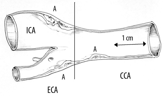
For ultrasound examination of intima-media complex, a USG Logiq 7 (GE) with a 7–14-MHz linear wideband transducer was used. Examinations were performed according to the standards accepted in the literature. The intima-media thickness (IMT) was evaluated bilaterally in 2 locations with 3 measurements, using the anterior approach, about 1.5 cm below the bifurcations of common carotid arteries. Measurements were made on one-centimeter-long segments on the distal walls of arteries visualized in the long axis, avoiding potential atherosclerotic lesions. The method of measurement is presented in Figure 1.
Figure 1.
The scheme of intima-media complex measurement: assessment was carried out in the posterior wall of the common carotid artery (CCA), below its bifurcation into internal (ICA) and external (ECA) carotid artery excluding atherosclerotic sections.
Test results were correlated with age of the subjects and duration of diabetes and subsequently, using student’s t-test, compared in particular age groups. The level of statistical significance was accepted at p<0.05.
Results
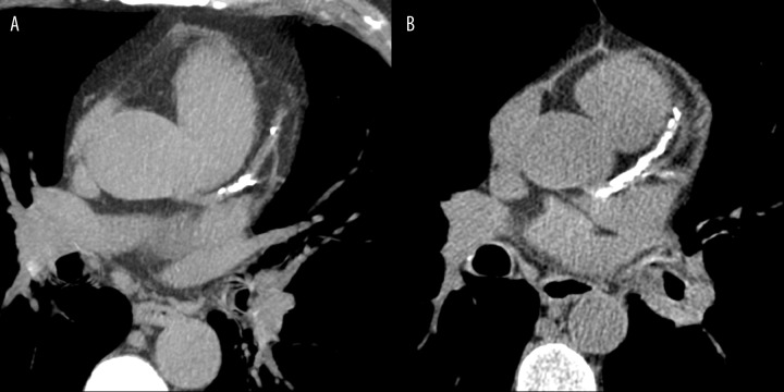
No coronary artery calcifications were observed in the subgroup aged below 28 years. In group I, calcifications were noted in 1 case (9%), in group II in 2 subjects (17%), in group III in as many as 18 (85%) patients and in group IV in 10 persons, which constituted 90% (Figure 2).
Figure 2.
Calcified atherosclerotic plaques in the left coronary artery – varying extent of lesions. (A) single calcification in the anterior descending branch – CS=253, (B) massive changes within the same vessel and diagonal branches – CS=2490.
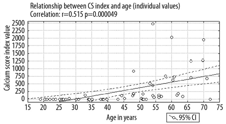
Detailed data regarding CS index values in particular age groups are presented in Table 2 and Figure 3.
Table 2.
Mean CS values for every age group.
| Age group | Mean | Median | Minimum | Maximum | SD | ||
|---|---|---|---|---|---|---|---|
| 18–30 I | CS | 12 | 2.2 | 0 | 0 | 27 | 7.8 |
| CS >0 | 1 | 27 | – | – | – | – | |
| 31–45 II | CS | 12 | 20 | 0 | 0 | 129 | 47 |
| CS >0 | 2 | 120.5 | 120.5 | 112 | 129 | 12 | |
| 46–60 III | CS | 21 | 394 | 115 | 0 | 2469 | 670 |
| CS >0 | 18 | 460 | 147.5 | 1 | 2469 | 704 | |
| >60 IV | CS | 11 | 691 | 683 | 0 | 1950 | 605 |
| CS >0 | 10 | 760 | 692 | 86 | 1950 | 591 | |
Figure 3.
Dependence of CS and age (individual values), correlation: r=0.515, p=0.000049.
CS – signifies the mean coronary artery calcification index value in the entire study group.
CS>0 – signifies the mean coronary artery calcification index value in patients with calcifications.
The correlation coefficient for CS index and patient age was over 0.5 (p<0.001). The correlation between the duration of diabetes and the value of CS index was weaker (r=0.3; p<0.05). Notably, there is a significant dispersion in the results of CSI measurements. A significant percentage of patients have index values of 0 or ≤250, both in the group with diabetes diagnosed less than 5 years prior as well as in the group treated for more than 20 years. Sporadically, CS values of over 2000 were noted including one patient with diabetes diagnosed within less than 5 years before.
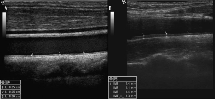
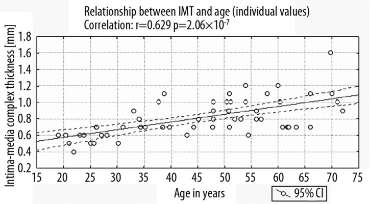
Good-quality images of intima-media complex were obtained in all studied cases. The lowest values were demonstrated in group I – mean IMT of 0.57 mm. In groups II–IV, mean IM thickness was higher and amounted to: II – 0.76 mm; III – 0.92 mm; IV – 0.93 mm (Figure 4).
Figure 4.
The intima-media complex assessment. IMT thickness of 0.6 mm (A), 1.3 mm (B).
IMT values were presented in detail for each group in Table 3 and Figure 5.
Table 3.
Mean IMT values for every age group.
| Age group | Mean IMT value (mm) | Median | Minimum | Maximum | SD |
|---|---|---|---|---|---|
| 18–30 (I) | 0.57 | 0.6 | 0.4 | 0.7 | 0.09 |
| 31–45 (II) | 0.76 | 0.7 | 0.5 | 1.1 | 0.17 |
| 46–60 (III) | 0.92 | 0.9 | 1.2 | 0.6 | 0.17 |
| >60 (IV) | 0.93 | 0.9 | 1.6 | 0.7 | 0.28 |
Figure 5.
Dependence of IMT and age (individual values), correlation: r=0.629, p=2.06×10⁻⁷.
The observed increase in intima-media complex thickness correlated with age. The correlation coefficient r was 0.63 (p<0.001). Linear correlation coefficient with regard to the means for the studied age groups was equally high.
Intima-media thickness depends on the duration of diabetes, but this association is weak. The degree of correlation between IM thickness and duration of diabetes was lower than the degree of correlation between IMT and patient age, and amounted to: r=0.35 (p<0.01).
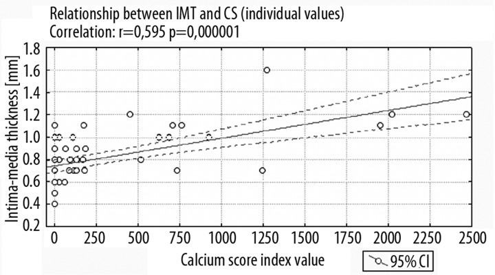
A wide range of IMT values was observed in the group of people without calcifications in coronary arteries. In the group of subjects with intima-media thickness higher than or equal to 1 mm, the percentage of people with calcium score of 0 was 19%. Even though in a substantial number of cases the CS value was 0, the relationship between intima-media complex thickening and calcium score index was apparent (Figure 6) and greater than the correlation between IMT values and the duration of diabetes (correlation coefficients of 0.59 and 0.35 respectively), but lower than the correlation between IMT value and age (correlation coefficient −0.63).
Figure 6.
Dependence of IMT and CS (individual values), correlation: r=0.595, p=0.000001.
Discussion
Prophylaxis of complications of angiodegenerative processes, especially atherosclerosis, should be based on assessment and potential modifications of risk factors for their development. One of the methods of such evaluation is the Framingham scale developed on the basis of one of the best-documented prospective studies on ischemic heart disease. Technological progress enabling visualization of coronary artery calcifications allowed for the evaluation of their prognostic value and potential application of CS index as an additional parameter improving risk assessment based on traditional scales. Numerous studies have demonstrated the prognostic significance of coronary artery calcification index calculated according to the Agatston algorithm applied in this study [5–9].
The data from the literature indicate that age is the main factor influencing the presence of coronary artery calcifications and that calcium score index increases in proportion to the number of traditional risk factors for a cardiovascular event and correlates with the presence of hemodynamically significant coronary artery stenoses [10].
As shown by the data published by Hoff and Pletcher [11,12], in the elderly calcifications may appear even without the presence of identified, additional risk factors. Calcified plaque occurs less often in people less than 30 years old independently of such proatherotic risk factors as smoking, obesity, hypercholesterolemia, hypertension or diabetes, which raises doubts as to the value of CS as an early prognostic test for the occurrence of angiodegeneration.
The relationship between the extent of coronary artery calcifications and duration of diabetes was demonstrated on larger populations, together with the fact that in this group of patients atherosclerotic lesions are more frequent and more intensive in comparison healthy subjects of the same age group, which the presented study confirmed. At the same time, in the diabetic group, absence of coronary artery calcifications is associated with low short-term risk of death and CS index may be a useful tool in risk assessment of occurrence of silent myocardial infarction in patients with stable, uncomplicated type 2 diabetes [13,14].
Many studies conducted in the past showed that the intima-media complex thickness correlates well with the extent of vascular degenerative processes. Atherosclerosis causes thickening of IMT, leading to atherosclerotic plaque formation and complete effacement of vascular wall echo-structure [15].
In this study, a strong correlation between intima-media thickness and age was observed, also in relation to the means for the studied age groups. Under normal conditions, intima-media thickness ranges from 0.6 to 1.0 mm. Many authors consider IMT of over 1 mm or even 1.2 mm as abnormal. Mean IMT value accepted in various scientific papers may also depend on the method of taking measurements and consistently rises in patients burdened with risk factors. In the work presented here, IMT value was measured on the distal wall, avoiding visible atherosclerotic plaques, according to the protocol accepted in the literature as an experts’ consensus [15]. Mean IMTs for all age groups were higher than in the cross-sectional data published by Touboul et al. (in the age group of 30–49 years: 0.63–0.73 vs. 0.76 in our group; in the age group of 50–59 years: 0.7–0.78 vs. 0.92 in our group; in the age group over 60 years: 0.79–0.85 vs. 0.93 in our group).
In the conducted study, an increase in IMT value was observed also in a group of people without calcifications in coronary arteries. In 19% of patients with intima-media complex thickness higher than or equal to 1 mm, calcium score value was 0. However, the relationship between the degree of intima-media complex thickening and calcium score index value was apparent and greater than the correlation between IMT values and duration of diabetes, but lower than the correlation between IMT and age. Obtained results suggest that IMT and CS values relate to different aspects of the degenerative process – IMT value may be more accurate in determining the extent of lesions in the vascular wall at an early stage of angiodegenerative process, especially in younger patients.
In this work, intima-media thickness is related to the duration of diabetes but this relationship is weak. The degree of correlation between IM thickness and duration of diabetes was lower than the association with the age of the subjects. The cause for this weaker correlation, aside from previously mentioned difficulties with defining the exact time of disease onset in case of patients with type 2 diabetes, may be intensive therapy of type 1 diabetes, which slows down the rate of IMC thickening [16].
Conclusions
In diabetic men, coronary artery calcification (CS) index remains low until the age of 45 despite the burden of the disease and intima-media thickness correlates well with age (r=0.6; p<0.05) and to a lesser extent with the duration of the disease (r=0.35; p<0.05). IMT assessment may be a useful tool in the evaluation of angiodegenerative predispositions, also in people below 30 years of age.
References:
- 1.Rosamond W, Flegal K, Friday G, et al. Heart Disease and Stroke Statistics – 2007 Update A Report From the American Heart Association Statistics Committee and Stroke Statistics Subcommittee. Circulation. :2006. doi: 10.1161/CIRCULATIONAHA.106.179918. [DOI] [PubMed] [Google Scholar]
- 2.Berenson GS, Wattigney WA, Tracy RE, et al. Atherosclerosis of the aorta and coronary arteries and cardiovascular risk factors in persons aged 6 to 30 years and studied at necropsy (The Bogalusa Heart Study) Am J Cardiol. 1992;70(9):851–58. doi: 10.1016/0002-9149(92)90726-f. [DOI] [PubMed] [Google Scholar]
- 3.Li S, Chen W, Srinivasan SR, et al. Childhood cardiovascular risk factors and carotid vascular changes in adulthood: the Bogalusa Heart Study. JAMA. 2003;290(17):2271–76. doi: 10.1001/jama.290.17.2271. [DOI] [PubMed] [Google Scholar]
- 4.Strong JP, Malcom GT, McMahan CA, et al. Prevalence and extent of atherosclerosis in adolescents and young adults: implications for prevention from the Pathobiological Determinants of Atherosclerosis in Youth Study. JAMA. 1999;281(8):727–35. doi: 10.1001/jama.281.8.727. [DOI] [PubMed] [Google Scholar]
- 5.Becker CR, Kleffel T, Crispin A, et al. Coronary artery calcium measurement: agreement of multirow detector and electron beam CT. AJR Am J Roentgenol. 2001;176(5):1295–98. doi: 10.2214/ajr.176.5.1761295. [DOI] [PubMed] [Google Scholar]
- 6.Carr JJ, Crouse JR, III, Goff DC, et al. Evaluation of subsecond gated helical CT for quantification of coronary artery calcium and comparison with electron beam CT. AJR Am J Roentgenol. 2000;174(4):915–21. doi: 10.2214/ajr.174.4.1740915. [DOI] [PubMed] [Google Scholar]
- 7.Daniell AL, Wong ND, Friedman JD, et al. Concordance of coronary artery calcium estimates between MDCT and electron beam tomography. AJR Am J Roentgenol. 2005;185(6):1542–45. doi: 10.2214/AJR.04.0333. [DOI] [PubMed] [Google Scholar]
- 8.Knez A, Becker C, Becker A, et al. Determination of coronary calcium with multi-slice spiral computed tomography: a comparative study with electron-beam CT. Int J Cardiovasc Imaging. 2002;18(4):295–303. doi: 10.1023/a:1015536705455. [DOI] [PubMed] [Google Scholar]
- 9.Stanford W, Thompson BH, Burns TL, et al. Coronary artery calcium quantification at multi-detector row helical CT versus electron-beam CT. Radiology. 2004;230(2):397–402. doi: 10.1148/radiol.2302020901. [DOI] [PubMed] [Google Scholar]
- 10.Rumberger JA, Schwartz RS, Simons DB, et al. Relation of coronary calcium determined by electron beam computed tomography and lumen narrowing determined by autopsy. Am J Cardiol. 1994;73(16):1169–73. doi: 10.1016/0002-9149(94)90176-7. [DOI] [PubMed] [Google Scholar]
- 11.Hoff JA, Chomka EV, Krainik AJ, et al. Age and gender distributions of coronary artery calcium detected by electron beam tomography in 35,246 adults. Am J Cardiol. 2001;87(12):1335–39. doi: 10.1016/s0002-9149(01)01548-x. [DOI] [PubMed] [Google Scholar]
- 12.Pletcher MJ, Tice JA, Pignone M, et al. What does my patient’s coronary artery calcium score mean? Combining information from the coronary artery calcium score with information from conventional risk factors to estimate coronary heart disease risk. BMC Med. 2004;2:31. doi: 10.1186/1741-7015-2-31. [DOI] [PMC free article] [PubMed] [Google Scholar]
- 13.Anand DV, Lim E, Hopkins D, et al. Risk stratification in uncomplicated type 2 diabetes: prospective evaluation of the combined use of coronary artery calcium imaging and selective myocardial perfusion scintigraphy. Eur Heart J. 2006;27(6):713–21. doi: 10.1093/eurheartj/ehi808. [DOI] [PubMed] [Google Scholar]
- 14.Raggi P, Shaw LJ, Berman DS, et al. Prognostic value of coronary artery calcium screening in subjects with and without diabetes. J Am Coll Cardiol. 2004;43(9):1663–69. doi: 10.1016/j.jacc.2003.09.068. [DOI] [PubMed] [Google Scholar]
- 15.Touboul PJ, Hennerici MG, Meairs S, et al. Mannheim carotid intima-media thickness consensus (2004–2006). An update on behalf of the Advisory Board of the 3rd and 4th Watching the Risk Symposium, 13th and 15th European Stroke Conferences, Mannheim, Germany, 2004, and Brussels, Belgium, 2006. Cerebrovasc Dis. 2007;23(1):75–80. doi: 10.1159/000097034. [DOI] [PubMed] [Google Scholar]
- 16.Nathan DM, Lachin J, Cleary P, et al. Intensive diabetes therapy and carotid intima-media thickness in type 1 diabetes mellitus. N Engl J Med. 2003;348(23):2294–303. doi: 10.1056/NEJMoa022314. [DOI] [PMC free article] [PubMed] [Google Scholar]


