Abstract
Dengue fever is an important public health problem in Jamaica and has various serious manifestations, which if not identified and treated at the appropriate time can lead to dire consequences. Bacterial co-infections have been seen in clinical practice but may be thought of as simply coincidental. This review highlights the importance of bacteria in exacerbating dengue infections and the importance of looking for co-infection in patients with certain clinical manifestations. It also provides the reader with a scientific understanding of the immune pathogenesis of dengue and bacterial co-infections.
Background
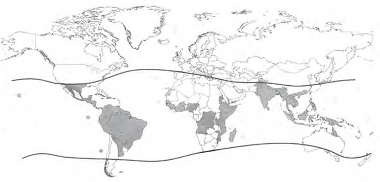
Dengue is a major medical problem in tropical regions (figure 1).1–3 In 2007, over 890 000 cases of dengue were reported to the WHO, in the Americans, 500 000 people with dengue haemorrhagic fever (DHF) require hospitalisation each year and without proper treatment the case death can be as high as 20%. It is endemic in over 100 countries worldwide.2 4 5 Dengue fever is now, as of 2009, classified into dengue fever and severe dengue. It is endemic in Jamaica and the Caribbean.6 7 Cases of dengue co-infection are seen but are sometimes just thought of as simply coincidence.
Figure 1.

WHO diagram showing distribution of dengue and Aedes aegypti. Dark areas: countries or areas at risk of dengue.
Case presentation
This is a report of a previously healthy 15-year-old man who was transferred to the University Hospital of the West Indies with a 1 day fever, diarrhoea and decreased responsiveness. The relative of the patient indicated that the he was well until he started having fever. There was no associated headache, no vomiting, no myalgia, no joint pain or rash.
He had an otherwise unremarkable history.
On examination, a young male was found drowsy looking, mucous membranes were pink and dry. He was anicteric, acyanotic and afebrile with conjuctival congestion.
The patient’s vitals were as follows: his blood pressure was 133/124 mm Hg, pulse 113 beats per min, respiratory rate 28 breaths per min, temperature 36.6C, saturation of oxygen was less than 95% on room air.
Respiratory system showed bronchovesicular sounds with basal crepitations and dullness on the left with decreased air entry.
Neurologic system showed a patient arousable to sternal rub, pupils were equal and reactive, fuduscopy was normal, muscle tone was decreased, kernig’s sign and neck rigidity were both negative.
The patient spent 12 days in the intensive care unit (ICU) and 5 days on the ward before discharge. He improved markedly.
During his stay in the ICU he was found to have rhabdomyolisis and a worsening of his respiratory symptoms characterised by a respiratory rate increase to 45 to 47 breaths per min on 15 liters of oxygen while in the ICU. This developed after the second day of ICU admission. He was intubated in ICU subsequently, due to his deterioration. Additionally, the patient developed hepatomegaly and renal dysfunction.1 The patient was found to have a liver enlargement of 12 centimeters and shifting dullness in his abdomen. The hepatomegaly decreased to 10 cm before discharge.
He was discharged to return to clinic however, he later developed respiratory problems due to intubation and was hospitalised for a short period at another hospital.
He was subsequently discharged and is well.
Investigations
On admission, his chest x-ray showed bilateral alveolar infiltrates with flattening of the right hemidiphragm. His x-ray report 6 days before discharge showed resolution of prior diffuse bilateral airspace opacification with a single thin walled cavitating lesion in the upper right zone. There was also blunting of the left pleural consistent with pleural effusion. There were no other significant findings.
The CT report of his brain was unremarkable.
His white blood cell count was 0.5×10⁹ cells/l on admission increasing to as much as 34.7×10⁹ cell/l 8 days after and decreasing to 16×109 cell/l thereafter. His platelets were 92×109 cell/l on admission. His platelets increased to within normal limits before discharge.
The patients’s total protein was 18.5 g/l, albumin 37.4 g/l, globulin 31.1 g/l, total bilirubin 17.8 umol/l, direct bilirubin 5.33 umol/l, alkalinephosphatase 148.2 U/l, γ glutamyl transferase 39.2 U/l, alanine aminotransferase 58.3 U/l, aspartate aminotransferase 134.5 U/l, creatinine phosphokinase 442 U/l, lactate dehydrogenase 428 U/l, sodium (Na+) 138.3 mmol/l, potassium (K+) 4.33 mmol/l, chloride Cl 98.8 mmol/l, blood urea nitrogen 9.6 mmol/d, creatinine 125 mol/d. Of note his liver parameters were elevated and his renal function was abnormal. His creatinine phoshokinase level was also elevated.
The patient’s dengue IgM was positive by dengue fever virus IgM Capture DxSelect ELISA. His hepatitis B surface antigen and hepatitis C screens were negative. The patient had two sputum cultures positive for Staphylococcus aureus, with sensitivities recorded to the following antibiotics, oxacillin, augmentin, gentamicin and erythomicin. The Kirby–Bauer disc diffusion method was used. Urine cultures and blood cultures were negative. S aureus control used was the S aureus ATCC strain 25923 and BD BBL rabbit coagulase plasma was used for the tube cogaulase test.
The patient had one sputum culture positive for coagulase negative Staphylococcus, after negative cultures for the S aureus. This was resistant to oxacillin by the Kirby–Bauer disc diffusion method.
Differential diagnosis
His problems were 1) pulmonary hypertension 2) rhabdomyolysis 3) haematuria 4) S aureus pneumonia and 5) pancytopenia.
Treatment
Ceftriaxone 2 grams intravenously once daily (od) and levofloxacin 750 mg intravenously od were commenced empirically and was subsequently switched to vancomicin and ciprofloxacin after getting culture reports.
Treatment included normal saline infusions, sodium bicarbonate- 75 mls in 500 mls of normal saline over 4 h, 10 mls of 10% Ca gluconate.
He was also treated with baralgin 1 g po od, panadol 1 g po four times daily, lasix 20 mg intravenously twice daily, lansoprazole 30 mg po od, bisolvon 8 mg po id and ventolin/atrovent nebulisers every 4 h. He was also given iron and folate supplements as well as blood transfusions.
The patient was discharged and will return to clinic for follow-up. All biochemical and haematologic parameters returned to normal.
Outcome and follow-up
The patient recovered completely and is being seen as an outpatient.
Discussion
In clinical and laboratory practice, cases of dengue virus and bacterial co-infection are seen, Lee et al observed that 5.5% of the patients among 774 patients presenting with DHF/dengue shock syndrome (DSS) showed bacteremia.8 9 Co-infection has been shown to worsen the outcome of dengue infection thus clinicians will find this review helpful as it highlights the clinical and scientific significance of dengue and bacterial co-infection.8
In the study by Lee et al, patients with prolonged fever, higher frequencies of acute renal failure, gastrointestinal bleeding, altered consciousness, unusual dengue manifestations and DSS needed to be assessed for possible co-infection.8 This patient had altered consciousness, rhabdomyolisis and renal impairment and thus fits Lee et al’s findings, however the patient did not have a prolonged course of fever only intermittent episodes over the course of illness.1 8 A prolonged course of fever has been shown to be an independent risk factor for co-infection.8 Interestingly, Lee et al also did not find a statistically significant association with raised white blood cell count.8 This is important to note as our case report showed an increasing white blood cell count, which is expected in a bacterial infection. This is important as it informs us that white blood cell count elevation is not a good indicator of bacterial co-infection in dengue patients.
Immunological mechanisms may explain the reason that bacterial co-infection can lead to a worse outcome in dengue co-infection. In particular, cytokines can be implicated. Tumour necrosis factor α, α interferon (IFNα), interleukin-1 (IL-1), IL-8, IL-12, MIP-1, RANTES, migratory inhibitory factor, human cytotoxic factor are cytokines known to be secreted in dengue infection, these are secreted by macrophages and other cells of the immune system.10–20 In one study, increased levels of plasma IL-10 and soluble tumour necrosis factor receptor-II correlated with the degree of thrombocytopaenia, in the same study higher levels of hepatic transaminases were correlated with increase in Il-2.21 This shows the significance of cytokines in the pathogenesis of dengue.
Lipopolysaccharide (LPS) produced by Gram negative bacteria mediate enhancement of virus replication, synergistic IFNα production and general enhancement of cytokine production.11
Gram positive bacteria do not produce LPS, however, they produce other molecules which may result in cytokine mediated pathogenesis. S aureus was found to express high levels of IL-6 and IL-12 in a study by Bost et al.22 This may be part of its pathogenesis in dengue as IL-12 can promote T lymphocytes and natural killer cells to secrete IFNγ which can cause macrophages and, again T lymphocytes to secrete a milieu of cytokines.22
This worsening of outcome due to an aberrant cytokine cascade can thus be seen as the reason, because severe dengue can be seen in primary infection and not just secondary infection, as in this case. This may be so especially in cases of co-infection as explained by Lee et al and others seen in this report. In this case, this patient had no previous history of dengue and had a severe presentation with a bacterial infection, thus the worse outcome of co-infection of S aureus and dengue is not just coincidence and could possibly lead to severe dengue.
Conclusion
Dengue virus and bacterial co-infection should thus be considered in the clinical management of patient with dengue fever or severe dengue. This could help the start of important antibiotics which can be life saving.9 23 It is also important to note that co-infections with Salmonella typhi, Shigella sonnei, hepatitis viruses, flu virus, chikungunya virus, malaria, leptospira, as well as other organisms have been noted in the literature.24–31 In these cases the authors have noted the importance of the clinical implications of co-infection and it is important to note this may not just be treated as a coincidence as it may have important implications for treatment as has been implied for other tropical diseases such as malaria.26–30 32 Finally, Clarke et al noted that the body’s response to a cytokine cascade leads to illness and pathology in malaria and other infections, again emphasising the importance of cytokines in worsening the outcome of co-infection.33
Learning points.
Severe dengue can occur in primary infection.
Co-infection can worsen the outcome of dengue infection.
Patients with prolonged fever, higher frequencies of acute renal failure, gastrointestinal bleeding, altered consciousness, unusual dengue manifestations and DSS needed to be assessed for possible co-infection.
Increased white blood cell count is not an accurate indicator of bacterial infection in dengue and bacterial co-infections.
Acknowledgments
The Consultants, Residents and staff of the Microbiology Department are graciously thanked. The authors would like to also thank Professor John Lindo.
Footnotes
Competing interests: None.
Patient consent: Obtained.
References
- 1.Dengue: Guidelines for diagnosis, treatment, prevention and control [homepage on the internet]. World Health Organisation and Special Programme for Research and Training in Tropical Diseases. WHO 2009. [cited January 2012]. Available from: http://whqlibdoc.who.int/publications/2009/9789241547871_eng.pdf. [Google Scholar]
- 2.George R. Unusual manifestations of dengue. JPOG 1999. [Google Scholar]
- 3.Gibbons RV, Vaughn DW. Dengue: an escalating problem. BMJ 2002;324:1563–6. [DOI] [PMC free article] [PubMed] [Google Scholar]
- 4.WHO Report on Global Surveillance of Epidemic-prone Infectious Diseases – Dengue and dengue haemorrhagic fever [homepage available on the internet]. Global Alert and Response [cited January 2012]. Available from: http://www.who.int/csr/resources/publications/dengue/CSR_ISR_2000_1/en/ (accessed 20 Jan 2012).
- 5.Dengue and dengue haemorrhagic fever [homepage available on the internet]. WHO Media centre March 2009 [cited January 2012]. Available from: http://www.who.int/mediacentre/factsheets/fs117/en/ (accessed 20 Jan 2012).
- 6.Brown MG, Salas RA, Vickers IE, et al. Dengue virus serotypes in Jamaica, 2003-2007. West Indian Med J 2011;60:114–9. [PubMed] [Google Scholar]
- 7.Prince HE, Yeh C, Lapé-Nixon M. Primary and probable secondary dengue virus (DV) infection rates in relation to age among DV IgM-positive patients residing in the United States mainland versus the Caribbean islands. Clin Vaccine Immunol 2012;19:105–8. [DOI] [PMC free article] [PubMed] [Google Scholar]
- 8.Lee IK, Liu JW, Yang KD. Clinical characteristics and risk factors for concurrent bacteremia in adults with dengue hemorrhagic fever. Am J Trop Med Hyg 2005;72:221–6. [PubMed] [Google Scholar]
- 9.Chai LY, Lim PL, Lee CC, et al. Cluster of Staphylococcus aureus and dengue co-infection in Singapore. Ann Acad Med Singap 2007;36:847–50. [PubMed] [Google Scholar]
- 10.Martina BE, Koraka P, Osterhaus AD. Dengue virus pathogenesis: an integrated view. Clin Microbiol Rev 2009;22:564–81. [DOI] [PMC free article] [PubMed] [Google Scholar]
- 11.Chen YC, Wang SY. Activation of terminally differentiated human monocytes/macrophages by dengue virus: productive infection, hierarchical production of innate cytokines and chemokines, and the synergistic effect of lipopolysaccharide. J Virol 2002;76:9877–87. [DOI] [PMC free article] [PubMed] [Google Scholar]
- 12.Chen LC, Lei HY, Liu CC, et al. Correlation of serum levels of macrophage migration inhibitory factor with disease severity and clinical outcome in dengue patients. Am J Trop Med Hyg 2006;74:142–7. [PubMed] [Google Scholar]
- 13.Chaturvedi UC, Agarwal R, Elbishbishi EA, et al. Cytokine cascade in dengue hemorrhagic fever: implications for pathogenesis. FEMS Immunol Med Microbiol 2000;28:183–8. [DOI] [PubMed] [Google Scholar]
- 14.Huang YH, Liu CC, Wang ST, et al. Activation of coagulation and fibrinolysis during dengue virus infection. J Med Virol 2001;63:247–51. [DOI] [PubMed] [Google Scholar]
- 15.Lei HY, Yeh TM, Liu HS, et al. Immunopathogenesis of dengue virus infection. J Biomed Sci 2001;8:377–88. [DOI] [PubMed] [Google Scholar]
- 16.Halstead SB. Neutralization and antibody-dependent enhancement of dengue viruses. Adv Virus Res 2003;60:421–67. [DOI] [PubMed] [Google Scholar]
- 17.Nielsen DG. The relationship of interacting immunological components in dengue pathogenesis. Virol J 2009;6:211. [DOI] [PMC free article] [PubMed] [Google Scholar]
- 18.Huan YL, Huang KJ, Lin YS, et al. Immunopathogenesis of dengue hemorrhagic fever. Am J Infect Dis 2008;4:1–9. [Google Scholar]
- 19.Huang KJ, Yang YC, Lin YS, et al. The dual-specific binding of dengue virus and target cells for the antibody-dependent enhancement of dengue virus infection. J Immunol 2006;176:2825–32. [DOI] [PubMed] [Google Scholar]
- 20.Rothman AL. Dengue: defining protective versus pathologic immunity. J Clin Invest 2004;113:946–51. [DOI] [PMC free article] [PubMed] [Google Scholar]
- 21.Libraty DH, Endy TP, Houng HS, et al. Differing influences of virus burden and immune activation on disease severity in secondary dengue-3 virus infections. J Infect Dis 2002;185:1213–21. [DOI] [PubMed] [Google Scholar]
- 22.Bost KL, Ramp WK, Nicholson NC, et al. Staphylococcus aureus infection of mouse or human osteoblasts induces high levels of interleukin-6 and interleukin-12 production. J Infect Dis 1999;180:1912–20. [DOI] [PubMed] [Google Scholar]
- 23.Araújo SA, Moreira DR, Veloso JM, et al. Fatal Staphylococcal infection following classic Dengue fever. Am J Trop Med Hyg 2010;83:679–82. [DOI] [PMC free article] [PubMed] [Google Scholar]
- 24.Sudjana P, Jusuf H. Concurrent dengue hemorrhagic fever and typhoid fever infection in adult: case report. Southeast Asian J Trop Med Public Health 1998;29:370–2. [PubMed] [Google Scholar]
- 25.Charrel RN, Abboud M, Durand JP, et al. Dual infection by dengue virus and Shigella sonnei in patient returning from India. Emerging Infect Dis 2003;9:271. [DOI] [PMC free article] [PubMed] [Google Scholar]
- 26.Perez MA, Gordon A, Sanchez F, et al. Severe coinfections of dengue and pandemic influenza A H1N1 viruses. Pediatr Infect Dis J 2010;29:1052–5. [DOI] [PMC free article] [PubMed] [Google Scholar]
- 27.Javed Y, Wasim J, Shaheer S, et al. Dengue Fever with Hepatitis E and Hepatitis A infection. J Pak Med Assoc 2009;59:176–7. [PubMed] [Google Scholar]
- 28.Charrel RN, Brouqui P, Foucault C, et al. Concurrent dengue and malaria. Emerging Infect Dis 2005;11:1153–4. [DOI] [PMC free article] [PubMed] [Google Scholar]
- 29.Jeevan MK, Rajendran R, Thangaratham PS, et al. Dual infection by dengue virus and Plasmodium vivax in Alappuzha District, Kerala, India. Jpn J Infect Dis 2006;59:211–2. [PubMed] [Google Scholar]
- 30.Kaur H, John M. Mixed infection due to leptospira and dengue. Indian J Gastroenterol 2002;21:206. [PubMed] [Google Scholar]
- 31.Chahar HS, Bharaj P, Dar L, et al. Co-infections with chikungunya virus and dengue virus in Delhi, India. Emerging Infect Dis 2009;15:1077–80. [DOI] [PMC free article] [PubMed] [Google Scholar]
- 32.Singhsilarak T, Phongtananant S, Jenjittikul M, et al. Possible acute coinfections in Thai malaria patients. Southeast Asian J Trop Med Public Health 2006;37:1–4. [PubMed] [Google Scholar]
- 33.Clark IA, Alleva LM, Budd AC, et al. Understanding the role of inflammatory cytokines in malaria and related diseases. Travel Med Infect Dis 2008;6:67–81. [DOI] [PubMed] [Google Scholar]


