Abstract
A 10-year old spayed, female Labrador retriever, with an 8-month history of weight loss, increased heart rate, and hyperactivity, was diagnosed with hyperthyroidism and a thyroid neoplasm. Thyrotoxic heart disease is documented in this case.
A 10-year-old, spayed female, Labrador retriever was referred to the Small Animal Clinic, Veterinary Teaching Hospital, Western College of Veterinary Medicine. The dog had a 2-year history of renal insufficiency, increased serum urea and creatinine, proteinuria, and low urine specific gravity. She had an 8-month history of weight loss, increased heart rate, hyperactivity, anxiety, and excessive panting. Blood analyses performed by the referring veterinarian revealed a markedly elevated thyroxine 4 (T4) level; increased alanine aminotransferase (ALT) and alkaline phosphatase (ALP) activities; increased urea, creatinine, and glucose levels; and decreased albumin. A urinalysis revealed hyposthenuria and proteinuria, and an increased urine protein to creatinine ratio. The tentative clinical diagnosis was hyperthyroidism and, possibly, immune-mediated glomerular disease.
Physical examination revealed a thin body condition with extensive muscle wasting and tachycardia with a 2/6 systolic murmur. A firm mass on the ventral aspect of the neck was noted. Thoracic radiographs revealed megaesophagus and a soft tissue density in the cranioventral cervical area that began just caudal to the larynx and extended caudad for 7.5 to 10 cm, appearing to wrap around the trachea. Abdominal radiographs showed nothing remarkable. Echocardiography demonstrated cardiac ventricular dilation and hypertrophy, moderate mitral valve regurgitation, endocardiosis, and dilation of the mitral valve annulus. Fine-needle aspirates of the ventral cervical mass contained neoplastic thyroid tissue.
Blood pressure measurements revealed hypertension (192/100 mmHg). The dog was treated with propranolol (Dom-Propranolol; Dominion Pharmacal, Montreal), 10 mg, PO, q24h. Blood pressure and heart rate measurements taken 5 d posttreatment did not change from pretreatment values.
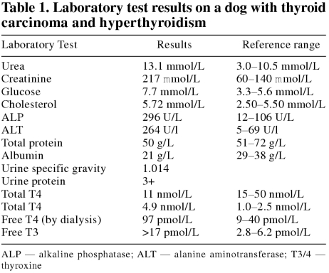
The results from the laboratory analyses (Table 1) and fine needle aspirates confirmed hyperthyroidism, secondary to a thyroid tumor.
Table 1.
One week later, general anesthesia was induced with valium (Diazepam; Sabex, Boucherville, Quebec), 7.5 mg, IV, and oxymorphone (Numorphan; Dupont Pharma, Mississauga, Ontario), 2.25 mg, IV, and maintained with isoflurane (Technilab). The ventral cervical area was clipped and prepped for surgery to remove the thyroid gland and associated tumor. A vascular mass was identified; it invaded into the extrinsic muscles of the larynx. A small amount of the neoplasm was adherent to the larynx and clean surgical margins were not achieved. Radioactive iodine treatment was scheduled, to treat any remaining neoplastic tissue and undetected metastases.
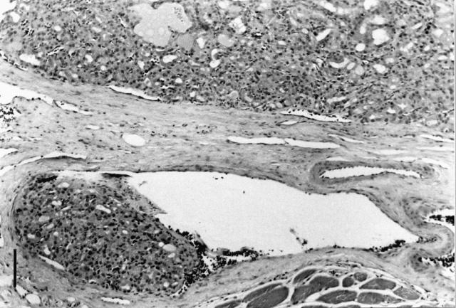
The diagnosis of thyroid carcinoma was confirmed by microscopic examination of biopsies of the gland, and there was invasion of skeletal muscle and blood vessels by neoplastic cells (Figure 1).

Figure 1. Numerous neoplastic cells present within a blood vessel bordering the thyroid carcinoma; bar = 100 μm.
Eight days after surgery, the dog developed pneumonia. She was treated with amoxicillin (Nu-Amoxi; NuPharm, Richmond Hill, Ontario), 250 mg, PO, q8h; orbifloxacin (Orbax; Schering-Plough, Pointe-Claire, Quebec), 68 mg, PO, q24h; nebulization; and coupage. The pneumonia improved; however, the dog remained depressed and anorectic. The serum T4 was 8 pmol/L (reference range, 15 to 50 pmol/L), confirming hypothyroidism; thyroid supplementation with thyroxine (Soloxine; Daniels Pharmaceuticals, St. Petersburg, Florida, USA), 100 mg, PO, q12h, was implemented. The propranolol therapy was discontinued as the heart rate and blood pressure had decreased. The dog was discharged on day 20 and scheduled to return within 6 mo for the radioactive iodine treatment. Upon return, the serum and urinalysis confirmed advanced renal failure, which precluded radioactive iodine treatment, and the dog was lost to follow-up.
Thyroid gland neoplasia represents approximately 1.2% of all canine tumors, carcinomas occurring more often than adenomas (1,2). There is an increased prevalence in older dogs (9 to 9.6 y old) and both sexes are represented (1). Boxers, beagles, and golden retrievers appear to be at the greatest risk for developing thyroid carcinomas (1,3). Unilateral tumors are about twice as frequent as bilateral, and a palpable cervical enlargement is the predominant clinical abnormality that is observed (1,3). Thyroid carcinomas are often rapidly growing, with invasion of adjacent tissues, such as the trachea, larynx, esophagus, and jugular vein. Laryngeal paralysis and megaesophagus associated with a thyroid carcinoma have been reported (1,2). Pulmonary and lymphatic metastases at the time of initial diagnosis have been confirmed in 35% of dogs (1). Unlike thyroid tumors in the cat, which are typically small and functional, canine thyroid tumors are usually malignant, large, solid masses, of which 90% do not secrete excess thyroid hormone (5). The clinical signs of hyperthyroidism, including weight loss, polyphagia, weakness, fatigue, heat intolerance, and nervousness, are infrequently detected (4). The dog reported here was unique in that it had hyperthyroid-induced heart disease, tachycardia, 2/6 heart murmur, hypertension, and ventricular hypertrophy. Unfortunately, the survival time following thyroidectomy was not determined; the presence of chronic renal failure would likely shorten the survival time. Treatment options for a thyroid carcinoma include surgical excision of affected lobe(s), surgery combined with cobalt or iodine-131 radiotherapy, and chemotherapy with doxorubicin, cyclophosphamide, and vincristine (1). Radioactive iodine therapy controls the thyrotoxic state, and may cause tumors to shrink; survival time of dogs treated with radioactive iodine is 7 to 20 mo (5). Surgical debulking of the primary tumor is recommended, as it may extend life expectancy (1,5). Survival time following surgical excision alone is reported to be approximately 7 mo (1).
Footnotes
Acknowledgments
The author thanks Drs. Dorothy Middleton and Bruce Grahn for their help with this case. CVJ
Pauli Bezzola will receive an animalhealthcare.ca fleece vest courtesy of the CVMA.
Dr. Bezzola's current address is Carolina Veterinary Specialists, 501 Nicholas Road, Greensboro, North Carolina 227409 USA.
References
- 1.Klein MK, Powers BE, Withrow SJ, et al. Treatment of thyroid carcinoma in dogs by surgical resection alone: 20 cases (1981–1989). J Am Vet Med Assoc 1995;206:1007–1009. [PubMed]
- 2.Harari J, Patterson JS, Rosenthal RC. Clinical and pathological features of thyroid tumors in 26 dogs. J Am Vet Med Assoc 1986; 188:1160–1163. [PubMed]
- 3.Capen CC. The endocrine glands. In: Jubb KVF, Kennedy PC, Palmer NC. Pathology of Domestic Animals. 4th ed. San Diego: Academic Pr, 1993:323–325.
- 4.Capen CC. Endocrine system. In: McGavin MD, Carlton WW, Zachary JF. Thomson's Special Veterinary Pathology. 3rd ed. St. Louis: Mosby, 2001:301–302.
- 5.Nelson RW. Disorders of the thyroid gland. In: Nelson RW, Couto CG. Small Animal Internal Medicine. 2nd ed. St. Louis: Mosby, 1998:729–732.


