Sir,
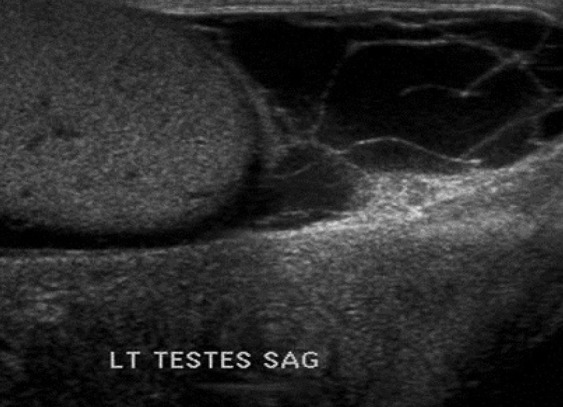
A scrotal pyocele is a rarely described urologic emergency that must be recognized and treated quickly to prevent testicular damage or Fournier's gangrene. We present a 74 year-old man with four days of left-sided scrotal pain and swelling. Physical examination revealed edema, erythema, and tenderness on the left side of the scrotum extending onto his penis proximally. A scrotal ultrasound was obtained [Figure 1] demonstrating a large scrotal pyocele. Urology surgically drained the pyocele without performing an orchiectomy.
Figure 1.

Scrotal ultrasound showing a normal appearing testicle with a scrotal free fluid with newly developed septations and echogenic debris and thickened scrotal skin without evidence of free air within the scrotal wall
Scrotal pyoceles are purulent collections within the potential space between the visceral and parietal tunica vaginalis surrounding the testicle.[1] They are commonly associated with acute epididymo-orchitis.[1] The presentation of scrotal pyoceles is subacute onset of pain and swelling, which may mimic other pathology. The imaging modality of choice to diagnose a scrotal pyocele is ultrasound.[2] Internal echoes within the pyocele fluid collection typically represent cellular debris. Other sonographic findings include loculations, septae, and fluid-fluid or air-fluid levels in the tunica vaginalis external to the testicle.[3] By contrast, a hydrocele will appear on ultrasound as a simple fluid with an anechoic region that collects anterior and lateral to the testis. If the fluid contains internal echoes on ultrasound, the diagnosis of hematocele (most common in the setting of trauma) or pyocele may be made.[3,4] Fournier's gangrene is the most concerning complication of a scrotal pyocele. If there are concerns for Fournier's, computed tomography is recommended to delineate the extent of disease and facilitate surgical planning.[5] Early diagnosis using ultrasound, therefore, will help prevent the development of sepsis and preserve a functional outcome.[2,6] Treatment requires broad spectrum antibiotics and surgical drainage. Many patients, however, ultimately require orchiectomy.[1,6]
ACKNOWLEDGMENT
The views expressed in this article are those of the author(s) and do not necessarily reflect the official policy or position of the Department of the Navy, Department of Defense or the United States Government.
REFERENCES
- 1.Slavis SA, Kollin J, Miller JB. Pyocele of the scrotum: Consequence of spontaneous rupture of testicular abscess. Urology. 1989;33:313–6. doi: 10.1016/0090-4295(89)90274-4. [DOI] [PubMed] [Google Scholar]
- 2.Rizvi SA, Ahmad I, Siddiqui MA, Zaheer S, Ahmad K. Role of Color Doppler Ultrasonography in Evaluation of Scrotal Swellings: Patter of disease in 120 patients with review of the literature. Urol J. 2011;8:60–5. [PubMed] [Google Scholar]
- 3.Ragheb D, Higgins JL. Ultrasonography of the scrotum: Techniques, anatomy, and pathologic entities. J Ultrasound Med. 2002;21:171–85. doi: 10.7863/jum.2002.21.2.171. [DOI] [PubMed] [Google Scholar]
- 4.Akin E, Khati NJ, Hill MC. Ultrasound of the scrotum. Ultrasound Q. 2004;20:181–200. doi: 10.1097/00013644-200412000-00004. [DOI] [PubMed] [Google Scholar]
- 5.Lee C, Henderson SO. Emergency surgical complications of genitourinary infections. Emerg Med Clin N Am. 2003;21:1057–74. doi: 10.1016/s0733-8627(03)00067-1. [DOI] [PubMed] [Google Scholar]
- 6.Butler JM, Chambers J. An unusual complication of epididymo-orchitis: Scrotal pyocele extending into the inguinal canal mimicking a strangulated inguinal hernia. J Emerg Med. 2008;35:379–84. doi: 10.1016/j.jemermed.2007.02.029. [DOI] [PubMed] [Google Scholar]


