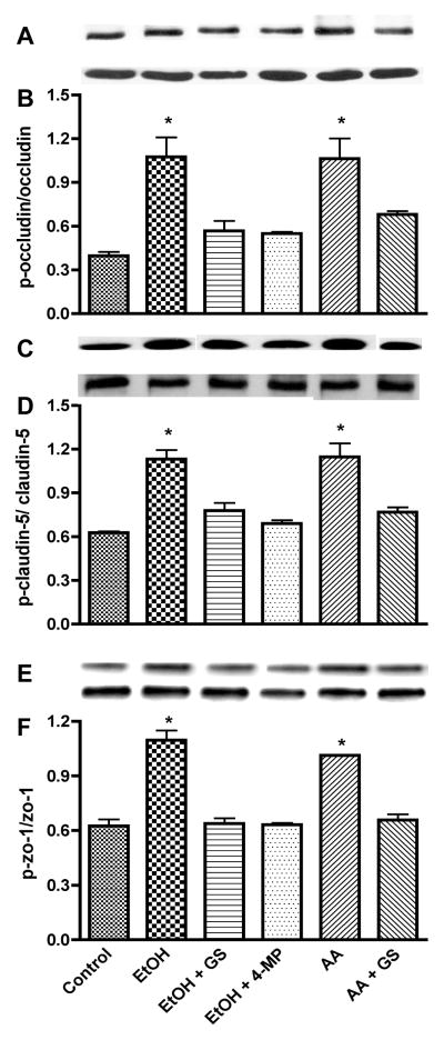
Figure 6.
Lysate proteins derived from human BMVEC treated with EtOH for 48 hr or with AA for 2 hr in the presence or absence of 4-MP or GS were subjected to Western blot analysis after immunoprecipitation. Representative immunoreactive bands of (A) occludin-phosphotyrosine, (B) total occludin, (C) ratio of occludin-phosphotyrosine/total occludin, (D) claudin-5-phosphotyrosine, (E) total claudin-5, (F) ratio of claudin-5-phosphotyrosine/total claudin-5, (G) ZO-1-phosphotyrosine, (H) total ZO-1, (I) ratio of ZO-1-phosphotyrosine/total ZO-1. * indicates statistical differences (p < 0.01) compared with control. Respective inhibitor did not change the expression of phosphorylated TJ protein of the basal controls.

