Abstract
This Classic Article is a reprint of the original work by F.W. Holdsworth, Dislocation and fracture-dislocation of the pelvis. An accompanying biographical sketch of F.W. Holdsworth is available at DOI 10.1007/s11999-012-2422-4. Reproduced and adapted with permission and copyright © of the British Editorial Society of Bone and Joint Surgery. Holdsworth FW. Dislocation and fracture-dislocation of the pelvis. J Bone Joint Surg Br. 1948;30:461–466.
This paper is based upon a study of fifty cases of disruption of the pelvic ring. The cases occurred during the years 1937 to 1946 in the Accident Department of the Sheffield Royal Infirmary and its associated hospitals.
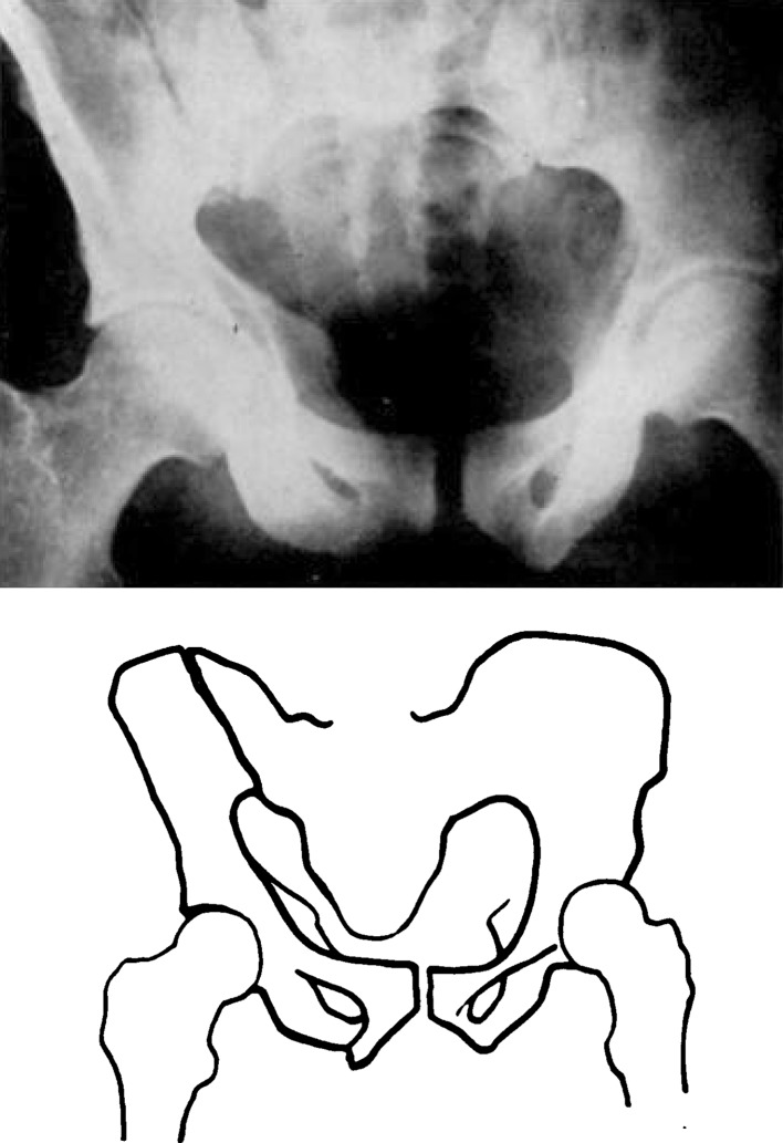
Two types of disruption of the pelvic ring—The injury occurs in two varieties : 1) dislocation of the sacro-iliac joint; 2) fracture of the ilium or sacrum adjacent to the sacro-iliac joint. In both types there is separation of the symphysis pubis, or fracture of both pubic rami. In both varieties there is displacement of one-half of the pelvis outwards, or outwards and upwards. Associated with this displacement there is often rotation of the large pelvic fragment in the sagittal plane (Figs. 4 and 6). This injury occurs with some frequency and it is surprising that only a few series have been reported in the literature. Most reports are confined to isolated cases, to particular methods of reduction, or to visceral complications of the lesion, and it is for this reason that a complete follow-up of a comparatively large series may be of interest.
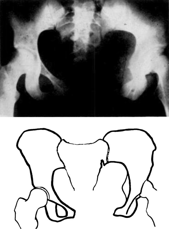
Fig. 4.
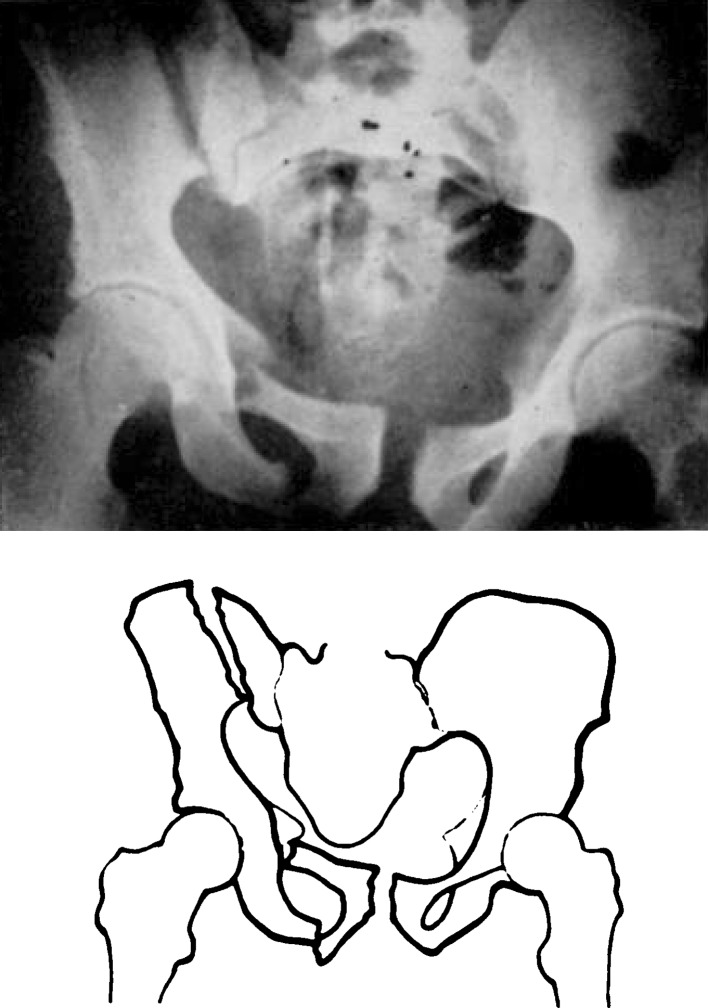
Fig. 6.
Mechanism of Injury and Displacement
Taylor (1942) reported twenty-two cases of dislocation of the symphysis pubis, or hind-quarter dislocation, and was of opinion that the injury was sustained by indirect violence, the leg acting as a long lever dislocating the pelvis. Most authors, however, consider the lesion to be the result of direct violence—a view which is strongly supported by this series. In all cases there was severe direct violence, either an antero-posterior crush, or a torsional injury where the sacrum was fixed and one-half of the pelvis was forced back by direct antero-posterior violence to one ilium. For instance, in Case 42 (Fig. 4) a woman was run over by a heavy motor-car, the wheel passing over the front of the pelvis, resulting in complete disruption of the pubis and the sacro-iliac joint. In Case 30, a steel erector, aged twenty-seven years, was standing on a girder with his sacrum against a vertical post, when the cage of a travelling crane moved back and struck his right ilium. He sustained separation of the pubis and fracture through the ilium.
Pubic injury with sacro-iliac dislocation is more common than pubic injury with fracture of the ilium or sacrum. Of the forty-two patients in this series who survived and were traced, there was sacro-iliac dislocation in twenty-seven, and fracture of the ilium or sacrum near the joint in fifteen. Both injuries, however, result in the same type of displacement. The pubic bones are separated and the pelvis is opened out at the posterior injury “ like the leaves of a book ” (Watson-Jones 1938). Associated with the outward rotation there is often upward displacement of the injured side and sometimes rotation (Fig. 6).
Visceral injuries and retroperitoneal haemorrhage—Most authors emphasize the severity and danger of visceral complications, particularly lesions of the bladder and urethra. These, however, appear to be rare. In this series, only four cases of injury to the bladder or urethra occurred. Two had large extra-peritoneal tears in the bladder and both died. Two had tears of the urethra and survived. No other visceral complications occurred. The most frequent complication was retroperitoneal haemorrhage, severe enough to be alarming, and in four cases causing death. The bleeding probably arises from tearing of the iliolumbar artery, and it tracks forward towards the anterior superior spine. When severe it may cause an alarming fall of blood-pressure. There appears to be no possibility of controlling the haemorrhage by surgery or pressure; the best treatment is to maintain the blood-pressure at not more than 100 mm. of mercury by controlled transfusion, until the bleeding ceases. Bleeding severe enough to cause alarm has occurred in eight patients, four of whom died.
Reduction and Immobilisation
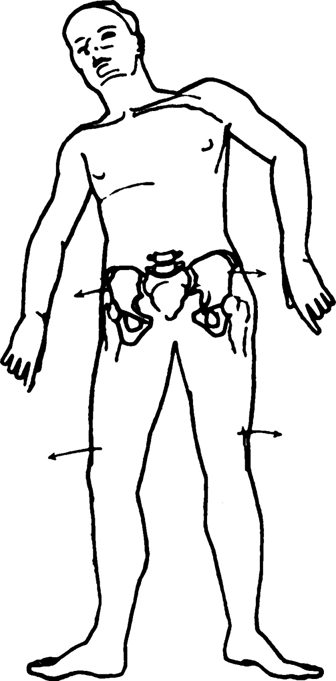
The method of reducing the deformity and maintaining the position should be determined by proper recognition of the displacement. Outward rotation of the large fragment of the pelvis is maintained by the outward roll of the leg and extension of the hip (Fig. 1). Many methods of reduction have been described, most of which do not recognise the importance of the position of the legs. Two methods only are really rational—the method of lateral recumbency and plaster of Watson-Jones (1938, 1943) and the sling method originated by Astley Cooper (1842) and developed by Bohler (1935). Lateral recumbency and plaster is theoretically sound but difficult in practice and, in this series, the sling method has been used exclusively (Figs. 2 and 3). The method is simple, safe, and efficient. The patient is placed upon a bed with fracture boards and an overhead frame; a firm canvas sling, spread by rods, is placed under the pelvis. The sling extends from above the iliac crest to below the greater trochanters. The rods are attached to cords which pass over pulleys in the overhead frame, the cords from the left side crossing over to the pulleys on the right and vice versa. The ends are attached to weights, the total of all four weights being sufficient just to raise the patient from the bed. Each leg is then placed upon a firmly bandaged Braun’s frame, so that the hips are flexed. The limbs are kept in this position by weight extensions of eight pounds, attached by strapping extensions. The patient is then supported on a back rest. If, in addition to separation of the pelvic halves, there is upward displacement of one half, skeletal traction is applied to the limb of the affected side with sufficient weight to pull the fragment into position. Frequently no anaesthetic is required for this procedure but, if rotational displacement of the fragment is also present, then manipulation is necessary. The patient is anaesthetised, placed upon the sling and turned on his side. The rotated fragment is pushed forwards; the patient is then rolled on his back and the sling and frames are adjusted.
Fig. 1.
The pelvic dislocation is opened out anteriorly by extension at the hip joint, and by outward rolling of the lower limbs.
Fig. 2.
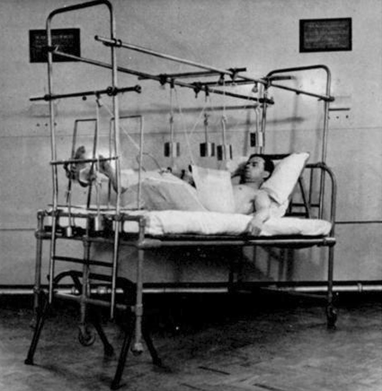
Fig. 3.
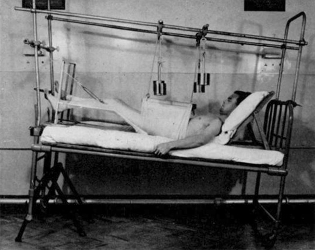
Fracture-dislocation of the pelvis immobilised in sling and frames with skeletal traction applied to the left leg.
The degree of compression by the pelvic sling can be adjusted by altering the obliquity of the ropes. Control radiographs are taken and the ropes are adjusted so that the pubic symphysis is kept in position. Reduction is usually complete and the position can be maintained with little difficulty (Figs. 4–7). Nursing in the apparatus is easy. The patient can be lifted for the use of bed-pans by pulling on the weights and, if the lower end of the sling is turned back, the bed-pan can easily be slid into place. Most patients are quite comfortable in the apparatus, though in the very old there may be some mental confusion in the early stages of treatment.
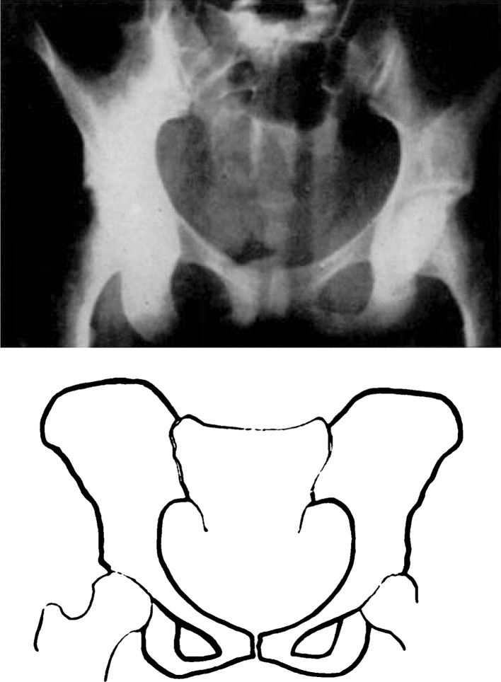
Fig. 5.
Gross separation of one half of the pelvis with wide separation of the symphysis and dislocation of one sacro-iliac joint (Fig. 4). Same case after reduction by the pelvic sling (Fig. 5).
Fig. 7.
Fracture-dislocation of the pelvis with double pubic fracture, and fracture of the ilium near the sacro-iliac joint (Fig. 6). Same case after reduction by the pubic sling (Fig. 7).
Fixation is maintained for twelve weeks but, after six weeks, the traction weights are removed and graduated leg exercises are allowed. Weight-bearing and active rehabilitation is started after twelve weeks.
Assessment of End-Results
The ultimate object of treatment of every injury is full restoration of function. Restoration of the anatomical position in any fracture is easy to assess and may or may not be important, but function is all important and, as Nicol has pointed out: “The assessment of function is based upon criteria which vary from one surgeon to another and are subject to individual judgment rather than precise measurement.” Therefore, following his lead, capacity for work has been used in this investigation as the criterion of recovery of function. Most patients were engaged at the time of accident in heavy industrial activity so that return to pre-accident work, or similar heavy work such as labouring, joinering, or bricklaying, formed a reasonable standard of full recovery. In women the ability to perform all household work has been considered heavy work. The average period of follow-up after accident is five years and no case has been assessed less than two years after the accident.
On the whole the end-results are good. It will be seen from Table 1 that the prognosis in dislocations of the sacro-iliac joint is not so good as in the case of fractures near the joint. About half the cases of sacro-iliac dislocation were able to return to reasonably heavy work but nearly all cases of fracture were able to do so. All patients complained of pain in the pubis, sometimes lasting as long as two years but eventually disappearing. Even in the best cases there was often aching in the back after prolonged effort. In sacro-iliac dislocations this pain was severe; it was localised to the joint and it prevented the patient from performing anything but the lightest of work. All patients who were free from pain showed a full range of spine and hip movements with complete pelvic stability and no limitation of straight leg raising. In the case of patients who had pain, spinal movements were good but there was definite limitation of straight leg raising on the affected side, which was often very marked.
Table 1.
Dislocations and fracture-dislocations of the pelvis
| Total number of cases | 50 | ||
| Died | 6 | ||
| Untraced | 2 | ||
| Complications | |||
| Rupture of the bladder | 2 | Died 2 | |
| Rupture of the urethra | 2 | Died 0 | |
| Retroperitoneal haemorrhage | 8 | Died 4 | |
| End-result in forty-two traced cases | |||
| Fracture or dislocation of pubis with | Total | At heavy work | Painful |
| 1) Dislocation of sacro-iliac joint | 27 | 12 | 15 |
| 2) Fracture of ilium or sacrum | 15 | 13 | 2 |
These figures appear to be significant. The frequency of persistent sacro-iliac pain after dislocation of the joint suggests that early sacro-iliac fusion should be considered in such cases. Certainly it appears to be indicated in patients with severe localised pain persisting for one to two years after injury. The pain in such cases is probably due to unsound ankylosis of the sacro-iliac joint. The strong anterior ligaments are torn from the front of the sacrum and ilium and the remnants may curl within the joint, preventing true restoration of position. Whatever the cause, however, there is no doubt about the residual pain and its localisation to the joint, and fusion seems to offer the best prospect of relief.
Summary and Conclusions
Fifty dislocations and fracture-dislocations of the pelvis have been reviewed.
Complications were unusual. Two patients with rupture of the bladder died; two with rupture of the urethra survived. Of eight patients with retroperitoneal haemorrhage four died; the treatment advised is controlled blood transfusion maintaining a blood-pressure of not more than 100 mm.
Two types of pelvic disruption should be distinguished: 1) pubic injury with sacro-iliac dislocation; 2) pubic injury with fracture near the sacro-iliac joint. The first is twice as common as the second.
In each type, displacement is maintained by extension of the hip and outward roll of the limb. This may be controlled by the Watson-Jones plaster method but the pelvic sling technique is preferred and was used in all cases in this series.
The prognosis in fracture-dislocations is very good; nearly all patients went back to heavy work.
The prognosis in sacro-iliac dislocations is not so good; only half the patients went back to heavy work and there was often persistent sacro-iliac pain. Sacro-iliac arthrodesis is advised in those cases.
Discussion
Dr H. Earle Gonwell (Birmingham, Ala.): Mr Holdsworth has presented an excellent series of radiographs and sketches showing the various locations of dislocation and fracture-dislocation of the pelvis, especially those about the sacro-iliac joint. I would point out that in 1930, and 1933, Noland and I reported in the Journal of the American Medical Association, and in Surgery, Gynecology, and Obstetrics, 183 fractures of the pelvis observed over a period of eleven years in which there were fifty dislocations of the sacro-iliac joint. Since that time I have observed over a period of fifteen years 240 cases of fractures of the pelvis in which there was about the same percentage of dislocations and fracture-dislocations of this type.
Such injuries are in most instances of a severe nature, and contrary to Mr Holdsworth’s observation I have found a far greater proportion of soft tissue complications involving the bladder and urethra, retro-peritoneal haemorrhage, and intra-abdominal involvement, which demanded immediate general surgical as well as orthopaedic consultation.
The plaster cast treatment with its various techniques, of which there is more than one, has been generally discarded in the States. The pelvic sling, with traction to the lower extremities, has been my choice of treatment for over a quarter of a century. After reduction has been carried out (a general anaesthetic being preferred) skeletal traction is applied through the lower end of the femur on the injured side with simple adhesive traction on the uninjured side, supplemented by the pelvic sling.
Function is the criterion of a good result, and reduction of these fractures and dislocations is not always necessary to bring about good function; reduction should not be carried out forcibly to the sacrifice of the patient’s general condition.
Arthrodesis is probably indicated in very few cases. It is difficult to make this decision until the patient has progressed sufficiently in convalescence. Moreover, it has been my experience that patients with persistent pain over the sacro-iliac areas also have complications in the low spine and that if arthrodesis is necessary it should include the lumbo-sacral as well as the sacro-iliac joint. I do feel that a simple back support during convalescence is of much value in relieving pain. Conservatism is indicated in most cases. Mr Holdsworth is to be commended for bringing this subject before us.
Footnotes
Paper read at the combined meeting of American, Canadian, and British Orthopaedic Associations in Quebec, June 1948.
Richard A. Brand MD (✉) Clinical Orthopaedics and Related Research, 1600 Spruce Street, Philadelphia, PA 19103, USA e-mail: dick.brand@clinorthop.org
References
- Bohler L. The Treatment of Fractures. 4. Bristol: John Wright & Sons; 1935. [Google Scholar]
- Cooper Sir Astley. A Treatise on Fractures and Dislocations of the Joints. London: Churchill; 1842. [Google Scholar]
- Watson-Jones R. British Journal of Surgery. 1938;25:773. doi: 10.1002/bjs.18002510008. [DOI] [Google Scholar]
- Watson-Jones R. Fractures and Joint Injuries. 3. Edinburgh: Livingstone; 1943. p. 377. [Google Scholar]


