Abstract
The current understanding of ciliate phylogeny is mainly based on analyses of a single gene, the small subunit ribosomal RNA (SSU-rDNA). However, phylogenetic trees based on single gene sequence are not reliable estimators of species trees, and SSU-rDNA genealogies are not useful for resolution of some branches within Ciliophora. Since congruence between multiple loci is the best tool to determine evolutionary history, we assessed the usefulness of alpha-tubulin gene, a protein-coding gene that is frequently sequenced, for ciliate phylogeny. Here, we generate alpha-tubulin gene sequences of 12 genera and 30 species within the order Euplotida, one of the most frequently encountered ciliate clades with numerous apparently cosmopolitan species, as well as four genera within its putative sister order Discocephalida. Analyses of the resulting data reveal that: 1) the alpha-tubulin gene is suitable phylogenetic marker for euplotids at the family level, since both nucleotide and amino acid phylogenies recover all monophyletic euplotid families as defined by both morphological criteria and SSU-rDNA trees; however, alpha-tubulin gene is not a good marker for defining species, order and subclass; 2) for seven out of nine euplotid species for which paralogs are detected, gene duplication appears recent as paralogs are monophyletic; 3) the order Euplotida is non-monophyletic, and the family Uronychiidae with sequences from four genera, is non-monophyletic; and 4) there is more genetic diversity within the family Euplotidae than is evident from dargyrome (geometrical pattern of dorsal “silverline system” in ciliates) patterns, habit and SSU-rDNA phylogeny, which indicates the urgent need for taxonomic revision in this area.
Introduction
Current studies on the relationships within the phylum Ciliophora are almost exclusively based on SSU-rDNA phylogenies [1]–[7]. These single gene analyses provided resolution for a number of important questions on the phylogenetic relationships within this group, but there are problems. The overall picture emerging from these studies confirmed the monophyly of most classes defined by morphological criteria; however, relationships among these classes vary with different taxon sampling (for example [3], [8]–[10]). Moreover, while some previous investigations based on SSU-rDNA alone resolved assignments of some taxa with ambiguous morphological classification (for example [4], [11], [12]), relationships within some orders/families containing a large number of taxa remain problematic [6], [13].
In recent years, other gene markers, including LSU-rDNA gene, ITS region, tubulins, phosphoglycerate kinase, actin, DNA Polymerase α, Hsp 70, etc., have been used to reconstruct ciliate phylogenies [14]–[22]. Traditionally, protein gene markers are considered more suitable alternatives to SSU-rDNA than LSU-rDNA gene and ITS region for two reasons. First, protein markers are less sensitive to differences in compositional bias, which can lead to artifacts in tree construction [23]–[25]. In addition, sequences of multiple unlinked loci have different histories, as opposed to linked SSU-rDNA, LSU-rDNA, ITS regions, and are necessary to estimate species trees [26]. However, protein-coding genes can possess paralogs that might bias phylogenetic trees [27], [28]. Among these protein-coding genes, alpha-tubulin is one of the mostly used gene makers for ciliated phylogeny [8], [11], [13], [15], [20], [21], [29], and its duplication in ciliates has been previously studied only with sparse taxon sampling [15], [30]. Therefore, alpha-tubulin is a promising candidate for testing whether protein-coding genes are suitable for phylogeny construction of ciliates.
Previous alpha-tubulin phylogenies showed that most classes could be well distinguished with high support [8], [15], [20], while subclasses appeared to be non-monophyletic [13], [21], [29]. In our recent study [13], we characterized alpha-tubulin gene from 15 genera covering all families of the order Urostylida, but we were unable to determine if alpha-tubulin gene is suitable for classification of lower level taxa since urostylid families are not well defined morphologically [31], [32] and their monophyly is rejected by both SSU-rDNA and alpha-tubulin phylogenies. Therefore, a group with well-defined families or genera is needed to test the ability of alpha-tubulin to resolve phylogeny for lower level taxa. The order Euplotida, one of the most frequently encountered ciliate clades with numerous putatively cosmopolitan species [33]–[37], is a good choice because most morphological families within this order are recovered robustly in SSU-rDNA analyses [38]–[40].
Here, we increase sampling of alpha-tubulin gene sequences from 30 taxa within the order Euplotida, including multiple morphospecies from four out of five families, as well as four species of its putative sister order Discocephalida. Our main aims are to: 1) assess the suitability of alpha-tubulin for circumscribing lower level taxa; 2) estimate phylogenetic relationships within the order Euplotida using two-gene combined, SSU-rDNA and alpha-tubulin trees; and 3) characterize patterns of molecular evolution among euplotid alpha-tubulin paralogs.
Results
Different Species with Same Amino Acid Sequences
Although species contained diverse alpha-tubulin sequences, we found that some euplotid species (e.g. Uronychia multicirrus and U. sinica; Euplotes sp.-GZJJM2009121510, Euplotoides parawoodruffi and Euplotopsis sp.-GZJJM2009121508; Euplotopsis encysticus and Euplotes cf. antarcticus) share identical amino acid sequences, revealing the high level of functional constraint on this protein (Fig. S1). Alpha-tubulin gene sequences of U. multicirrus and U. sinica are different from each other at 56 sites, all of which are third codon positions (Fig. S1). Similarly, 69 residues, 3 first codon positions and 66 third codon positions, are different between alpha-tubulin gene sequences of Euplotopsis encysticus and Euplotes cf. antarcticus (Fig. S1). There are totally 124 polymorphic nucleotide sites between Euplotes sp.-GZJJM2009121510, Euplotoides parawoodruffi and Euplotopsis sp.-GZJJM2009121508. And among these sites, 11 are in first codon positions, only one is in second codon position, and the remaining 112 sites are in third codon positions (Fig. S1).
Intraspecific Variation
Multiple clones have been sequenced from ten species and two populations of the morphospecies Diophrys parappendiculata and Euplotes sinicus are sampled (Table 1). No paralogs are detected in two populations of D. parappendiculata. Only one sequence was found in E. sinicus population II, while two paralogs are present in population I, with the first being identical to the sequence of population I. (Table 2).
Table 1. Euplotid Species for Which Alpha-Tubulin Genes Were Sequenced in the Present Work.
| Taxa | Sampling localityLocation | DNA sample | GenBank No. | Taxa | Sampling localityLocation | DNA sample | GenBank No. |
| Aspidisca aculeata | Daya Bay (22°43′N; 114°32′E), Guangdong | one cell | JQ736724 | Euplotes sinicus popI | Qingdao (36°03′N; 120°20′E), Shandong | one cell | JQ73674-JQ736748 |
| Aspidisca leptaspis P1-P3 | Qingdao (36°03′N; 120°20′E), Shandong | 20 cells | JQ736687-JQ736691 | Euplotes sinicus popII | Daya Bay (22°43′N; 114°32′E), Guangdong | 20 cells | JQ736734, |
| Aspidisca magna | Gaoqiao (21°31′N; 109°45′E), Guangdong | one cell | JQ736722 | Euplotes sp.-GZJJM2009121510 | Zhanjiang (21°27′N; 110°35′E), Guangdong | one cell | JQ736721 |
| Aspidisca orthopogon P1-P2 | Qingdao (36°03′N; 120°20′E), Shandong | 3 cells | JQ736740, JQ736741 | Euplotoides parawoodruffi | Daya Bay (22°43′N; 114°32′E), Guangdong | 10 cells | JQ736737 |
| Aspidisca steini | Gaoqiao (21°31′N; 109°45′E), Guangdong | 2 cells | JQ736723 | Euplotopsis encysticus | Daya Bay (22°43′N; 114°32′E), Guangdong | 4 cells | JQ736725 |
| Apodiophrys ovalis P1-P3 | Dameisha (22°59′N; 114°30′E), Guangdong | one cell | JQ736709-JQ736717 | Euplotopsis raikovi | Qingdao (36°03′N; 120°20′E), Shandong | 12 cells | JQ736749 |
| Diophryopsis hystrix P1-P3 | Qingdao (36°03′N; 120°20′E), Shandong | one cell | JQ736694-JQ736698 | Euplotopsis sp.-GZJJM2009121508 | Zhanjiang (21°27′N; 110°35′E), Guangdong | one cell | JQ736720 |
| Diophrys apoligothix | Qingdao (36°03′N; 120°20′E), Shandong | several cells | JQ736742 | Gastrocirrhus monilifer | Qingdao (36°03′N; 120°20′E), Shandong | 4 cells | JQ918351 |
| Diophrys scutum P1-P2 | Qingdao (36°03′N; 120°20′E), Shandong | 5 cells | JQ736692, JQ736693 | Gastrocirrhus sp.-GZCXM2008122201 | Daya Bay (22°43′N; 114°32′E), Guangdong | 8 cells | JQ736708 |
| Diophrys parappendiculata popI | Daya Bay (22°43′N; 114°32′E), Guangdong | 9 cells | JQ736739 | Heterodiophrys zhui | Dameisha (22°59′N; 114°30′E), Guangdong | several cells | JQ736718 |
| Diophrys parappendiculata popII | Daya Bay (22°43′N; 114°32′E), Guangdong | 5 cells | JQ918349 | Leptoamphisiella vermis | Qingdao (36°03′N; 120°20′E), Shandong | several cells | JQ736707 |
| Discocephalus rotatorius | Dameisha (22°59′N; 114°30′E), Guangdong | several cells | JQ736719 | Moneuplotes minuta | Qingdao (36°03′N; 120°20′E), Shandong | one cell | JQ736750 |
| Euplotes cf. antarcticus | Daya Bay (22°43′N; 114°32′E), Guangdong | 30 cells | JQ736735 | Paradiophrys zhangi | Qingdao (36°03′N; 120°20′E), Shandong | 4 cells | JQ736743, JQ736744 |
| Euplotes balteatus | Qingdao (36°03′N; 120°20′E), Shandong | 20 cells | JQ736702 | Pseudoamphisiella elongate | Qingdao (36°03′N; 120°20′E), Shandong | 4 cells | JQ918350 |
| Euplotes charon | Qingdao (36°03′N; 120°20′E), Shandong | one cell | JQ736751 | Prodiscocephalus borrori | Qingdao (36°03′N; 120°20′E), Shandong | one cell | JQ918352 |
| Euplotes neaplolitanus P1-P3 | Daya Bay (22°43′N; 114°32′E), Guangdong | 4 cells | JQ736726-JQ736733 | Uronychia multicirrus | Daya Bay (22°43′N; 114°32′E), Guangdong | 10 cells | JQ736738 |
| Euplotes petzi | Qingdao (36°03′N; 120°20′E), Shandong | 3 cells | JQ736703-JQ736705 | Uronychia sinica | Mangrove in Shenzhen (22°37′N; 114°04′E 1), Guangdong | 15 cells | JQ736736 |
| Euplotes rariseta | Qingdao (36°03′N; 120°20′E), Shandong | 31 cells | JQ736749 | Uronychia sp.-SXL2007102501 | Hangzhou (30°16′N; 120°10′E 1), Zhejiang | 8 cells | JQ918348 |
Table 2. Intraspecific Distances Between/Among α-Tubulin Clones and Between/Among Paralogs.
| Taxa | Comparisonof Paralog(s) | Clone names | N | R/S | d | dA |
| Apodiophrys ovalis | 1 | Clone 1–5 | 5 | 1.6/2 | 0.337±0.109 | 0.927±0.328 |
| 2 | Clone 6 | 1 | – | – | – | |
| 3 | Clone 7–9 | 3 | 0.7/2 | 0. 249±0. 135 | 0.180±0.176 | |
| 1, 2, 3 | 3.3/91.7# | 10.265±1.106 | 0.398±0.178 | |||
| Aspidisca leptaspis | 1 | Clone 1 | 1 | – | – | – |
| 2 | Clone 2–4 | 3 | 0/0 | 0.000 | – | |
| 3 | Clone 5 | 1 | – | – | – | |
| 1, 2, 3 | 10.3/55# | 6.317±0.789 | 1.948±0.596 | |||
| Aspidisca orthopogon | 1 | Clone 1 | 1 | – | – | – |
| 2 | Clone 2 | 1 | – | – | – | |
| 1, 2 | 7/51# | 6.255±0.855 | 1.785±0.680 | |||
| Diophryopsis hystrix | 1 | Clone 1 | 1 | – | – | – |
| 2 | Clone 2 | 1 | – | – | – | |
| 3 | Clone 3, 4 | 2 | 0/7 | 0.658±0.241 | 0.000±0.000 | |
| 4 | Clone 5 | 1 | – | – | – | |
| 1, 2, 3, 4 | 7/64.3# | 7.452±0.859 | 0.911±0.421 | |||
| Diophrys scutum | 1 | Clone 1 | 1 | – | – | – |
| 2 | Clone 2 | 1 | – | – | – | |
| 1, 2 | 5/18 | 2.183±0.450 | 0.808±0.414 | |||
| Diophrys parappendiculata* | 1 | Clone 1, 2 | 2 | 0/0 | 0.000 | 0.000±0.000 |
| Euplotes neaplolitanus | 1 | Clone 1 | 1 | - | – | – |
| 2 | Clone 2, 3 | 2 | 1/3 | 0.468±0.227 | 0.270±0.260 | |
| 3 | Clone 4–8 | 5 | 1.2/0.4 | 0.150±0. 074 | 0.324±0.180 | |
| 1, 2, 3 | 6/69.3# | 7.988±0.852 | 1.284±0.540 | |||
| Euplotes petzi | 1 | Clone 1–3 | 3 | 3/10.3 | 0.792±0.245 | 0.878±0.381 |
| Euplotes sinicus ** | 1 | Clone 1, 2, 5 | 3 | 3/5 | 0.784±0.176 | 0.188±0.019 |
| 2 | Clone 3, 4 | 2 | 2/5 | 0.729±0.270 | 0.545±0.365 | |
| 1, 2 | 2/24# | 3.763±0.564 | 1.219±0.524 | |||
| Paradiophrys zhangi | 1 | Clone 1, 2 | 2 | 3/5 | 0.999±0.345 | 0.718±0.479 |
NOTE.- N, number of clones; d, number of nucleotide substitutions per site calculated using Tamura-Nei model; dA, number of amino acid substitutions per site calculated using Dayhoff model; R/S, number of replacement site substitutions/number of synonymous substitutions among clones.
Fixed between paralogs.
Euplotes sinicus population I: Clone 1–4; E. sinicus population II: Clone 5.
The intraspecific variation among putative orthologs for these species ranges between 0% (Aspidisca leptaspis P2, Paradiophrys zhangi) and 0.792% (Euplotes petzi) (Table 2). Among these ten species, paralogs appear absent in four: Euplotes petzi, Diophrys parappendiculata, Paradiophrys irmgard and P. zhangi. For these, average pairwise difference among clones within each of these taxa is low (0.000–0.999%; Table 2). Ratios of replacement substitutions to silent substitutions are 3/10.3, 0/0, 0/0, and 3/5 for E. petzi, D. parappendiculata, P. irmgard, and P. zhangi, respectively. Paralogs are detected for remaining six species: Apodiophrys ovalis, Aspidisca leptaspis, A. orthopogon, Euplotes neapolitanus, Diophrys scutum and Diophryopsis hystrix (Table 2). Among these six species, there are more synonymous site substitutions than replacement substitutions both within paralogs and fixed between paralogs. Synonymous substitutions appear less frequent than replacement substitutions for E. neapolitanus paralog P3 (1.2/0/4), but the small numbers here suggest that experimental error may be contributing factor (Table 2). The average pairwise amino acid difference among clones of a specific paralog is 0.000% (Aspidisca leptaspis P2, Diophrys parappendiculata and P. irmgard) to 0.792% (E. petzi P1), and that between/among paralogs of a specific species is from 0.398% (Apodiophrys ovalis) to 1.948% (Aspidisca leptaspis) (Table 2).
Phylogenetic Trees Inferred from Alpha-Tubulin Nucleotide (Atub_n74, Atub_n52) and Amino acid Sequences (Atub_aa)
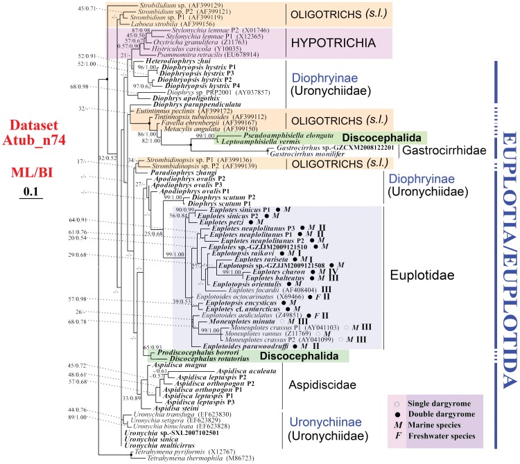
We analyzed three different alpha-tubulin datasets: nucleotides from 74 taxa (Atub_n74), nucleotides from a subset of 52 taxa (Atub_n52, only the paralog with shortest branch length in Atub_n74 is selected) and 70 amino acid sequences (Atub_aa). Within the class Spirotrichea, Hypotrichia appears as monophyletic in all analyses (Figs. 2, S2 and S3). For example, Stylonychia, Oxytrichia, Histriculus and Psammomitra always fall into the same clade. Oligotrichia is shown to be monophyly in Figure 2 (Atub_n74) and Figure S2 (Atub_aa). The other subclass/order level taxa, i.e. Choreotrichia, Euplotia/Euplotida and Discocephalida, are not monophyletic, which may reflect limited taxon sampling (Figs. 2, S2, S3, Table 3).
Figure 2. Best tree of the Spirotrichea inferred by Maximum likelihood of Dataset Atub_n74.
Species newly sequenced in the present study are shown in bold type. Bootstrap values for branches of the ML tree and posterior probability values for BI tree, respectively, are given on nodes. Fully supported (100%/1.00) branches are marked with solid circles. The scale bar corresponds to 10 substitutions per 100 nucleotide positions. Dargyrome patterns and natural habitats are given after species name of euplotids by symbols. Clades I-IV for euplotids were designated according to Petroni [42] and Yi et al. [38].
Table 3. Support for Major Clades of Spirotrichean Species in Analyses Based on Five Datesets.
| Alpha-tubulin | Two-gene combined (52), Atub-SSU | SSU-rDNA (52), SSU | |||
| Nucleotide (74), Atub_n74 | Nucleotide (52), Atub_n52 | Amino acid (70), Atub_aa | |||
| Supported clades | |||||
| Aspidiscidae/Aspidisca | 33%/0.89 | 40% | 45%/0.79 | 98% | 40% |
| Euplotida/Euplotidae | 99%/1.00 | 98% | 66%/0.66 | 100% | 98% |
| Gastrocirrhidae/Gastrocirrhus | 100%/1.00 | 100% | 1.00/100% | 100% | 100% |
| Moneuplotes | 68%/0.78 | nm | 66%/0.99 | 99% | nm |
| Weak hypothesis | |||||
| Choreotrichia | nm | nm | nm | 95% | 93% |
| Discocephalida | nm | nm | nm | nm | nm |
| Hypotrichia | 57%/0.90 | 52% | 37%/nm | nm | nm |
| Oligotrichia | nm | nm | 27%/0.98 | 95% | 91% |
| Uronychiidae | nm | nm | nm | nm | nm |
| Diophrys-complex | nm | nm | nm | nm | nm |
| Euplotes | nm | nm | nm | nm | nm |
| Euplotoides | nm | nm | nm | 100% | 100% |
| Euplotopsis | nm | m | nm | nm | nm |
| Uronychia | nm | nm | nm | 100% | 100% |
| Dataset statistics | |||||
| Number of lineages | 59 | 49 | 44 | 49 | 48 |
NOTE.-nm = nonmonophyletic.
Within the order Euplotida, monophyly of the family Gastrocirrhidae is supported with highest bootstrap values in all alpha-tubulin trees (Figs. 2, S2 and S3), though only two species are sequenced. Similarly, the family Euplotidae appears to be monophyletic with variable support values (66%–99% ML, 0.66–1.00 BI; Figs. 2, S2, S3). Within this family, Euplotes, Euplotoides and Euplotopsis are always non-monophyletic, and monophyly of the genus Moneuplotes is found only in Datasets Atub_n74 (Fig. 2) and Atub_aa (Fig. S2).
Relationships among species in the family Euplotidae do not always corresponding to dargyrome patterns or natural habitats. For example, species possessing single dargyrome always cluster with each other, while those of double dargyrome fall into several clades (Figs. 2, S2, S3). Three stable clades (Clade I-III) found in previous phylogenetic analyses inferred from SSU-rDNA sequences [38], [41], [42] are not recovered here. In our analyses (Figs. 2, S2, S3), monophyly of members of Clade I (Euplotopsis raikovi, Euplotes rariseta) is never found. Species of Clade II (Euplotoides parawoodruffi, E. octocarinatus, E. aediculatus) fall into different clades, and Euplotes focardii is always apart from the other three species of Clade III (Moneuplotes minuta, M. crassus, M. vannus) in all our alpha-tubulin trees (Figs. 2, S2, S3).
Taxa among the family Aspidiscidae group together but with low support values (33%–45% ML, 0.79–0.89 BI) (Figs. 2, S2, S3). Within this clade, Aspidisca steini diverges first from remaining species, followed by A. magna (Figs. 2, S2, S3). The family Uronychiidae is the only non-monophyletic family out of two families with sequenced samples from multiple genera, and the monophyly of it is only shown in Dataset Atub_n52 (Fig. S3); In contrast, Uronychia setigera, U. transfuga and U. binucleata always cluster together (Figs. 2, S2, S3). The Diophrys-complex (viz. Apodiophrys, Diophrys, Diophryopsis and Heterodiophrys) species appear in several clades, and their relationships are distinct in trees based on different datasets (Figs. 2, S2, S3).
Within the order Discocephalida, two families (viz. Pseudoamphisiellidae and Discocephalidae) are included. The Pseudoamphisiellidae (Pseudoamphisiella and Leptoamphisiella) form a monophyletic group as do the Discocephalidae (Discocephalus and Prodiscocephalus), indicating the monophyly of these two families. However, sister relationship between these two families is never recovered (Figs. 2, S2, S3).
In our phylogenetic trees (Figs. 2, S2, S3), nine species, viz. Apodiophrys ovalis, Aspidisca leptaspis, A. orthopogon, Diophryopsis hystrix, Diophrys scutum, Euplotes neapolitanus, E. sinicus, Strombidinopsis sp., Stylonychia lemnae, have paralogs. Among them, paralogs of Aspidisca leptaspis and A. orthopogon do not cluster together in analyses of Atub_n74 (Fig. 2) and Atub_aa (Fig. S2), respectively; however, they are always in Aspidisca-clade.
Phylogenetic Analyses Inferred from Two-Gene Combined Dataset (ATUB-SSU) and SSU-rDNA Dataset (SSU)
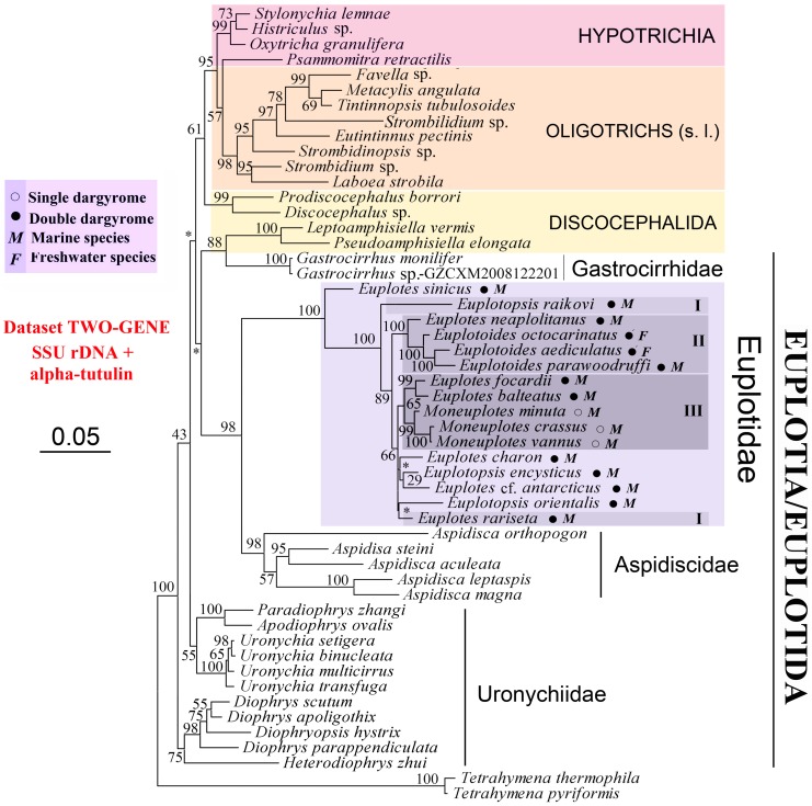
Topologies of two-gene combined tree (Fig. 3) and SSU-rDNA tree (Fig. S4) are nearly the same. There are several notable differences from analyses of alpha-tubulin alone (Fig. 2, Figs. S2, S3) including: 1) the monophyly of Oligotrichia and Choreotrichia; and 2) the non-monophyly of Hypotrichia and Discocephalida. Species of Euplotida cluster into a clade in SSU-rDNA tree with no support (Fig. S4), and fall into different clades in two-gene combined tree (Fig. 3). Similar to the alpha-tubulin analyses (Figs. 2, S2, S3), three out of four euplotid families, i.e. Euplotidae, Gastrocirrhidae and Aspidiscidae, are monophyletic in SSU-rDNA (Fig. S4) and two-gene combined trees (Fig. 3), though only several species of one genus are sequenced in the last two families, respectively. As shown in the alpha-tubulin trees (Figs. 2, S2, S3), Clade I as determined by previous investigations [38], [41], [42] does not appear in two-gene combined tree (Fig. 3) and SSU-rDNA tree (Fig. S4). However, Euplotes focardii, E. balteatus, two species not included in Petroni et al. [42] and Yi et al. [38], and three Moneuplotes species group together and form Clade III (Figs. 3, S4).
Figure 3. Best tree of the Spirotrichea inferred by two-gene combined sequences (Atub-SSU).
Bootstrap values for branches of the ML tree is given on nodes. The scale bar corresponds to 5 substitutions per 100 nucleotide positions. Dargyrome patterns and natural habitats are given after species name of euplotids by symbols. Clades I-IV for euplotids were designated according to Petroni [42] and Yi et al. [38].
Nodes of two-gene combined tree (Fig. 3) are better supported than SSU-rDNA tree (Fig. S4). There are 43 and 41 supported nodes (Bootstrap values >50%) in two-gene combined tree (Fig. 3) and SSU-rDNA tree (Fig. S4), respectively. Among them, support values of 28 nodes for these two trees are more than 90%, but more nodes are fully supported in two-gene combined tree (13 for combined tree vs. 10 for SSU-rDNA tree). Moreover, the two-gene combined tree (Fig. 3) posses more monophyletic taxa as predicted by morphology (e.g. spirotrichean subclasses and euplotid families, genera) than other trees (Table 3).
Discussion
Is Alpha-Tubulin a Suitable Marker for Inferring Ciliate Phylogeny?
The topologies of trees inferred from of ciliate proteins may be confounded by the many paralogs present in these lineages [14], [18] However, in the present investigation, alpha-tubulin gene paralogs of euplotid species are not very divergent from one another, and only paralogs of two of the nine species are non-monophyletic (Figs. 2, S2, S3). Similarly, with samples of five species from three ciliate classes, Israel et al. [15] also found that alpha-tubulin gene paralogs in any given taxon appear to be most closely related to each other or to a sequence from a congener than to others These data indicate that only recent paralogs of alpha-tubulin are retained, and thus gene duplication may not pose a substantial problem in defining ciliate clades [30]. However, alpha-tubulin is not always a good marker for studying relationships at the level of species or below given the high level of amino acid conservation among sequences (Fig. S1). Moreover, it is possible that a combination of gene duplication followed by concerted evolution and differential extinction of some alpha-tubulin paralogs has obscured the evolutionary history in some part of the ciliate tree [15].
The best way to evaluate the quality of one gene marker for tree construction is to look for its congruence with species tree inferred by morphology [14] and by other gene markers. We follow the criterion as a modified one given by Budin and Philippe [14], which is to assess the recovery of the monophyletic groups unquestionably supported by both morphology and SSU-rDNA trees. The monophyly of the family Euplotidae recovered by SSU-rDNA trees is consistently reconstructed in our three alpha-tubulin trees. In recent study, only three out of eight genera (Moneuplotes, Gastrocirrhus and Aspidisca), with alpha-tubulin gene sequences from several species, are monophyletic (Figs. 2, S2, S3). And for the other five genera, only species within Uronychia appear to be monophyletic. Same situation occurs in SSU-rDNA analyses [38]–[40], [43]–[45]. Therefore, according to the important criterion of accepted monophyletic groups, the reliability of alpha-tubulin is comparable to that of SSU-rDNA at the genus and family levels.
In the present investigation, only the subclass Hypotrichia, which contains only four genera, is monophyletic in three alpha-tubulin gene trees (Figs. 2, S2, S3). However, with more samples of alpha-tubulin gene from the Hypotrichia, monophyly of Hypotrichia was rejected by previous investigation [13]. For the other four subclasses for which we have sufficient taxon sampling, Oligotrichia is monophyletic (Figure 2, Atub_n74; Figure S2, Atub_aa), and others are not monophyletic. Therefore, alpha-tubulin might not be a good gene marker for examine relationships among high level taxa.
Ambiguous Assignment of Discocephalida
The phylogenetic position of the sister taxa Prodiscocephalus and Discocephalus is not stable in our analyses as that their position varies in different trees (Figs. 2, 3, S2, S3, S4). This corresponds to the variable classification schemes for this clade based on morphological and morphogenetic characters [31], [46]–[52]. For example, Prodiscocephalus and Discocephalus were regarded as genus-complex, family, suborder and order in previous investigations, and were also considered as members of Euplotidae/Euplotida [31], [46], [49], Sporadotrichina, Hypotrichida [48], Oxytrichia [51], [52], Stichotrichia [50] and so on. A relationship between pseudoamphisiellids and discocephalids is only revealed in SSU-rDNA tree (Fig. S4) and the sister relationship of these two groups is not rejected by AU test of Atub_aa (p = 0.152) and Atub-SSU (p = 0.241).
Phylogenetic Relationships within the Order Euplotida
Multi-gene analyses are proving useful as a means of placing some taxa within phylogenetic trees where morphological evidence and single gene analyses have not been successful [53]–[56]. Our results also show that two-gene combined tree is better than single gene trees for most clades (Table 3). However, relationship among four euplotid genera was not resolved by any of our analyses, including two-gene combined tree (Figs. 2, 3, S2, S3, S4). Inclusion of more taxa, especially species within the family Certesiidae, coupled with more genes are likely necessary to resolve sister relationships among euplotid families.
All six genera of the family Uronychiidae have been sequenced in the present study, revealing that this family is not monophyletic (Figs. 2, 3, S2, S3, S4). This result is consistent with some previous investigations inferred from SSU-rDNA sequences [45], [57], [58], though in some other SSU-rDNA trees this family appears monophyletic [38]–[40], [44]. Therefore, it is too early to infer whether this family should be further defined before more gene information is available. Similarly, the Diophrys-complex contains five genera (Diophrys, Diophryopsis, Paradiophrys, Heterodiophrys and Apodiophrys) but due to variable positions in different trees, it is difficult to infer their related relationships. However, similar to previous SSU-rDNA investigations [38]–[40], [44], the genus Diophrys seems to be non-monophyletic.
The family Euplotidae is composed of Euplotes-complex, and was divided into several genera or groups based on different morphological characters [34], [59], [60] or SSU-rDNA trees [38], [41], [42]. However, these classifications are not consistent with one another. For example, based on cortical structure, endosymbionts, morphometric data, morphogenetic patterns, and ecology, Euplotes-complex was separated into four genera (i.e. Euplotes, Euplotopsis, Euplotoides and Moneuplotes) by Borror and Hill [34]. Previous SSU-rDNA trees [38], [39], [41], [42], [44] and our analyses based on SSU-rDNA plus the two-gene combined trees demonstrate the monophyly of Moneuplotes and Euplotoides, but reject the monophyly of the other two genera (Figs. 3, S4). Similarly, the three species groups (i.e., single-, double-, or multiple- dargyrome types) defined according to dargyrome patterns (dorsal silverline system) by Gates and Curds [60] are not always monophyletic in molecular phylogenetic trees (our investigation [38], [42]), indicating the presence of more complexity within this group than is evident from dargyrome patterns. Moreover, the three well resolved clades (Clade I-III) repeatedly shown in previous SSU-rDNA trees [38], [39], [41], [42] are not always present in our trees (Figs. 2, 3, S2, S3, S4) nor are they supported by morphological characters, which indicates that these well resolved clades in SSU-rDNA gene trees may not capture valid taxonomic relationships. Finally, clades within Euplotidae are inconsistent with respect to morphology and habit, since the two freshwater species and the marine forms are interdigitated (Figs. 2, 3, S2, S3, S4).
Evolutionary Patterns in Duplicated Alpha-Tubulin
Among seven euplotid species for which paralogs are detected, duplicated alpha-tubulin genes of all taxa show some changes in the amino acid sequence following duplication (Table 2). Compared to those of Paramecium tetraurelia, which has much longer macronuclear chromosomes, there are bigger amino acid distances between paralogs of euplotid species. This elevated level of sequence divergence is similar to patterns in proteins from other ciliates with gene-sized macronuclear chromosomes (Israel et al. 2002, Zufall et al. 2006), and supports the hypothesis that genome processing is associated with increased protein diversification as proposed by previous investigations [30], [61], [62].
Materials and Methods
No specific permits were required for the described field studies. All locations are not privately-owned or protected in any way, and none endangered or protected species was involved.
Collection and Identification of Ciliates
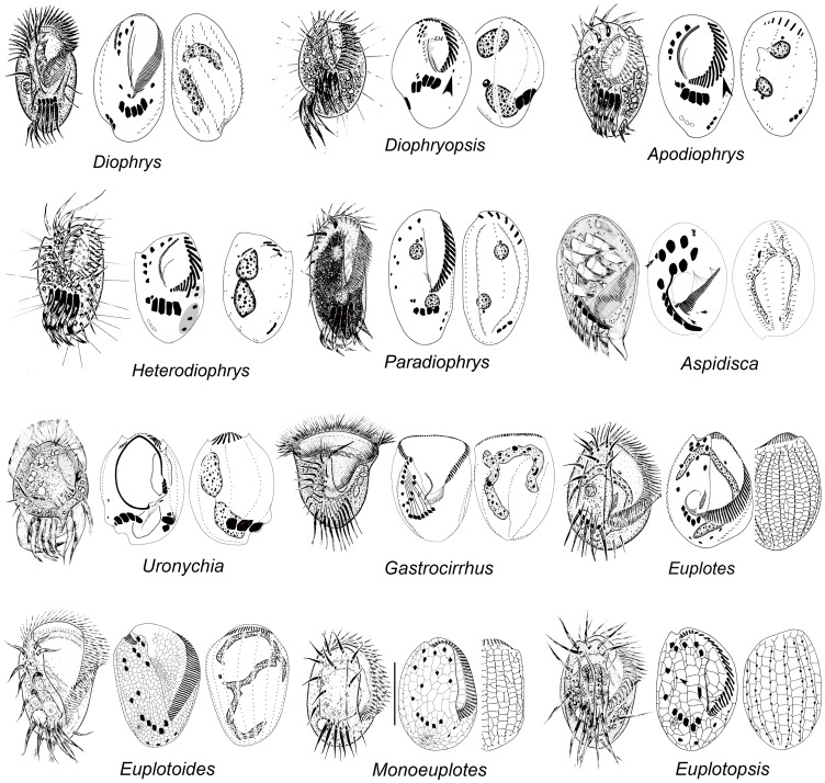
We isolated genomic DNAs from 34 morphospecies samples (Fig. 1) from China. Exact collection localities, sample information and GenBank accession numbers of sequenced alpha-tubulin genes are listed in Table 1. All isolates were identified by the methods of Shen et al. [37]. Terminology and systematic classification used in the current paper follow Lynn [31]. The term dargyrome used in the present paper is here defined as in previous reference [38], and refers to the overall geometrical pattern of the dorsal argyrome or “silverline system” in some euplotid ciliates. This pattern consists of net- or web-like structure revealed by silver impregnation methods, which is of great taxonomic importance at generic or specific level [59].
Figure 1. Representative euplotid species from live material and after protargol impregnation.
Extraction and Sequencing of DNA
Genomic DNA was extracted according to methods described in Yi et al. [63]. All DNA samples are extracted from one to several cells of one population, except for that there are two DNA samples for Diophrys parappendiculata and Euplotes sinicus, which are from two populations, respectively (Table 1). The PCR amplifications of the alpha-tubulin genes were performed using a TaKaRa ExTaq DNA Polymerase Kit (TaKaRa Biomedicals, Japan). Primers used for partial alpha-tubulin gene amplification were Tub-1 (5′-AAG GCT CTC TTG GCGTAC AT-3′) and the reverse primer Tub-2 (5′-TGATGC CTT CAA CAC CTT CTT-3′) [11]. PCR conditions were: 5 min initial denaturation (95°C), followed by 35 cycles of 1 min at 95°C, 1 min at 56°C and 1.5 min at 72°C, with a final extension of 15 min (72°C). The amplicons were directly sequenced using the same primers. However, if paralogs were detected in a sample, then it was purified using the TIANgel Midi Purification Kit and inserted into a pUCm-T vector. Two to nine clones were selected and sequenced by Invitrogen (Shanghai, China). Though it is impossible to detect all paralogs of investigated species due to interpretation of direct sequencing and limited clone samples, these sequences provide an estimate of paralog diversity.
Data Analyses
Sequence divergence between paralogs of ciliates is not clear. In the present investigation, we follows criterion of previous study [15], which defines sequences that diverge by more than 2% as paralogs, considering sequences errors produced by repeated PCRs and cloning [64]. Under this approach, recent paralogs may be confounded with allelic diversity and some paralogs may be missed, but this should not substantially bias our interpretations.
Five data sets were included in phylogenetic analyses: (1) Atub_n74: alpha-tubulin nucleotide sequences including first two codon positions (74 sequences in total); (2) Atub_aa: alpha-tubulin amino acid (70 sequences in total); (3) Atub-SSU: two-gene combined dataset including all euplotid species available (the paralog with shortest branch length is selected for alpha-tubulin) and other spirotrichean species of Dataset Atub_n74 except for Discocephalus ehrenbergi and Histriculus histrio for SSU-rDNA, and D. rotatorius and H. cavicola for alpha-tubulin (52 sequences in total); (4) SSU: SSU-rDNA sequences including all taxa in Dataset Atub-SSU (52 sequences in total); (5)Atub_n52: alpha-tubulin nucleotide sequences with first two codon positions including all taxa in Dataset Atub-SSU (52 sequences in total). For phylogenetic analyses, 27 sequences of alpha-tubulin genes from GenBank were used in addition to ones newly sequenced in the present study. The sequences were aligned using the ClustalW implemented in BIOEDIT 7.0.0 [65], and further modified manually using BIOEDIT. Final alignments used for subsequent phylogenetic analyses included 710 positions (Atub_n74), 355 positions (Atub_aa), 2,303 positions (Atub-SSU) and 1,593 positions (SSU), respectively. GTR + I + C was the best fitted model for nucleotide dataset (Atub_n74) selected by AIC as implemented in MrModeltest v2 [66], and Blosum62+I+G was the best one for amino acid dataset (Atub_aa) selected by AIC as implemented in ProtTest 1.4 [67]. Maximum likelihood analyses, and 1,000 bootstrap replicates, were conducted using RaxML-HPC v7.2.7 [68]. A Bayesian inference (BI) analysis was performed with MrBayes 3.1.2 [69] using the GTR+I+G model selected by MrModeltest 2 [66] under the AIC criterion. Markov chain Monte Carlo (MCMC) simulations were run with two sets of four chains using the default settings: chain length 1,500,000 generations, with trees sampled every 100 generations. The first 3,000 trees were discarded as burn-in. The remaining trees were used to generate a consensus tree and to calculate the posterior probabilities (PP) of all branches using a majority-rule consensus approach. Phylogenetic trees were visualized with TreeView v1.6.6 [70] and MEGA 4 [71].Congruence of different data partitions (in this case genes) was tested with both the incongruence length difference (ILD) test [72] and Shimodaira-Hasegawa (S-H) test [73] as implemented in PAUP*4.0b 10. PAUP* 4.0b 10 was used to generate constraint trees, and resulting trees were compared with unconstrained ML tree using the approximately unbiased (AU) test [74] as implemented in CONSEL package [75].
Supporting Information
Identical alpha-tubulin amino acid sites with different nucleotide sequences of Uronychia multicirrus and U. sinica (A); Euplotopsis encysticus and Euplotes cf. antarcticus (B); Euplotes sp.-GZJJM2009121510, Euplotoides parawoodruffi and Euplotopsis sp.-GZJJM2009121508 (C). A dot indicates a base that is identical to the first species. Solid circles highlight different first codon positions among/between species, and pentagram highlights different second codon position among species.
(TIF)
Best tree of the Spirotrichea inferred by Maximum likelihood of alpha-tubulin amino acid sequences (Atub_aa). Species newly sequenced in the present study are shown in bold type. Bootstrap values for branches of the ML tree and posterior probability values for BI tree, respectively, are given on nodes. Fully supported (100%/1.00) branches are marked with solid circles. The scale bar corresponds to 1 substitutions per 100 nucleotide positions. Dargyrome patterns and natural habitats are given after species name of euplotids by symbols.
(TIF)
Best tree of the Spirotrichea inferred by Maximum likelihood of Dataset Atub_n52. The scale bar corresponds to 1 substitution per 100 nucleotide positions.
(TIF)
Best tree of the Spirotrichea inferred by SSU-rDNA sequences (SSU). Bootstrap values for branches of the ML tree is given on nodes. The scale bar corresponds to 5 substitutions per 100 nucleotide positions. Dargyrome patterns and natural habitats are given after species name of euplotids by symbols.
(TIF)
Acknowledgments
Our deepest gratitude goes to Ms. Feng Gao, Jie Huang and Yan Zhao, Laboratory of Protozoology, OUC for gene sequencing.
Footnotes
Competing Interests: The authors have declared that no competing interests exist.
Funding: This work was supported by the ‘The Natural Science Foundation of China’ (Project No. 31030059, 41006098) to W.S. and Z.Y., China Postdoctoral Science Foundation (20110491623) to Z.Y., and two National Science Foundation awards (DEB 0816828 and DEB RUI:0919152) to L.A.K. The funders had no role in study design, data collection and analysis, decision to publish, or preparation of the manuscript.
References
- 1.Miao M, Shao C, Song W. Evolution of discocephalid ciliates: all molecular, morphological and ontogenetic data support a sister group of discocephalids and pseudoamphisiellids (Protozoa, Ciliophora), with establishment of a new suborder Pseudoamphisiellina subord. n. Sci China Ser C-Life Sci. 2011;54:634–641. doi: 10.1007/s11427-011-4192-8. [DOI] [PubMed] [Google Scholar]
- 2.Lynn DH, Sogin ML. Assessment of phylogenetic relationships among ciliated protists using partial ribosomal RNA sequences derived from reverse transcripts. BioSystems. 1988;21:249–254. doi: 10.1016/0303-2647(88)90020-2. [DOI] [PubMed] [Google Scholar]
- 3.Lynn DH, Small EB. A revised classification of the phylum Ciliophora Doflein, 1901. Rev Soc Mex Hist Nat 47 65–78. 1997.
- 4.Stoeck T, Foissner W, Lynn DH. Small-subunit rRNA phylogenies suggest that Epalxella antiquorum (Penard, 1922) Corliss, 1960 (Ciliophora, Odontostomatida) is a member of the Plagyopylea. J Eukaryot Microbiol. 2007;54:436–42. doi: 10.1111/j.1550-7408.2007.00283.x. [DOI] [PubMed] [Google Scholar]
- 5.Strüder-Kypke MC, Wright AD, Foissner W, Chatzinotas A, Lynn DH. Molecular phylogeny of litostome ciliates (Ciliophora, Litostomatea) with emphasis on free-living haptorian genera. Protist. 2006;157:261–78. doi: 10.1016/j.protis.2006.03.003. [DOI] [PubMed] [Google Scholar]
- 6.Schmidt SL, Bernhard D, Schlegel M, Foissner W. Phylogeny of the Stichotrichia (Ciliophora; Spirotrichea) reconstructed with nuclear small subunit rRNA gene sequences: discrepancies and accordances with morphological data. J Eukaryot Microbiol. 2007;54:201–9. doi: 10.1111/j.1550-7408.2007.00250.x. [DOI] [PubMed] [Google Scholar]
- 7.Yi Z, Dunthorn M, Song W, Stoeck T. Increasing taxon sampling using both unidentified environmental sequences and identified cultures improves phylogenetic inference in the Prorodontida (Ciliophora, Prostomatea). Mol Phylogenet Evol. 2010;57:937–941. doi: 10.1016/j.ympev.2010.08.001. [DOI] [PubMed] [Google Scholar]
- 8.Li L, Shao C, Song W, Lynn D, Chen Z, et al. Does Kiitricha (Protista, Ciliophora, Spirotrichea) belong to Euplotida or represent a primordial spirotrichous taxon? With suggestion to establish a new subclass Protohypotrichia. Int J Syst Evol Microbiol. 2009;59:439–446. doi: 10.1099/ijs.0.65801-0. [DOI] [PubMed] [Google Scholar]
- 9.Li L, Stoeck T, Shin MK, Al-Rasheid K, Al-Khedhairy A, et al. Protocruzia, a highly ambiguous ciliate (Protozoa; Ciliophora): Very likely an ancestral form for Heterotrichea, Colpodea or Spirotrichea? With reevaluation of its evolutionary position based on multigene analyses. Science China Series C-Life Sciences. 2010;53:131–138. doi: 10.1007/s11427-010-0012-9. [DOI] [PubMed] [Google Scholar]
- 10.Cho B, Park J, Xu K, Choi J. Morphology and molecular phylogeny of Trimyema koreanum n. sp., a ciliate from the hypersaline water of a solar saltern. J Eukaryot Microbiol. 2008;55:417–426. doi: 10.1111/j.1550-7408.2008.00340.x. [DOI] [PubMed] [Google Scholar]
- 11.Yi Z, Song W, Stoeck T, Al-Rasheid K, Al-Khedhairy A, et al. Phylogenetic analyses suggest that Psammomitra (Ciliophora, Urostylida) should represent an urostylid family, based on SSrRNA and alpha-tubulin gene sequence information. Zool J Linn Soc. 2009;157:227–236. [Google Scholar]
- 12.Foissner W, Stoeck T. Morphology, ontogenesis and molecular phylogeny of Neokeronopsis (Afrokeronopsis) aurea nov. subgen., nov. spec. (Ciliophora: Hypotricha), a new African flagship ciliate confirms the CEUU hypothesis. Acta Protozoologica. 2008;47:1–33. [PMC free article] [PubMed] [Google Scholar]
- 13.Yi Z, Song W. Evolution of the order Urostylida (Protozoa, Ciliophora): new hypotheses based on multi-gene information and identification of localized incongruence. PLoS ONE. 2011;6:e17471. doi: 10.1371/journal.pone.0017471. doi: 10.1371/journal.pone.0017471. [DOI] [PMC free article] [PubMed] [Google Scholar]
- 14.Budin K, Philippe H. New insights into the phylogeny of eukaryotes based on ciliate Hsp70 sequences. Mol Biol Evol. 1998;15:943–56. doi: 10.1093/oxfordjournals.molbev.a026010. [DOI] [PubMed] [Google Scholar]
- 15.Israel RL, Kosakovsky Pond SL, Muse SV, Katz LA. Evolution of duplicated alpha-tubulin genes in ciliates. Evolution. 2002;56:1110–22. doi: 10.1111/j.0014-3820.2002.tb01425.x. [DOI] [PubMed] [Google Scholar]
- 16.Miao M, Warren A, Song W, Wang S, Shang H, et al. Analysis of the internal transcribed spacer 2 (ITS2) region of scuticociliates and related taxa (Ciliophora, Oligohymenophorea) to infer their evolution and phylogeny. Protist. 2008;159:519–533. doi: 10.1016/j.protis.2008.05.002. [DOI] [PubMed] [Google Scholar]
- 17.Hewitt EA, Muller KM, Cannone J, Hogan DJ, Gutell R, et al. Phylogenetic relationships among 28 spirotrichous ciliates documented by rDNA. Mol Phylogenet Evol. 2003;29:258–267. doi: 10.1016/s1055-7903(03)00097-6. [DOI] [PubMed] [Google Scholar]
- 18.Hoffman DC, Prescott DM. Phylogenetic relationships among hypotrichous ciliates determined with the macronuclear gene encoding the large, catalytic subunit of DNA polymerase alpha. J Mol Evol. 1997;45:301–10. doi: 10.1007/pl00006234. [DOI] [PubMed] [Google Scholar]
- 19.Dalby AB, Prescott DM. The scrambled actin I gene in Uroleptus pisces. Chromosoma. 2004;112:247–254. doi: 10.1007/s00412-003-0270-4. [DOI] [PubMed] [Google Scholar]
- 20.Tourancheau AB, Villalobo E, Tsao N, Torres A, Pearlman RE. Protein coding gene trees in ciliates: comparison with rRNA-based phylogenies. Mol Phylogenet Evol. 1998;10:299–309. doi: 10.1006/mpev.1998.0534. [DOI] [PubMed] [Google Scholar]
- 21.Gong Y, Xu K, Zhan Z, Yu Y, Li X, et al. Alpha-tubulin and small subunit rRNA phylogenies of Peritrichs are congruent and do not support the clustering of Mobilids and Sessilids (Ciliophora, Oligohymenophorea). J Eukaryot Microbiol. 2010;57:265–272. doi: 10.1111/j.1550-7408.2010.00472.x. [DOI] [PubMed] [Google Scholar]
- 22.Sun P, Clamp J, Xu D. Analysis of the secondary structure of ITS transcripts in peritrich ciliates (Ciliophora, Oligohymenophorea): Implications for structural evolution and phylogenetic reconstruction. Mol Phylogenet Evol. 2010;56:242–251. doi: 10.1016/j.ympev.2010.02.030. [DOI] [PubMed] [Google Scholar]
- 23.Galtier N, Gouy M. Inferring phylogenies from DNA sequences of unequal base compositions. Proc Natl Acad Sci U S A. 1995;92:11317–11321. doi: 10.1073/pnas.92.24.11317. [DOI] [PMC free article] [PubMed] [Google Scholar]
- 24.Leipe DD, Gunderson JH, Nerad TA, Sogin ML. Small subunit ribosomal RNA+ of Hexamita inflata and the quest for the first branch in the eukaryotic tree. Mol Biochem Parasitol. 1993;59:41–48. doi: 10.1016/0166-6851(93)90005-i. [DOI] [PubMed] [Google Scholar]
- 25.Hasegawa M, Hashimoto T. Ribosomal RNA trees misleading? Nature. 1993;361:23. doi: 10.1038/361023b0. [DOI] [PubMed] [Google Scholar]
- 26.Maddison W. Gene trees in species trees. Syst Biol. 1997;46:523–536. [Google Scholar]
- 27.Fitch WM. Distinguishing homologous from analogous proteins. Syst Zool. 1970;28:132–163. [PubMed] [Google Scholar]
- 28.Goodman M, Czelusniak J, Moore G, Romero-Herrera A, Matsuda G. Fitting the gene lineage into its species lineage, a parsomony strategy illustrated by cladograms constructed from globin sequences. Syst Zool. 1979;19:99–113. [Google Scholar]
- 29.Snoeyenbos-West OL, Salcedo T, McManus GB, Katz LA. Insights into the diversity of choreotrich and oligotrich ciliates (Class: Spirotrichea) based on genealogical analyses of multiple loci. Int J Syst Evol Microbiol. 2002;52:1901–13. doi: 10.1099/00207713-52-5-1901. [DOI] [PubMed] [Google Scholar]
- 30.Zufall RA, McGrath CL, Muse SV, Katz LA. Genome architecture drives protein evolution in ciliates. Mol Biol Evol. 2006;23:1681–1687. doi: 10.1093/molbev/msl032. [DOI] [PubMed] [Google Scholar]
- 31.Lynn DH. The Ciliated Protozoa. New York: Pergamon Press. 2008.
- 32.Berger H. Monograph of the Urostyloidea (Ciliophora, Hypotricha). Monographiae Biologicae. 2006;85:1–1304. [Google Scholar]
- 33.Schlegel M, Kramer M, Hahn K. Taxonomy and phylogenetic relationship of eight species of the genus Euplotes (Hypotrichida, Ciliophora) as revealed by enzyme electrophoresis. Eur J Protistol. 1988;24:22–29. doi: 10.1016/S0932-4739(88)80005-1. [DOI] [PubMed] [Google Scholar]
- 34.Borror AC, Hill BF. The order Euplotida (Ciliophora): taxonomy, with division of Euplotes into several genera. J Eukaryot Microbiol. 1995;42:457–466. [Google Scholar]
- 35.Bernhard D, Stechmann A, Foissner W, Ammermann D, Hehn M, et al. Phylogenetic relationships within the class Spirotrichea (Ciliophora) inferred from small subunit rRNA gene sequences. Mol Phylogenet Evol. 2001;21:86–92. doi: 10.1006/mpev.2001.0997. [DOI] [PubMed] [Google Scholar]
- 36.Song W, Wilbert N, Al-Rasheid K, Warren A, Shao C, et al. Redescriptions of two marine hypotrichous ciliates, Diophrys irmgard and D. hystrix (Ciliophora, Euplotida), with a brief revision of the genus Diophrys. J Eukaryot Microbiol. 2007;54:283–96. doi: 10.1111/j.1550-7408.2007.00266.x. [DOI] [PubMed] [Google Scholar]
- 37.Shen Z, Yi Z, Warren A. The morphology, ontogeny, and small subunit rRNA gene sequence analysis of Diophrys parappendiculata n. sp. (Protozoa, Ciliophora, Euplotida), a new marine ciliate from coastal waters of southern China. J Eukaryot Microbiol. 2011;58:242–248. doi: 10.1111/j.1550-7408.2011.00536.x. [DOI] [PubMed] [Google Scholar]
- 38.Yi Z, Song W, Clamp J, Chen Z, Gao S, et al. Reconsideration of systematic relationships within the order Euplotida (Protista, Ciliophora) using new sequences of the gene coding for small-subunit rRNA and testing the use of combined data sets to construct phylogenies of the Diophrys-complex. Mol Phylogenet Evol. 2009;50:599–607. doi: 10.1016/j.ympev.2008.12.006. [DOI] [PubMed] [Google Scholar]
- 39.Jiang J, Zhang Q, Warren A, Al-Rasheid K, Song W. Morphology and SSUrRNA gene-based phylogeny of two marine Euplotes species, E. orientalis spec. nov.and E. raikovi Agamaliev, 1966 (Ciliophora, Euplotida). Eur J Protistol. 2010;46:121–132. doi: 10.1016/j.ejop.2009.11.003. [DOI] [PubMed] [Google Scholar]
- 40.Jiang J, Song W. Two new Diophrys-like genera and their type species, Apodiophrys ovalis n. g., n. sp. and Heterodiophrys zhui n. g., n. sp. (Ciliophora: Euplotida), with notes on their molecular phylogeny. J Eukaryot Microbiol. 2010;57:354–361. doi: 10.1111/j.1550-7408.2010.00482.x. [DOI] [PubMed] [Google Scholar]
- 41.Schwarz M, Zuendorf A, Stoeck T. Morphology, ultrastructure, molecular phylogeny, and autecology of Euplotes elegans Kahl, 1932 (Hypotrichida; Euplotidae) isolated from the Anoxic Mariager Fjord, Denmark. J Eukaryot Microbiol. 2007;54:125–136. doi: 10.1111/j.1550-7408.2007.00243.x. [DOI] [PubMed] [Google Scholar]
- 42.Petroni G, Dini F, Verni F, Rosati G. A molecular approach to the tangled intrageneric relationships underlying phylogeny in Euplotes (Ciliophora, Spirotrichea). Mol Phylogenet Evol. 2002;22:118–30. doi: 10.1006/mpev.2001.1030. [DOI] [PubMed] [Google Scholar]
- 43.Miao M, Song W, Chen Z, Al-Rasheid KA, Shao C, et al. A unique euplotid ciliate, Gastrocirrhus (Protozoa, Ciliophora): assessment of its phylogenetic position inferred from the small subunit rRNA gene sequence. J Eukaryot Microbiol. 2007;54:371–8. doi: 10.1111/j.1550-7408.2007.00271.x. [DOI] [PubMed] [Google Scholar]
- 44.Jiang J, Zhang Q, Hu X, Shao C, Al-Rasheid K, et al. Two new marine ciliates, Euplotes sinicus sp. nov. and Euplotes parabalteatus sp. nov., and a new small subunit rRNA gene sequence of Euplotes rariseta (Ciliophora, Spirotrichea, Euplotida). Int J Syst Evol Microbiol. 2010;60:1241–1251. doi: 10.1099/ijs.0.012120-0. [DOI] [PubMed] [Google Scholar]
- 45.Lynn DH, Strüder-Kypke M. Phylogenetic position of Licnophora, Lechriopyla, and Schizocaryum, three unusual ciliates (phylum Ciliophora) endosymbiotic in echinoderms (phylum Echinodermata). J Eukaryot Microbiol. 2002;49:460–8. doi: 10.1111/j.1550-7408.2002.tb00229.x. [DOI] [PubMed] [Google Scholar]
- 46.Corliss JO. The ciliated protozoa: characterization, classification and guide to the literature. Oxford: Pergamon Press. 1979.
- 47.Jankowski AW. Revision of the order Hypotrichida Stein, 1859. Generic catalogue, phylogeny, taxonomy. Proc. Acad. Sci. USSR. 1979;86:48–85. [Google Scholar]
- 48.Wicklow BJ. The Discocephalina (n. subord.): ultrastructure, morphogenesis and evolutionary implications of a group of endemic marine interstital hypotrichs (Ciliophora, Protozoa). Protistologica. 1982;18:299–330. [Google Scholar]
- 49.Small EB, Lynn DH. Phylum Ciliophora Doflein, 1901. In: JJ Lee, SH Hutner, ED Bovee, eds (1985) An Illustrated Guide to the Protozoa Lawrence, Kansas: Soc. Protozool. Special Publ., Allen Press. p. 1985. pp. 393–575.
- 50.Shao C, Song W, Li L, A W, Al-Rasheid K, et al. Systematic position of Discocephalus-like ciliates (Ciliophora: Spirotrichea) inferred from SSU rDNA and ontogenetic information. Int J Syst Evol Microbiol. 2008;58:2962–2972. doi: 10.1099/ijs.0.65781-0. [DOI] [PubMed] [Google Scholar]
- 51.de Puytorac P, Batisse A, Deroux G, Fleury A, Grain J, et al. Proposition d’une nouvelle classification du phylum des protozoaires Ciliophora Doflein, 1901. C R Acad Sci Paris. 1993;316:716–720. (in French). [PubMed] [Google Scholar]
- 52.Tuffrau M, Fleury A. Classe des Hypotrichea Stein, 1859. Traite de Zoologie. 1994;2:83–151. [Google Scholar]
- 53.Parfrey LW, Grant J, Tekle YI, Lasek-Nesselquist E, Morrison HG, et al. Broadly sampled multigene analyses yield a well-resolved eukaryotic tree of life. Syst Biol. 2010;59:518–533. doi: 10.1093/sysbio/syq037. [DOI] [PMC free article] [PubMed] [Google Scholar]
- 54.Tekle YI, Grant J, Anderson OR, Nerad TA, Cole JC, et al. Phylogenetic placement of diverse amoebae inferred from multigene analyses and assessment of clade stability within ‘Amoebozoa’ upon removal of varying rate classes of SSU-rDNA. Mol Phylogenet Evol. 2008;47:339–352. doi: 10.1016/j.ympev.2007.11.015. [DOI] [PubMed] [Google Scholar]
- 55.Yoon YS, Grant J, Tekle YI, Wu M, Chaon BC, et al. Broadly sampled multigene trees of eukaryotes. BMC Evolutionary Biology. 2008;8:14. doi: 10.1186/1471-2148-8-14. [DOI] [PMC free article] [PubMed] [Google Scholar]
- 56.Nikolaev SI, Berney C, Fahrni J, Bolivar I, Polet S, et al. The twilight of Heliozoa and rise of Rhizaria, an emerging supergroup of amoeboid eukaryotes. Proc. Natl. Acad. Sci. USA. 2004;101:8066–8071. doi: 10.1073/pnas.0308602101. [DOI] [PMC free article] [PubMed] [Google Scholar]
- 57.Chen Z, Song W. Phylogenetic positions of Uronychia transfuga and Diophrys appendiculata (Euplotida, Hypotrichia, Ciliophora) within hypotrichous ciliates inferred from small subunit ribosomal RNA gene sequences. Eur J Protistol. 2001;37:291–301. [Google Scholar]
- 58.Chen Z, Song W. Phylogenetic positions of Aspidisca steini and Euplotes vannus within the order Euplotida (Hypotrichia: Ciliophora) inferred from complete small subunit ribosomal RNA gene sequences. Acta Protozoologica. 2002;41:1–9. [Google Scholar]
- 59.Curds RC. A guide to the species of Euplotes (Hypotrichida, Ciliatea). Bull Br Mus Nat Hist (Zool) 1975;28:3–61. [Google Scholar]
- 60.Gates MA, Curds CR. The dargyrome of the genus Euplotes. Bull Br Mus Nat Hist (Zool) 1979;35:127–200. [Google Scholar]
- 61.Zufall RA, Katz LA. Micronuclear and macronuclear forms of β-tubulin genes in the ciliate Chilodonella uncinata reveal Insights into genome processing and protein evolution. J Eukaryot Microbiol. 2007;54:275–282. doi: 10.1111/j.1550-7408.2007.00267.x. [DOI] [PubMed] [Google Scholar]
- 62.Katz LA, Bornstein J, Lasek-Nesselquist E, Muse SV. Dramatic diversity of ciliate histone H4 genes revealed by comparisons of patterns of substitutions and paralog divergences among eukaryotes. Mol Biol Evol. 2004;21:555–562. doi: 10.1093/molbev/msh048. [DOI] [PubMed] [Google Scholar]
- 63.Yi Z, Lin X, Warren A, Al-Rasheid K, Song W. Molecular phylogeny of Nothoholosticha (Protozoa, Ciliophora, Urostylida) and systematic relationships of the Holosticha-complex. Syst Biodiv. 2010;8:149–155. [Google Scholar]
- 64.Riley JL, Katz LA. Widespread distribution of extensive chromosomal fragmentation in ciliates. Mol Biol Evol. 2001;18:1372–7. doi: 10.1093/oxfordjournals.molbev.a003921. [DOI] [PubMed] [Google Scholar]
- 65.Hall TA. BioEdit: a user-friendly biological sequence alignment editor and analysis program for Windows 95/98/NT. Nucleic Acids Symp Ser. 1999;41:95–98. [Google Scholar]
- 66.Nylander JA. MrModeltest v2. : Evolutionary Biology Centre, Uppsala University. 2004.
- 67.Abascal F, Zardoya R, Posada D. ProtTest: Selection of best-fit models of protein evolution. Bioinformatics. 2005;21:2104–2105. doi: 10.1093/bioinformatics/bti263. [DOI] [PubMed] [Google Scholar]
- 68.Stamatakis A, Hoover P, Rougemont J. A rapid bootstrap algorithm for the RAxML web servers. Syst Biol. 2008;57:758–771. doi: 10.1080/10635150802429642. [DOI] [PubMed] [Google Scholar]
- 69.Ronquist F, Huelsenbeck JP. MRBAYES 3: Bayesian phylogenetic inference under mixed models. Bioinformatics. 2003;19:1572–1574. doi: 10.1093/bioinformatics/btg180. [DOI] [PubMed] [Google Scholar]
- 70.Page RDM. TREEVIEW: An application to view phylogenetic trees on personal computers. Comput Appl Biosci. 1996;12:357–358. doi: 10.1093/bioinformatics/12.4.357. [DOI] [PubMed] [Google Scholar]
- 71.Tamura K, Dudley J, Nei M, Kumar S. MEGA4: Molecular Evolutionary Genetics Analysis (MEGA) software version 4.0. Mol Biol Evol. 2007;24:1596–1599. doi: 10.1093/molbev/msm092. [DOI] [PubMed] [Google Scholar]
- 72.Farris JS, Källersjö M, Kluge AG, Bult C. Testing significance of incongruence. Cladistics. 1994;10:315–319. [Google Scholar]
- 73.Shimodaira H, Hasegawa M. Multiple comparisons of log-likelihoods with applications to phylogenetic inference. Mol Biol Evol. 1999;16:1114–1116. [Google Scholar]
- 74.Shimodaira H. An approximately unbiased test of phylogenetic tree selection. Syst Biol. 2002;51:492–508. doi: 10.1080/10635150290069913. [DOI] [PubMed] [Google Scholar]
- 75.Shimodaira H, Hasegawa M. CONSEL: For assessing the confidence of phylogenetic tree selection. Bioinformatics. 2001;17:1246–1247. doi: 10.1093/bioinformatics/17.12.1246. [DOI] [PubMed] [Google Scholar]
Associated Data
This section collects any data citations, data availability statements, or supplementary materials included in this article.
Supplementary Materials
Identical alpha-tubulin amino acid sites with different nucleotide sequences of Uronychia multicirrus and U. sinica (A); Euplotopsis encysticus and Euplotes cf. antarcticus (B); Euplotes sp.-GZJJM2009121510, Euplotoides parawoodruffi and Euplotopsis sp.-GZJJM2009121508 (C). A dot indicates a base that is identical to the first species. Solid circles highlight different first codon positions among/between species, and pentagram highlights different second codon position among species.
(TIF)
Best tree of the Spirotrichea inferred by Maximum likelihood of alpha-tubulin amino acid sequences (Atub_aa). Species newly sequenced in the present study are shown in bold type. Bootstrap values for branches of the ML tree and posterior probability values for BI tree, respectively, are given on nodes. Fully supported (100%/1.00) branches are marked with solid circles. The scale bar corresponds to 1 substitutions per 100 nucleotide positions. Dargyrome patterns and natural habitats are given after species name of euplotids by symbols.
(TIF)
Best tree of the Spirotrichea inferred by Maximum likelihood of Dataset Atub_n52. The scale bar corresponds to 1 substitution per 100 nucleotide positions.
(TIF)
Best tree of the Spirotrichea inferred by SSU-rDNA sequences (SSU). Bootstrap values for branches of the ML tree is given on nodes. The scale bar corresponds to 5 substitutions per 100 nucleotide positions. Dargyrome patterns and natural habitats are given after species name of euplotids by symbols.
(TIF)


