Abstract
The vestibular organs consist of complementary sensors: the semicircular canals detect rotations while the otoliths detect linear accelerations, including the constant pull of gravity. Several fundamental questions remain on how the vestibular system would develop and/or adapt to prolonged changes in gravity such as during long-term space journey. How do vestibular reflexes develop if the appropriate assembly of otoliths and semi-circular canals is perturbed? The aim of present work was to evaluate the role of gravity sensing during ontogeny of the vestibular system. In otoconia-deficient mice (ied), gravity cannot be sensed and therefore maculo-ocular reflexes (MOR) were absent. While canals-related reflexes were present, the ied deficit also led to the abnormal spatial tuning of the horizontal angular canal-related VOR. To identify putative otolith-related critical periods, normal C57Bl/6J mice were subjected to 2G hypergravity by chronic centrifugation during different periods of development or adulthood (Adult-HG) and compared to non-centrifuged (control) C57Bl/6J mice. Mice exposed to hypergravity during development had completely normal vestibulo-ocular reflexes 6 months after end of centrifugation. Adult-HG mice all displayed major abnormalities in maculo-ocular reflexe one month after return to normal gravity. During the next 5 months, adaptation to normal gravity occurred in half of the individuals. In summary, genetic suppression of gravity sensing indicated that otolith-related signals might be necessary to ensure proper functioning of canal-related vestibular reflexes. On the other hand, exposure to hypergravity during development was not sufficient to modify durably motor behaviour. Hence, 2G centrifugation during development revealed no otolith-specific critical period.
Introduction
The vestibular organs consist of complementary sensors: the semicircular canals detect angular accelerations while the otoliths detect linear accelerations, including the constant pull of gravity. Integration by the vestibulo-cerebellum of canal and otolith information to visual and proprioceptive inputs generates an estimate of the position and movements of the head in space [1]. In frog, most of the central vestibular neurons receive convergent monosynaptic inputs from a single semicircular canal and from the spatially complementary otolith [2]. In rat, convergence of macular and canal inputs concern about 80% of tested vestibular neurons [3]. In cat, 1/3 of studied neurons receive convergent inputs from vertical canals and otoliths and 1/5 receive convergent inputs from horizontal canals and otoliths [4]. The anatomical convergence and functional complementarities between canals and otoliths raise questions on how spatial refinements of vestibular microcircuits at the origin of the vestibulo-ocular reflex (VOR) occur during development and adapt throughout lifespan. As for other developing systems, ontogeny of the vestibular system depends on both genetics and experience [5]. Initial formation, pathfinding to specific targets and survival of inner ear neurons are based initially on genetic programs [6] during the 2 perinatal weeks [7]. Along the 3-neurons-arc that drives the VOR, connectivity is established around birth independently of vestibular activity [8]. Then, late activity-mediated maturation would refine its function [9], [10].
In that context, various experiments investigated the effects of an environmental alteration of gravity on the development of the vestibular system. Prenatal exposure of rats to weightlessness induced severe and often long lasting abnormalities on vestibular-related behaviors [7]. Also, increasing gravity during development induced non-lasting effects on the morphology and physiology of the vestibular periphery [11]–[13] however correlated to limited locomotor deficits [14]–[16]. Overall, a change in gravity-load for periods including development would modify both peripheral and central vestibular-related circuits, but the persistence of these alterations remains uncertain [7], [17] and the ontogeny of the oculomotor control was not examined.
Therefore, this study was aimed at investigating the role of the gravito-inertial information in the combined development of otolith-based and canal-based vestibular reflexes. In particular, the two following hypotheses were tested: i) tuning of canal-related vestibulo-ocular reflexes (VOR) need gravito-inertial information and ii) alteration of gravity during critical periods of development indefinitely impairs the acquisition of otolith-specific reflex and of canal-specific VOR. To examine these questions, the VOR presents many experimental advantages. First, VOR circuitry has been investigated in detail. Second, although body movements are drastically modified under altered gravity due to the loading/unloading of large body segments, eyes are mechanistically less affected. Finally, otolith-specific reflex (e.g. maculo-ocular reflex) can be evaluated using paradigms such as the off-vertical axis rotation test [18].
In order to alter the sensing of gravito-inertial forces during development, we used two types of paradigms. First, otoconia-deficient mice, which suffer from the absence of gravity-related modulation of otolith inputs [19] were used as a model of development without the vestibular sensing of gravity [20]. Second, the effect of an increase of gravity-load on the gaze stabilization was studied in mice centrifuged during adulthood, during the entire development, or for periods covering respectively the early onset or late maturation of the vestibular ontogeny [7].
Methods
Ethics Statement
All procedures used were in strict compliance with the European Directive 86/609/EEC on the protection of animals used for experimental purposes. CNRS review board and the Direction Departementale des Services Vétérinaires specifically approved this study; authorization number 75–1641.
Forty-six normal mice (C57Bl/6J - Charles River Laboratories, France), including 31 mice born and raised in 2G centrifuge were used in this study (see detailed procedure below). In addition, otoconia-deficient mice (inner ear defect, ied, see [20]) with a C3HeB/FeJ Background were initially purchased from the Centre De Transgénèse d’Orléans, France, and breeding was subsequently done in our institution. Ied mutation consists in a splicing defect on the chromosome 5 otop 1 gene, which leads to complete otoconial agenesis [20]–[22]. Thirteen ied mice aged 6 months were phenotyped during swimming task to ensure inner ear deficiency [23], [24]. As other otoconia-deficient strains, ied mice also presented a head deviation in frontal plane (or head-tilt, Figure S1; [20]).
Centrifuge Configuration and Housing Conditions
Hypergravity was produced using a centrifuge consisting in 1.4 m radius carousel with gondolas hanged on the periphery (maximum radius during rotation: 1.8 m) [25]. 2G vector at the center of the gondolas (±5% on the edges) was produced with 29.6 rpm rotational speed. C57Bl/6J mice were placed in standard cages located in the gondolas. Cages were supplied enough food and water for 3 weeks and were left undisturbed during the chronic centrifugation. When centrifugation duration exceeded 3 weeks, centrifugation was interrupted during minimal time needed for refilling food and water. Infra-red camera videos fixed on the centrifuge arms allowed a remote day and night control of the mice in their cages. All the environmental variables, except the gravity level, were the same as in standard housing. The cages containing control C57Bl/6J mice were placed in similar gondolas and in the same room as the centrifuged mice.
Description of the Groups of Centrifuged Mice
C57Bl/6J mice bred in the centrifuge had a previous experience of chronic centrifugation of at least eight weeks, and were all multiparous to reduce the risks of misbehavior against the pups during the centrifugation. The experiments involved four groups of C57Bl/6J mice centrifuged during different periods and duration of development or adulthood. For Developmental nomenclature “E10” indicates 10th embryonic day of development and “P10” 10th postnatal day of maturation.
-
-
Group Control (n = 8): C57Bl/6J mice were born and raised in the room of the centrifuge, but were never exposed to hypergravity (HG). Control mice were tested at age of 6 months.
-
-
Group Adult-HG (n = 7): C57Bl/6J mice born and raised in the centrifuge room were exposed to 50 days of 2G centrifugation between the age of 2–4 months. Mice were tested 1 month and 6 months after their return to normogravity.
-
-
Group Pre (E0–P10, prenatal exposition to HG; n = 8): C57Bl/6J mice were conceived and born in the centrifuge. The pups remained with their parents in the centrifuge until 10th postnatal day. Mice were then put in standard housing under normal gravity in the same room.
-
-
Group Post (P10–P30: postnatal exposition to HG; n = 8): C57Bl/6J mice reproduced in standard housing. The pups were transferred to the centrifuge with their parents at 10th postnatal day. Mice were left in the centrifuge until the 30th postnatal day, and then returned to standard housing in the centrifuge room.
-
-
Group Full (E0–P30: complete development in HG; n = 15): The conception and delivery occurred in the centrifuge but the C57Bl/6J mice remained in the centrifuge until 30th postnatal day.
The pups were weaned at the age of 30 days, they were weighed, individually tagged with RFID microchips and grouped 3 same sex mice per cage. They were left undisturbed until the age of 2 months when a set of behavioral tests [26] were carried out. Once the tests were completed, sets of mice were sacrificed for tissue analysis, while the other returned in standard housing for 5 months for a second series of tests.
Surgeries
Surgical preparation and postoperative care for head implant surgery have been described previously [27]. Gas anesthesia was induced using Isoflurane. A small custom-built head holder was then cemented (C&B Metabond) to the skull just anterior to the lambda landmark [28]. Following the surgery, animals were isolated and closely surveyed for 48 hours. Buprenorphine (0.05 mg/kg) was provided for postoperative analgesia and care was taken to avoid hypothermia and dehydration.
Vestibular Stimulations and Data Acquisition
Recorded eye and head position signals were sampled at 1 kHz, digitally recorded (CED power1401 MkII) under spike 2 environment and later exported into the Matlab (The MathWorks) programming environment for off-line analysis.
Mice were head-fixed at a ∼30° nose-down position to align the horizontal canals on yaw plane [29]–[31]. Because the precise orientation of the canals in C3HeB/FeJ background was never reported, we tested in a preliminary experiment the efficiency of the horizontal vestibulo-ocular reflex for different head-pitch angles (Figure S1, A) and found that a head pitch of 30° was about optimal. Accordingly, ied were tested following the same procedure as C57Bl6/J mice.
Mice were initially tested during sinusoidal rotations (0.2–2 Hz; 40°/s) in yaw plane. Rotations at constant velocities around vertical axis were also applied to ied and 50 days exposed groups (Full and Adult-HG groups) at 50°/s. Once yaw testing completed, the table was tilted 17° off-vertical axis with the mouse in a nose-down position. Off-vertical axis rotation (OVAR) responses were tested at constant velocities of 30 and 50°/s in randomized CW or CCW directions. For each paradigm at least 10 consecutives cycles were recorded.
Eye Movements Measurements during OVAR
The experimental set-up, apparatus, and method of data acquisition used to record eye movements were similar to those previously described [27], [32]. Animals were placed in a custom built Plexiglas tube secured on the superstructure of a vestibular stimulator. Eye movements were recorded using an infrared video system (ETL-200, ISCAN, Burlington MA). As eyes were recorded in the absence of light, 2% pilocarpine (Laboratoire Chauvin) was applied to keep the pupil size constant [33], [34]. In ied mice, an ocular instability in dark was occasionally observed at the onset of the experiment just after the animal was head-fixed (Figure S1, A, left trace). Instability could consist in horizontal, vertical, and torsion cyclic movements. Importantly, the ied mice are not blind as the eye instability was reduced in light (Figure S1, A, middle and left trace). In cases where instability did reappear during the experiment, the data were only taken after it abated, either spontaneously or after the animal was transiently put back in light until stabilization.
The measurements for transforming the 2D eye position into 3D coordinates is the most accurate when the eye is moving along a single axis [35]; see for discussion [31]. During OVAR stimulation the eye position however changes both horizontally and vertically with head-in-space rotation; in this situation the eye position calculated using calibration parameters determined for a particular head-in-space inclination (30° pitch, no roll) represents an approximation which accuracy decreases with the eye vertical eccentricity. To minimize this approximation, off-vertical angle was limited to 17°, so that the absolute head pitch actually varied between 13–47° and the absolute head roll between ±17°. In this range, the changes in initial eye position were shown to vary linearly horizontally by ∼5° and vertically by 5–10° (see figure 1–2 in [31]). We measured the error made by repeating the calibration procedures [36] for 5, 10 and 15° camera excursions at these extreme positions, and calculated a mean error of 5.3±0.84%. Because the vertical eye movements generated in present tests never exceeded 20° amplitude, measurements under these conditions have a confidence range close to 95%. Finally, no absolute zero position was defined such that horizontal and vertical signal processing consisted in a quantification of the relative change in eye position and velocity over time [37] which does not account for 2nd order effects [18].
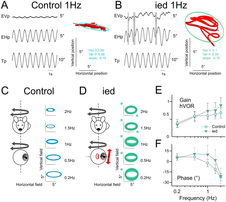
Figure 1. Horizontal angular vestibulo-ocular reflex in C57Bl/6J and ied mice.
A–B, Example of eye movements evoked in C57Bl/6J (A) or ied (B) mice during 1 Hz sinusoidal oscillation in horizontal. Shaded areas indicate quick phases. Plots present oculomotor fields. Red points are the eye position of the same traces. The ellipses present 95% of the horizontal and vertical eye positions. θ are horizontal and vertical variance; inclination of the ellipse was computed as the slope of the linear regression between vertical and horizontal eye positions. C–D Averaged oculomotor fields in C57Bl/6J (C) and ied (D) mice across tested frequencies. Line and surface of the ellipses present the mean and standard deviation of the population, respectively. The slopes of the ellipses are the mean slope of the individuals’ ellipses. Green asterisks indicate significantly larger response in ied compared to C57Bl/6J. E–F, Gain (E) and timing (F) of the horizontal component of eye movement responses for C57Bl/6J and ied populations. Asterisk indicates statistical difference with p<0.05. Tp, Table position; EVp, Eye Vertical position; EHp, Eye Horizontal position. In this and following figures, plots present mean ± SD.
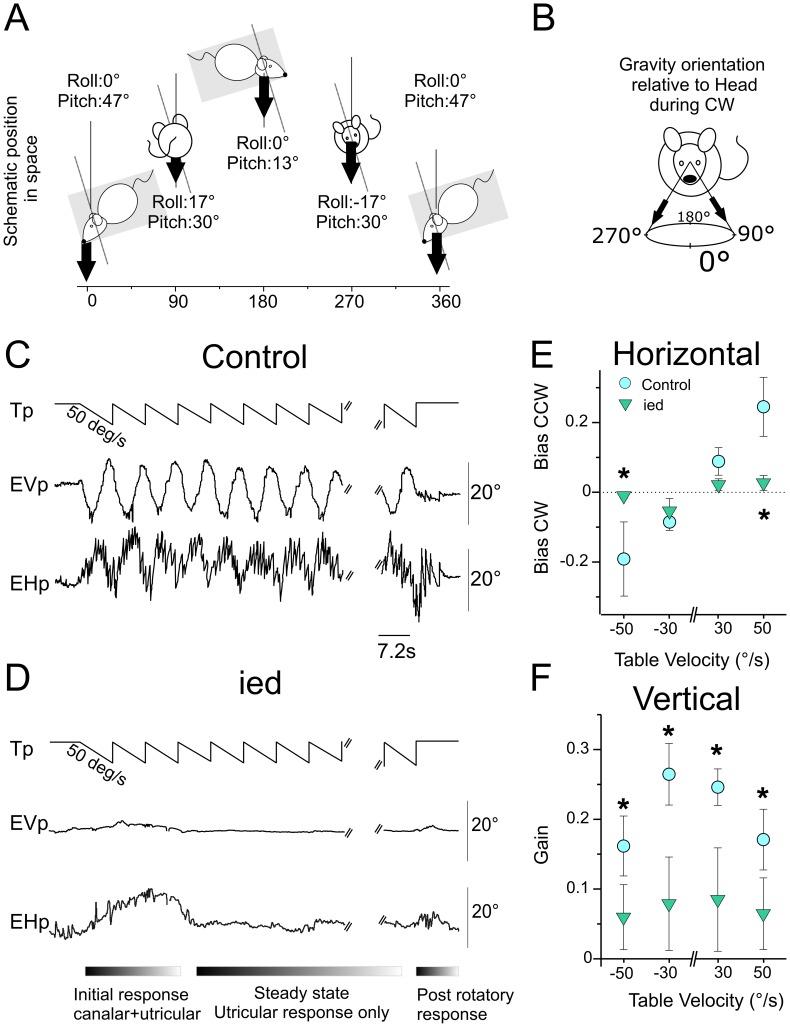
Figure 2. Maculo-ocular reflex in C57Bl/6J and ied mice.
A, Left panel: Scheme of spatial displacement of the mouse during counter-clockwise rotation at constant velocity. Mouse was head-fixed 30° nose down; pitch of table was 17°. Roll and Pitch angles variations during rotations are reported for every ¼ cycle. B, Orientation of the gravity in head-fixed coordinates during clockwise rotation. C, Eye movements observed during 50°/s constant velocity off-vertical axis rotation in counter clockwise direction. Vertical eye position changed periodically with table rotation, reaching maximal elevation and depression when head roll was maximal (±8.5°). Horizontal eye movements also consisted in periodical modulation of the eye position on which was superimposed a horizontal nystagmus in compensatory direction (left for CW; right for CCW rotations). D, in ied mice, absence of otoconia resulted in the absence of horizontal and vertical movements during the steady-state. E, F, horizontal bias (E) and vertical gain (F) for C57Bl/6J and ied mice during off vertical axis in all tested conditions. In controls, horizontal bias increased with increasing table velocity. Vertical velocity gains decreased with increasing table velocity. Plots illustrate the absence of modulation of horizontal bias and vertical gain in ied mice. Asterisks indicate significantly larger response in C57Bl/6J compared to ied mice. Tp, Table position; EVp, Eye Vertical position; EHp, Eye Horizontal position.
Data Analysis
Analysis procedures for horizontal angular vestibulo-ocular reflex (aVOR) have already been reported elsewhere [27]. Briefly, horizontal and vertical eye and head movement data were digitally low pass-filtered (cut-off frequency: 40 Hz), and position data were differentiated to obtain velocity traces. Segments of data with saccades were excluded from analysis. For horizontal sinusoidal rotations, at least 10 cycles were analyzed for each frequency. VOR gain and phase were determined by the least-squares optimization of the equation (a):
| (a) |
where EHv(t) is eye horizontal velocity, g (gain) is constant value, HHv (t) is head horizontal velocity, td is the dynamic lag time (in msec) of the eye movement with respect to the head movement, and Cte is an offset. td was used to calculate the corresponding phase (ϕ°) of eye velocity relative to head velocity. The Variance-Accounted-For (VAF) of each fit was computed as
where var represents variance, est represents the modeled eye velocity, and EHv represents the actual eye horizontal velocity. VAF values were typically between 0.70–1, where a VAF of 1 indicates a perfect fit to the data. Trials for which the VAF was less than 0.5 were excluded from the analysis.
To analyze the oculomotor fields, vertical position was plotted as a function of horizontal position. The distribution of horizontal and vertical eye positions were then computed as an ellipse with half-axis set at 1.96*standard deviation (ellipse therefore represents 95% confidence interval of the distribution). Inclination of the ellipse was set as the slope of the linear regression between vertical and horizontal eye positions.
Time constant of the aVOR was measured in response to angular rotation at constant velocity (50°/s). The horizontal slow phase velocity decay was fitted to an exponential curve (f(x) = a*exp(b*x)) using Matlab cftool function and the time constant τ was then calculated as τ = −1/b.
To analyze maculo-ocular reflex (MOR), horizontal and vertical eye movements recorded during OVAR were processed separately. For horizontal OVAR responses, quick-phases were identified and removed [32]. During rotations, the velocity of horizontal slow phases is modulated (modulation, μ) around a constant bias (β). Both parameters (μ and β) were calculated from the sinusoidal fit of eye horizontal slow-phase velocity using the least-squares optimization of the equation (b):
| (b) |
where SP(t) is slow-phase velocity, β is the steady-state bias slow phase velocity, μ is the modulation of eye velocity, f0 is the frequency of table rotation.
To analyze vertical OVAR responses, the eye vertical position signal (EVp) was analyzed using a Fast Fourier Transform (equation (c)). Amplitude of vertical eye movements (EVa) was then calculated at table frequency (f0) (equation (d)). Eye velocity was computed following equation (e):
| (c) |
| (d) |
| (e) |
The gain of vertical eye movement was defined as the ratio between the eye velocity and the table velocity. The phase was calculated as the difference between the phase angles of the Fast Fourier Transform of eye and table signals as given by the Matlab angle function.
Statistics
Statistical processing of all results was carried out using the Statistica 7.1 software (StatSoft France). Due to variable sampling size, influence of hypergravity (HG) on the VOR and MOR was tested by comparing controls (no HG) to developmental groups (Pre, Post and Full) and adults exposed to hypergravity (Adult-HG) groups using the non-parametric ANOVA Kruskal-wallis, followed by a post-hoc analysis. Comparison between ied and controls was achieved separately using the same procedure. For sake of clarity, the following text reports only the two-by-two post-hoc comparison between controls and each hypergravity-exposed group. All numbers presented in text and figures are mean ± standard deviation.
Results
VOR Responses in Controls C57Bl/6J versus Otoconia-deficient Mice
During horizontal sinusoidal rotation, aVOR consisted in control mice in horizontal eye movements and little vertical eye movements (Fig. 1A). In contrast, large vertical eye movements were observed in ied mice. Figure 1B shows in ied an example of the horizontal eye movements and of the abnormal vertical response evoked during 1 Hz aVOR. In control mice, the oculomotor fields could be represented as ellipses with a constant ratio between vertical and horizontal axis of about 0.15 at all frequencies (Fig. 1C). The inclination of the eye during sinusoidal movement (slope of the regression on Fig. 1A, B) was in range ±0.13, with a mean of −0.008±0.062. Figure 1C illustrates the regular relation between horizontal and vertical oculomotor fields across tested frequencies and the homogeneity of the aVOR responses among control mice. In ied, vertical eye fields were significantly larger than in controls (Fig. 1D; p<0.01 at all frequencies). Horizontal eye fields were also larger (p<0.019 at 1.5 and 2 Hz). The oculomotor fields of ied population therefore consisted in broader ellipses with a vertical/horizontal ratio in range 0.34 (at 1.5 Hz)–0.63 (at 0.2 Hz). Overall, slopes of the regression revealed that there was no consistency in the vertical responses observed in ied mice (range: −0.19– +0.7; mean 0.054±0.174; p>0.161 compared to controls at all frequencies). When only horizontal components were considered, the gain (p = 0.032) and the phase (p = 0.0052) of the horizontal aVOR were different compared to controls. Thus in general the horizontal aVOR in ied was qualitatively intact, but quantitatively impaired and perturbed by larger vertical eye displacements than observed in controls.
Maculo-ocular Reflex (MOR) in Controls C57Bl/6J versus Otoconia-deficient Mice
The maculo-ocular reflex of mice was tested using constant velocity rotations with the table angled 17° off-vertical axis (Fig. 2A). In an egocentric frame of reference, off-vertical axis rotation (OVAR) makes the gravity vector continuously rotating around the head in a 17° wide circle (Fig. 2B). In control mice, OVAR evoked distinct horizontal and vertical responses similar to those previously described in other lateral-eye species (rabbit: [38]; rat: [18]; gerbil: [37]). Because initial response is produced by both canals- and otoliths-related signals, the first cycles were discarded from the analysis (Fig. 2C and 2D, bottom). During the steady-state utricular-only response, the horizontal eye velocity was modulated around a constant bias. Both bias and modulation increased with the speed of rotation. In control mice, 50°/s rotations in CCW direction evoked a mean gain bias of 0.24±0.08 and a mean gain modulation of 0.09±0.04. The corresponding phase was −40.3°±6.11. Vertical responses decreased with increasing table velocity. The gain of vertical eye velocity during 50°/s CCW rotations was 0.17±0.04. The mean corresponding phase was −0.93°±2.8. At the end of rotation, post-rotatory responses comparable to those observed following yaw rotations were observed.
In ied mice, onset of rotation was often associated to erratic eye movements (Fig. 2D). In the absence of utricular modulation and while constant velocity rotation continued, the eyes stabilized and remained almost immobile until the rotation was stopped. Post-rotatory canal responses were then always present. Absence of MOR was observed in all ied mice, during both CW and CCW rotations and at all tested velocities (Fig. 2E and 2F).
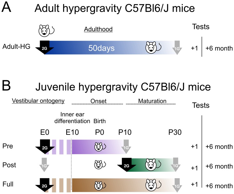
ied mice demonstrate that in the absence of detection of gravity by the otoliths (as revealed by the absence of MOR), even reflexes based on semi-circular canals such as horizontal aVOR might be impaired. Would a prolonged change in gravity affect both macula-ocular and canal-related reflexes? How do VOR develop if the appropriate assembly of otoliths and semi-circular canals signals is perturbed? In order to evaluate these questions the vestibular responses of C57Bl/6J mice exposed to hypergravity at the adult stage and/or at different periods of their development were tested. Figure 3 summarizes the centrifugation protocols and the timing of the experiments.
Figure 3. Scheme of hypergravity groups and timing of the experiments.
A, Adult-HG group was composed of two month old mice centrifuged at 2G during 50 days. Vestibulo-ocular responses were tested 1 and 6 months after return to normal gravity. B, developmental groups. Mice were conceived, born and raised in 2G centrifuge (50 days, group Full), or exposed 20 days to hypergravity during early onset (Pre) or late maturation (Post) periods of vestibular development. For all groups, “+1” and “+6” refer to recording made 1 and 6 months after return to normal gravity, respectively.
Long Term Effects of 50 Days of Exposure to Hypergravity in Adult C57Bl/6J Mice
To determine the effects of long term centrifugation on VOR, Adult C57Bl/6J adult mice were exposed to 50 days of 2G hypergravity (Fig. 3A). The aVOR and MOR were tested 1 and 6 months after the end of centrifugation.
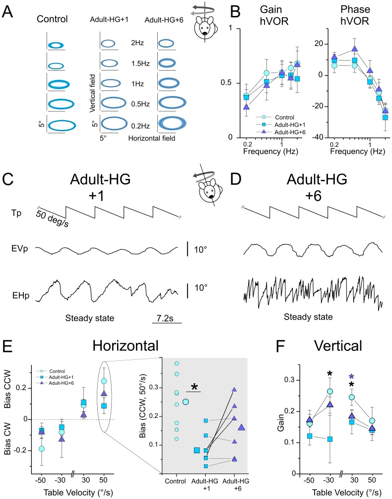
When tested with sinusoidal rotations the aVOR of centrifuged Adults-HG was normal compared to non-centrifuged adults (n = 8). First, oculomotor fields revealed no difference in the horizontal/vertical ratios as observed in the otoconia-deficient mice (Fig. 4A). In addition, both the gain and phase of horizontal aVOR were unchanged at all tested frequencies (up to 2 Hz; Fig. 4B).
Figure 4. Modification of the angular vestibulo-ocular and maculo-ocular reflexes in adult centrifuged C57Bl/6J mice.
A, Averaged oculomotor fields in non-centrifuged (control) and centrifuged (Adult-HG) mice across tested frequencies. Line and surface of the ellipses present the mean and standard deviation of the population, respectively. The slopes of the ellipses are the mean slope of the individuals’ ellipses. Oculomotor fields were not altered in adult-HG compared to control. B, Bode plots of horizontal VOR in dark gain and phase during horizontal sinusoidal rotations. There was no consistent effect of centrifugation on the responses to sinusoidal rotations. C–D, Raw traces showing steady state response of the same adult-HG mouse followed at time +1 month (C) and +6 months (D) after centrifugation. Note the absence of nystagmus on the horizontal traces and the strong reduction in the amplitude of the vertical movements at 1 month. E, horizontal bias (E) for non-centrifuged (control) and centrifuged (Adult-HG) mice in all tested conditions. Horizontal biases (E) were significantly affected in 50°/s CCW condition. Right panel presents individual (small symbols) and mean of the populations (large symbols). Solid lines indicate the evolution of individuals’ bias at +1 and +6 months after centrifugation (black: improved responses; dotted: no or little improvement in the responses). F, vertical gain (F) for non-centrifuged (controls) and centrifuged (Adult-HG) mice in all tested conditions. Tp, Table position; EVp, Eye Vertical position; EHp, Eye Horizontal position. Black or purple asterisks indicate p<0.05 between control and Adult-HG+1 or Adult-HG+6, respectively.
The MOR of Adults-HG was tested during OVAR stimulation (n = 7). As illustrated on figure 4C, the eye movements evoked in Adults-HG were strongly impaired. In particular, the horizontal compensatory nystagmus was found to be reduced or even absent. The modulation of horizontal eye position was periodic and followed cycles of rotations resulting in uncompensatory displacements (e.g. presence of CW directed slow-phases during CCW rotation). Furthermore, both horizontal and vertical modulations of the eye position were reduced in amplitude (compare traces in Figure 2C and 4C; mean vertical amplitude at 30°/s: 10.2°±2.85 in centrifuged adults vs. 17.0°±2.25 in controls). Horizontal gain bias was reduced 1 month after the end of centrifugation to 0.08±0.05 compared to 0.24±0.08 in controls (p<0.001; post-hoc analysis CW 50°/s, p = 0.042; CCW 50°/s rotations p<0.001; Fig. 4E). Vertical gains were also significantly reduced (Fig. 4F: p<0.001; post-hoc analysis CCW 30°/s rotations p = 0.011; CW 30°/s rotations p<0.001). Taken together, MOR responses were diminished by a factor 2 to 3 compared to control conditions. To investigate the persistence of these deficits, we recorded again the VOR and MOR 6 months after the adult mice returned to normo-gravity.
The responses to sinusoidal rotations and to yaw constant rotations remained normal during the 6 months following the end of the centrifugation (Fig. 4A and B). There was an improvement in the MOR during the same period. Figure 4D shows the evolution of the eye movements observed in the same animal as in Figure 4C. At 6 months, the horizontal eye movements were compensatory, although the nystagmus was often less pronounced than in controls. The mean horizontal gain bias of adult-HG improved (see right panel in Figure 4E). Recovery of MOR was quite heterogeneous between individuals (Fig. 4E, right panel). While the horizontal responses (thick lines) improved in some animals and reached the range of control values, other animals showed no or little improvement (thin lines). Overall, adult-HG MOR appeared strongly modified 1 month after the end of the centrifugation. During the following months, a substantial readaptation to normal gravity was observed, however we note that the quality of the recovery was quite heterogeneous among individuals.
In adult mice, 50 days of hypergravity altered durably the MOR, but not the horizontal aVOR. To assess how a change in gravity would affect the development and maturation of vestibulo-ocular circuitry, we then tested animals which developed in the centrifuge.
Effects of Hypergravity on the Acquisition of aVOR
To assess the developmental effects hypergravity, C57Bl/6J mice were raised under 2G during specific periods of development (Groups Pre and Post; fig. 3B) or entire development (group full).
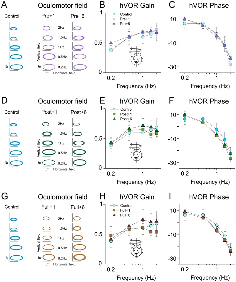
In groups Pre and Post centrifuged for only part of their development, or in group Full, no significant alterations were found during sinusoidal testing. Figure 5A, D, G present the oculomotor fields of the different populations of mice tested 1 and 6 months after end of centrifugation. No abnormal vertical movement was observed in any of the groups. In addition, both the gain (Fig. 5B, E, H) and phase (Fig. 5C, F, I) of the aVOR was normal compared to controls in all conditions. These results suggest that, unlike what was observed in otoconia-deficient mice, alteration of gravity during the development does not impair the spatial tuning of canal-related reflexes.
Figure 5. Angular vestibulo-ocular reflex is not modified following developmental exposure to hypergravity.
A, B, C: Averaged oculomotor fields (A), horizontal gain (B) and phase (C) in non-centrifuged (control) mice compared to mice centrifuged between E0–P10 (pre) tested 1 and 6 months after centrifugation. No significant differences were found. D, E, F: Averaged oculomotor fields (D), horizontal gain (E) and phase (F) in non-centrifuged (control) mice compared to mice centrifuged between P10–P30 (post) tested 1 and 6 months after centrifugation. No significant differences were found. G, H, I: Averaged oculomotor fields (G), horizontal gain (H) and phase (I) in non-centrifuged (control) mice compared to mice centrifuged between E0–P30 (full) tested 1 and 6 months after centrifugation. No significant differences were found.
Effects of Hypergravity on the Acquisition of Maculo-ocular Reflex
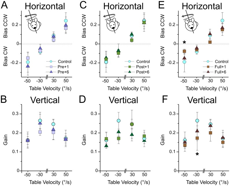
Does exposure to hypergravity during development induce long term alteration of MOR as observed in the adults after fifty days of hypergravity? One month after end of the centrifugation, we found no statistical differences in the OVAR responses of the Pre and the Post groups compared to controls. No significant change was observed on the eye horizontal component (Fig. 6A, C) or in the vertical component (Fig. 6B, D).
Figure 6. Maculo-ocular reflex in mice centrifuged during development.
A–B, horizontal bias (A) and vertical gain (B) for control and pre mice during off vertical axis rotation in all tested conditions. No significant differences were found at 1 and 6 months. C–D, horizontal bias (C) and vertical gain (D) for control and post mice during off vertical axis rotation in all tested conditions. No significant differences were found at 1 and 6 months. E–F, horizontal bias (E) and vertical gain (F) for control and full mice during off vertical axis rotation in all tested conditions. Asterisks indicate significant differences (p<0.05) between control and full+1 groups.
These results demonstrate that 3 weeks exposure to hypergravity before or after P10 had no measurable effect on the MOR. What happens when hypergravity covers the entire development? Again, we found no major difference in the MOR responses of the Full group. Extending hypergravity exposure to the entire development however had significant effects (Fig. 6E, 6F), but the variability in the responses was quite large. The reduction in vertical eye movements was comparable to the one observed for Adult-HG; however the horizontal eye movements were both qualitatively and quantitatively better, i.e. compensatory nystagmus was present and bias values were greater in Full group than in Adult-HG.
How did the MOR of the developmental groups evolved during the following months? As expected, both Pre and Post groups presented completely normal MOR (Fig. 6A–D). The group Full had recovered normal responses: vertical eye movements were not different from controls. For horizontal eye movements, improvement in the bias was observed in previously affected conditions and henceforth comparable to controls in all conditions.
These observations suggest that centrifugation during entire, but not specific parts, of development might have transitorily delayed acquisition of normal MOR. However after return to normal gravity, the juvenile mice show no long term impairment so that 2G hypergravity did not reveal any critical period during VOR ontogeny.
Discussion
Otoconial Agenesis Affects Vestibular Signal Processing and Gaze Stabilization
How does complete absence of otolith-related inputs affect vestibular processing? Observations made on different otoconia-deficient mice showed that the absence of modulation of the macular signals was detrimental for the development of vestibulo-motor circuitry. In the tilted mice, another otoconia-deficient strain, the impairment in aVOR was functionally compensated by an increase in the efficiency of the optokinetic reflex [39]. Our observation that many ied mice initially suffer from ocular instability (Figure S1) when put in dark also suggests that, in the absence of otolith information, visual inputs become instrumental for gaze stabilization. The most striking deficit in ied was that horizontal head movements triggered abnormal vertical eye movements. What is the origin of this impairment in the spatial tuning of aVOR? .
We have checked experimentally that the deficit was not due to a misalignment of the semi-circular canals during the rotation (Figure S1). The deficit could alternatively originate from the abnormal branching at the level of the semi-circular canals. For instance, otoconial agenesis could result in a mis-directed innervation of the cristae by otolith afferent axons during the development of the inner ear. However, no morphological or cytological abnormalities were reported in the cristae ampullares of ied mutants [20]. Similar observations were made in het [40] and tilted strain [21]. Most of the studies which investigated vestibular periphery or afferents in otoconia-deficient strains instead concluded that absence of modulation in the macular afferents was the main consequence of the mutation. Hence, it was demonstrated in Het mice that presence of otoconia was not required for the general formation and maintenance of synapses [41] or normal development of vestibular ganglia [42]. The resting discharge pattern of macular primary afferents was also unperturbed in otoconia-deficient mice (Head-tilt and tilted; [19]). Further characterization of the innervations and physiological properties of the semi-circular canals and afferents in ied would however be required to exclude the peripheral origin of the observed deficit.
Another hypothesis would be that the central branching or processing of vestibular information at the level of vestibular nuclei is responsible for the default in spatial tuning of aVOR. Ontogeny of central vestibulo-ocular circuitry depends initially on genetic [6]. Activity-mediated spatial refinements of connections and fine tuning of the temporal dynamics of aVOR occurs during a second developmental phase [9], [10]. It has been suggested in larval frog that otolith-driven responses in vestibular circuits could serve as a spatial reference frame for the refinement of canal-driven responses during early development [43]. Present results are compatible with this hypothesis, such that in the absence of gravity-related signals, central vestibular neurons would substitute otolith inputs with spatially non-matching canal inputs. Comparable process was suggested in labyrinthectomized frogs as a “basic reaction pattern” which substituted the missing utricular inputs with commissural and/or afferent signals. Functionally, the restoration of macula-ocular reflex gain also led to the impairment of its spatial-tuning [44].
At this stage, the peripheral and/or central origin of the deficit in the spatial tuning of aVOR observed in ied remains uncertain. Characterization of the electrophysiological responses in canal afferents and in central vestibular neurons would be required to sort out the cause for the abnormal eye movements. In addition, the same experimental paradigm could be applied on other otoconial mutant to confirm that it is the absence of modulation of the otolith-related signals which is the origin of this deficit and therefore exclude some other central neurologic ramifications.
Hypergravity Transiently Affects Macula-ocular Reflex in Juvenile and Adult Centrifuged Mice
How are vestibular organs and central integration of multisensory inputs affected by altered gravity? On ground, an alteration of gravity can be produced by mean of centrifuges [25]. Prenatal exposure of rats to hypergravity have been reported to affect sensory epithelium morphology [45], local connectivity [46], physiology of hair cells [13], glutamatergic neurotransmission between hair cell and afferent neuron [47] and peripheral vestibulocerebellar afferents [48]. Bouët and collaborators [14], [15], [49] reported in rats that development and maturation under 2G hypergravity induces postural and locomotor deficits which all recovered in about 3 weeks. There is currently little evidence of a permanent deficit following development under hypergravity. Sondag et al. [17] however showed that a 20 weeks long development of hamsters at 2.5G induces inabilities in swimming and air-righting reflexes even 8 months after return to normal gravity. Overall, an increase in gravity-load during development affects both peripheral and central vestibular-related circuits. Most of the changes however seem transitory and highly depend on the level of gravity-load imposed and on the length of exposure to HG.
Present results on VOR extend these previous experiments. We have shown that a 3 week long exposure to hypergravity during development does not alter the acquisition of vestibulo-ocular reflex in Pre and Post groups. However, a 50 days exposure of juvenile mice (Full group) induced a delay in the acquisition of macula-ocular reflex. This retard is compatible with a delayed development of the vestibular periphery [13], [45]–[47]. Another hypothesis could lie in the central integration of canal and otolith inputs: in particular, the central process known as “velocity storage” which provides a spatially referenced estimate of head velocity [50], [51] could be affected. Velocity storage normally allows compensating the deficiencies of peripheral vestibular sensors to accurately estimate rotation velocity, and to distinguish between a prolonged linear acceleration and a head tilt [52]–[54]. Shortening of aVOR time constant was observed in weightlessness [55], [56] or after sustained centrifugation [57] and is attributed to a decreased coupling between canalar and otolith inputs [57], [58]. The impairment we observed in the horizontal bias during OVAR is compatible with this hypothesis. More experiments will however be needed to determine how a 50 days long exposure to hypergravity affects the velocity storage mechanism.
Adult-HG mice presented the most striking and persistent alteration of macula-ocular reflex following exposure to hypergravity. In the absence of histological analysis of the effects of the prolonged centrifugation on the inner ear structures, we cannot exclude that the observed persistent alteration could reflect damage to the inner ears as a result of the chronic centrifugation. However, previous experiments on rats suggested that the vestibular sensory epithelium was qualitatively unaffected after a prolonged period of hypergravity [11], [17]. The deficits in adult-HG mice could alternatively reflect the difficulty of mature brains to adapt/readapt to successive alterations in gravity. As in other sensory systems, the plastic processes underlying adaptation to gravitational changes appear variable among individuals and more restricted in mature brain than during development [59].
A Vestibular Critical Period during Ontogeny of Motor Control?
The existence of a critical period during which the vestibular information are mandatory for the motor development is disputed [60]. It is supported by the permanent inability to swim and to perform surface righting in rats that matured in space [61], [62], and by the permanent motor deficits in mice centrifuged between P10–P30 [25], [26]. In rodents, deprivation experiments claim for an early need for vestibular inputs. The complete removal of vestibular organs in rats before P5 leads to permanent head-bobbing in adults [63], [64], and mice which congenitically lack all vestibular organs show a typical shaker/waltzer behaviour, head-bobbing and abnormal locomotion [29], [65].
Interestingly, specific unilateral lesion of the otolith in the adult guinea-pig [66] triggered a transient head-tilt and postural symptoms, which resembled those observed in otoconia-deficient mice. These symptoms markedly differed from the plane specific horizontal oscillations and circling observed after unilateral lesion of the horizontal canal [63] or unilateral vertical canal lesions [66]. These postural syndromes observed after selective vestibular impairment would suggest that the permanent head-bobbing and circling in vestibular-deprived mutants could reflect a critical need for the information coded by the vertical and horizontal semicircular canals, respectively, rather than by the otoliths. On the other hand the permanent oculomotor deficits we describe here in ied and the permanent head-tilt observed in otoconia-deficient strains could be the signature of an otolithical critical period.
The environmental removal of gravity during development is another way to test the hypothesis of an otolith-specific critical period. In rodents, transient changes in gravity have been tested by exposing animal to weightlessness (µG) during space journey. Experiments conducted on rats exposed to environmental deprivation of gravity during gestation concluded in a normal development in space. Hence, no change were found on ground in motor behavior such as walking (11 days µG between embryonic day E9–E20; [67]), righting response, negative geotaxis or rotating platform (5 days µG, E13–E18; [68], [69]). However, following 11 days in µG (E9–E20) vestibular-specific tests suggested a retarded ability to respond to gravistatic stimuli [70], abnormal water immersion responses [71], and increased bradychardia following roll stimulation [7].
In conclusion, genetic suppression of gravity-related signals indicated that otolith-related signals might be necessary to ensure a proper functioning of canal-related vestibular reflexes. It also suggested an otolithical critical period for motor control, which could be the pendant of a canalar critical period previously described. On the other hand, exposure to hypergravity during development was not sufficient to alter durably motor behaviour. However, in centrifuge the sensing of gravity is altered rather than suppressed as it would be during a space journey. More experiments with long (>2 month) exposure of mice to weightlessness will be needed to rule out the existence of critical period during vestibular development.
Supporting Information
Spontaneous oculomotor instability and determination of head pitch in otoconia-deficient mice. A, Spontaneous ocular instabilities in horizontal, vertical and torsion (not shown) were observed. Nystagmus was present in dark (left), diminished at light (center) and abated as the animal got accustomed to the apparatus (right panel). B, Gain of the horizontal VOR at light measured at different head pitch angle. Gain was found to be maximal at 30°. Note that oculomotor fields horizontal and vertical components did not vary according to head pitch, suggesting that the deficit observed in ied was not related to a misalignement of the semi-circular canals.
(TIF)
Acknowledgments
This work was funded by the Centre National d’Etudes Spatiales, the Agence Nationale de la Recherche (program ANR-09-BLAN-0148), CNRS PEPS and Neuro-IC programs. M. Bojados received fellowship from CNES and the regional PACA.
Footnotes
Competing Interests: The authors have declared that no competing interests exist.
Funding: This work was funded by the Centre National d’Etudes Spatiales, the Agence Nationale de la Recherche (program ANR-09-BLAN-0148), Centre National de la Recherche Scientifique “Projets Exploratoires Premier Soutien” and Neuro-IC programs. M. Bojados received a fellowship from the Centre National d’Etudes Spatiales and the regional Provence-Alpes-Côte d’Azur. The funders had no role in study design, data collection and analysis, decision to publish, or preparation of the manuscript.
References
- 1.Angelaki DE, Cullen KE. Vestibular system: the many facets of a multimodal sense. Annu Rev Neurosci. 2008;31:125–150. doi: 10.1146/annurev.neuro.31.060407.125555. doi: 10.1146/annurev.neuro.31.060407.125555. [DOI] [PubMed] [Google Scholar]
- 2.Straka H, Holler S, Goto F. Patterns of canal and otolith afferent input convergence in frog second-order vestibular neurons. J Neurophysiol. 2002;88:2287–2301. doi: 10.1152/jn.00370.2002. doi: 10.1152/jn.00370.2002. [DOI] [PubMed] [Google Scholar]
- 3.Bush GA, Perachio AA, Angelaki DE. Encoding of head acceleration in vestibular neurons. I. Spatiotemporal response properties to linear acceleration. J Neurophysiol. 1993;69:2039–2055. doi: 10.1152/jn.1993.69.6.2039. [DOI] [PubMed] [Google Scholar]
- 4.Uchino Y, Sasaki M, Sato H, Bai R, Kawamoto E. Otolith and canal integration on single vestibular neurons in cats. Exp Brain Res. 2005;164:271–285. doi: 10.1007/s00221-005-2341-7. doi: 10.1007/s00221-005-2341-7. [DOI] [PubMed] [Google Scholar]
- 5.Morishita H, Hensch TK. Critical period revisited: impact on vision. Curr Opin Neurobiol. 2008;18:101–107. doi: 10.1016/j.conb.2008.05.009. doi: 10.1016/j.conb.2008.05.009. [DOI] [PubMed] [Google Scholar]
- 6.Maklad A, Fritzsch B. Development of vestibular afferent projections into the hindbrain and their central targets. Brain Res Bull. 2003;60:497–510. doi: 10.1016/s0361-9230(03)00054-6. [DOI] [PMC free article] [PubMed] [Google Scholar]
- 7.Ronca AE, Fritzsch B, Bruce LL, Alberts JR. Orbital spaceflight during pregnancy shapes function of mammalian vestibular system. Behav Neurosci. 2008;122:224–232. doi: 10.1037/0735-7044.122.1.224. doi: 10.1037/0735-7044.122.1.224. [DOI] [PMC free article] [PubMed] [Google Scholar]
- 8.Glover JC. The development of vestibulo-ocular circuitry in the chicken embryo. J Physiol Paris. 2003;97:17–25. doi: 10.1016/j.jphysparis.2003.10.003. doi: 10.1016/j.jphysparis.2003.10.003. [DOI] [PubMed] [Google Scholar]
- 9.Fritzsch B. Molecular developmental neurobiology of formation, guidance and survival of primary vestibular neurons. Adv Space Res. 2003;32:1495–1500. doi: 10.1016/S0273-1177(03)90387-5. [DOI] [PubMed] [Google Scholar]
- 10.Straka H. Ontogenetic rules and constraints of vestibulo-ocular reflex development. Curr Opin Neurobiol. 2010;20:689–695. doi: 10.1016/j.conb.2010.06.003. doi: 10.1016/j.conb.2010.06.003. [DOI] [PubMed] [Google Scholar]
- 11.Wubbels RJ, Sondag HNPM, van Marle J, de Jong HAA. Effects of hypergravity on the morphological properties of the vestibular sensory epithelium. I. Long-term exposure of rats after full maturation of the labyrinths. Brain Res Bull. 2002;57:677–682. doi: 10.1016/s0361-9230(01)00778-x. [DOI] [PubMed] [Google Scholar]
- 12.Bruce LL. Adaptations of the vestibular system to short and long-term exposures to altered gravity. Adv Space Res. 2003;32:1533–1539. doi: 10.1016/S0273-1177(03)90392-9. [DOI] [PubMed] [Google Scholar]
- 13.Chabbert C, Brugeaud A, Lennan G, Lehouelleur J, Sans A. Electrophysiological properties of the utricular primary transducer are modified during development under hypergravity. Eur J Neurosci. 2003;17:2497–2500. doi: 10.1046/j.1460-9568.2003.02682.x. [DOI] [PubMed] [Google Scholar]
- 14.Bouët V, Gahéry Y, Lacour M. Behavioural changes induced by early and long-term gravito-inertial force modification in the rat. Behav Brain Res. 2003;139:97–104. doi: 10.1016/s0166-4328(02)00085-2. [DOI] [PubMed] [Google Scholar]
- 15.Bouët V, Borel L, Harlay F, Gahéry Y, Lacour M. Kinematics of treadmill locomotion in rats conceived, born, and reared in a hypergravity field (2 g). Adaptation to 1 g. Behav Brain Res. 2004;150:207–216. doi: 10.1016/S0166-4328(03)00258-4. doi: 10.1016/S0166-4328(03)00258-4. [DOI] [PubMed] [Google Scholar]
- 16.Nguon K, Ladd B, Sajdel-Sulkowska EM. Exposure to Altered Gravity During Specific Developmental Periods Differentially Affects Growth, Development, the Cerebellum and Motor Functions in Male and Female Rats. Adv Space Res. 2006;38:1138–1147. doi: 10.1016/j.asr.2006.09.007. doi: 10.1016/j.asr.2006.09.007. [DOI] [PMC free article] [PubMed] [Google Scholar]
- 17.Sondag HN, de Jong HA, Oosterveld WJ. Altered behaviour in hamsters conceived and born in hypergravity. Brain Res Bull. 1997;43:289–294. doi: 10.1016/s0361-9230(97)00008-7. [DOI] [PubMed] [Google Scholar]
- 18.Hess BJM, Dieringer N. Spatial Organization of the Maculo-Ocular Reflex of the Rat: Responses During Off-Vertical Axis Rotation. Eur J Neurosci. 1990;2:909–919. doi: 10.1111/j.1460-9568.1990.tb00003.x. [DOI] [PubMed] [Google Scholar]
- 19.Jones TA, Jones SM, Hoffman LF. Resting discharge patterns of macular primary afferents in otoconia-deficient mice. J Assoc Res Otolaryngol. 2008;9:490–505. doi: 10.1007/s10162-008-0132-0. doi: 10.1007/s10162-008-0132-0. [DOI] [PMC free article] [PubMed] [Google Scholar]
- 20.Besson V, Nalesso V, Herpin A, Bizot J-C, Messaddeq N, et al. Training and aging modulate the loss-of-balance phenotype observed in a new ENU-induced allele of Otopetrin1. Biol Cell. 2005;97:787–798. doi: 10.1042/BC20040525. doi: 10.1042/BC20040525. [DOI] [PubMed] [Google Scholar]
- 21.Ornitz DM, Bohne BA, Thalmann I, Harding GW, Thalmann R. Otoconial agenesis in tilted mutant mice. Hear Res. 1998;122:60–70. doi: 10.1016/s0378-5955(98)00080-x. [DOI] [PubMed] [Google Scholar]
- 22.Jones SM, Erway LC, Johnson KR, Yu H, Jones TA. Gravity receptor function in mice with graded otoconial deficiencies. Hear Res. 2004;191:34–40. doi: 10.1016/j.heares.2004.01.008. doi: 10.1016/j.heares.2004.01.008. [DOI] [PubMed] [Google Scholar]
- 23.Jones TA, Jones SM. Short latency compound action potentials from mammalian gravity receptor organs. Hear Res. 1999;136:75–85. doi: 10.1016/s0378-5955(99)00110-0. [DOI] [PubMed] [Google Scholar]
- 24.Yoder RM, Taube JS. Head direction cell activity in mice: robust directional signal depends on intact otolith organs. J Neurosci. 2009;29:1061–1076. doi: 10.1523/JNEUROSCI.1679-08.2009. doi: 10.1523/JNEUROSCI.1679-08.2009. [DOI] [PMC free article] [PubMed] [Google Scholar]
- 25.Jamon M, Serradj N. Ground-Based Researches on the Effects of Altered Gravity on Mice Development. Microgravity Science and Technology. 2008;21:327–337. doi: 10.1007/s12217-008-9098-0. [Google Scholar]
- 26.Bojados M, Jamon M. Exposure to hypergravity during specific developmental periods affects metabolism and vestibular reactions in adult mice C57BL/6JJ. European Journal of Neuroscience in press. 2011. [DOI] [PubMed]
- 27.Beraneck M, Cullen KE. Activity of vestibular nuclei neurons during vestibular and optokinetic stimulation in the alert mouse. J Neurophysiol. 2007;98:1549–1565. doi: 10.1152/jn.00590.2007. doi: 10.1152/jn.00590.2007. [DOI] [PubMed] [Google Scholar]
- 28.Oommen BS, Stahl JS. Eye orientation during static tilts and its relationship to spontaneous head pitch in the laboratory mouse. Brain Res. 2008;1193:57–66. doi: 10.1016/j.brainres.2007.11.053. doi: 10.1016/j.brainres.2007.11.053. [DOI] [PMC free article] [PubMed] [Google Scholar]
- 29.Vidal P-P, Degallaix L, Josset P, Gasc J-P, Cullen KE. Postural and locomotor control in normal and vestibularly deficient mice. J Physiol (Lond) 2004;559:625–638. doi: 10.1113/jphysiol.2004.063883. doi: 10.1113/jphysiol.2004.063883. [DOI] [PMC free article] [PubMed] [Google Scholar]
- 30.Calabrese DR, Hullar TE. Planar relationships of the semicircular canals in two strains of mice. J Assoc Res Otolaryngol. 2006;7:151–159. doi: 10.1007/s10162-006-0031-1. doi: 10.1007/s10162-006-0031-1. [DOI] [PMC free article] [PubMed] [Google Scholar]
- 31.Stahl JS, Oommen BS. Eye hyperdeviation in mouse cerebellar mutants is comparable to the gravity-dependent component of human downbeat nystagmus. Prog Brain Res. 2008;171:503–508. doi: 10.1016/S0079-6123(08)00672-9. doi: 10.1016/S0079-6123(08)00672-9. [DOI] [PubMed] [Google Scholar]
- 32.Beraneck M, McKee JL, Aleisa M, Cullen KE. Asymmetric recovery in cerebellar-deficient mice following unilateral labyrinthectomy. J Neurophysiol. 2008;100:945–958. doi: 10.1152/jn.90319.2008. doi: 10.1152/jn.90319.2008. [DOI] [PMC free article] [PubMed] [Google Scholar]
- 33.Iwashita M, Kanai R, Funabiki K, Matsuda K, Hirano T. Dynamic properties, interactions and adaptive modifications of vestibulo-ocular reflex and optokinetic response in mice. Neurosci Res. 2001;39:299–311. doi: 10.1016/s0168-0102(00)00228-5. [DOI] [PubMed] [Google Scholar]
- 34.van Alphen B, Winkelman BHJ, Frens MA. Three-dimensional optokinetic eye movements in the C57BL/6JJ mouse. Invest Ophthalmol Vis Sci. 2010;51:623–630. doi: 10.1167/iovs.09-4072. doi: 10.1167/iovs.09–4072. [DOI] [PubMed] [Google Scholar]
- 35.Stahl JS. Using eye movements to assess brain function in mice. Vision Res. 2004;44:3401–3410. doi: 10.1016/j.visres.2004.09.011. doi: 10.1016/j.visres.2004.09.011. [DOI] [PubMed] [Google Scholar]
- 36.Stahl JS, van Alphen AM, De Zeeuw CI. A comparison of video and magnetic search coil recordings of mouse eye movements. J Neurosci Methods. 2000;99:101–110. doi: 10.1016/s0165-0270(00)00218-1. [DOI] [PubMed] [Google Scholar]
- 37.Kaufman G, Weng T, Ruttley T. A rodent model for artificial gravity: VOR adaptation and Fos expression. J Vestib Res. 2005;15:131–147. [PubMed] [Google Scholar]
- 38.Maruta J, Simpson JI, Raphan T, Cohen B. Orienting otolith-ocular reflexes in the rabbit during static and dynamic tilts and off-vertical axis rotation. Vision Res. 2001;41:3255–3270. doi: 10.1016/s0042-6989(01)00091-8. [DOI] [PubMed] [Google Scholar]
- 39.Andreescu CE, De Ruiter MM, De Zeeuw CI, De Jeu MTG. Otolith deprivation induces optokinetic compensation. J Neurophysiol. 2005;94:3487–3496. doi: 10.1152/jn.00147.2005. doi: 10.1152/jn.00147.2005. [DOI] [PubMed] [Google Scholar]
- 40.Bergstrom RA, You Y, Erway LC, Lyon MF, Schimenti JC. Deletion mapping of the head tilt (het) gene in mice: a vestibular mutation causing specific absence of otoliths. Genetics. 1998;150:815–822. doi: 10.1093/genetics/150.2.815. [DOI] [PMC free article] [PubMed] [Google Scholar]
- 41.Hoffman LF, Ross MD, Varelas J, Jones SM, Jones TA. Afferent synapses are present in utricular hair cells from otoconia-deficient mice. Hear Res. 2006;222:35–42. doi: 10.1016/j.heares.2006.05.013. doi: 10.1016/j.heares.2006.05.013. [DOI] [PubMed] [Google Scholar]
- 42.Smith M, Yuan Wang X, Wolgemuth DJ, Murashov AK. Development of the mouse vestibular system in the absence of gravity perception. Brain Res Dev Brain Res. 2003;140:133–135. doi: 10.1016/s0165-3806(02)00591-6. [DOI] [PubMed] [Google Scholar]
- 43.Lambert FM, Beck JC, Baker R, Straka H. Semicircular canal size determines the developmental onset of angular vestibuloocular reflexes in larval Xenopus. J Neurosci. 2008;28:8086–8095. doi: 10.1523/JNEUROSCI.1288-08.2008. doi: 10.1523/JNEUROSCI.1288-08.2008. [DOI] [PMC free article] [PubMed] [Google Scholar]
- 44.Rohregger M, Dieringer N. Postlesional vestibular reorganization improves the gain but impairs the spatial tuning of the maculo-ocular reflex in frogs. J Neurophysiol. 2003;90:3736–3749. doi: 10.1152/jn.00561.2003. doi: 10.1152/jn.00561.2003. [DOI] [PubMed] [Google Scholar]
- 45.Wubbels RJ, van Marle J, Sondag HNPM, de Jong HAA. Effects of hypergravity on the morphological properties of the vestibular sensory epithelium. II. Life-long exposure of rats including embryogenesis. Brain Res Bull. 2002;58:575–580. doi: 10.1016/s0361-9230(02)00828-6. [DOI] [PubMed] [Google Scholar]
- 46.Gaboyard S, Sans A, Lehouelleur J. Differential impact of hypergravity on maturating innervation in vestibular epithelia during rat development. Brain Res Dev Brain Res. 2003;143:15–23. doi: 10.1016/s0165-3806(03)00069-5. [DOI] [PubMed] [Google Scholar]
- 47.Uno Y, Horii A, Uno A, Fuse Y, Fukushima M, et al. Quantitative changes in mRNA expression of glutamate receptors in the rat peripheral and central vestibular systems following hypergravity. J Neurochem. 2002;81:1308–1317. doi: 10.1046/j.1471-4159.2002.00933.x. [DOI] [PubMed] [Google Scholar]
- 48.Bruce LL, Burke JM, Dobrowolska JA. Effects of hypergravity on the prenatal development of peripheral vestibulocerebellar afferent fibers. Advances in Space Research. 2006;38:1041–1051. doi: 10.1016/j.asr.2006.03.002. [Google Scholar]
- 49.Bouët V, Wubbels RJ, de Jong HAA, Gramsbergen A. Behavioural consequences of hypergravity in developing rats. Brain Res Dev Brain Res. 2004;153:69–78. doi: 10.1016/j.devbrainres.2004.03.022. doi: 10.1016/j.devbrainres.2004.03.022. [DOI] [PubMed] [Google Scholar]
- 50.Angelaki DE, Hess BJ. Inertial representation of angular motion in the vestibular system of rhesus monkeys. I. Vestibuloocular reflex. J Neurophysiol. 1994;71:1222–1249. doi: 10.1152/jn.1994.71.3.1222. [DOI] [PubMed] [Google Scholar]
- 51.Moore ST, Clément G, Dai M, Raphan T, Solomon D, et al. Ocular and perceptual responses to linear acceleration in microgravity: alterations in otolith function on the COSMOS and Neurolab flights. J Vestib Res. 2003;13:377–393. [PubMed] [Google Scholar]
- 52.Yakusheva TA, Shaikh AG, Green AM, Blazquez PM, Dickman JD, et al. Purkinje cells in posterior cerebellar vermis encode motion in an inertial reference frame. Neuron. 2007;54:973–985. doi: 10.1016/j.neuron.2007.06.003. doi: 10.1016/j.neuron.2007.06.003. [DOI] [PubMed] [Google Scholar]
- 53.Angelaki DE, Yakusheva TA. How vestibular neurons solve the tilt/translation ambiguity. Comparison of brainstem, cerebellum, and thalamus. Ann N Y Acad Sci. 2009;1164:19–28. doi: 10.1111/j.1749-6632.2009.03939.x. doi: 10.1111/j.1749-6632.2009.03939.x. [DOI] [PMC free article] [PubMed] [Google Scholar]
- 54.Laurens J, Angelaki DE. The functional significance of velocity storage and its dependence on gravity. Exp Brain Res. 2011;210:407–422. doi: 10.1007/s00221-011-2568-4. doi: 10.1007/s00221-011-2568-4. [DOI] [PMC free article] [PubMed] [Google Scholar]
- 55.Oman CM, Pouliot CF, Natapoff A. Horizontal angular VOR changes in orbital and parabolic flight: human neurovestibular studies on SLS-2. J Appl Physiol. 1996;81:69–81. doi: 10.1152/jappl.1996.81.1.69. [DOI] [PubMed] [Google Scholar]
- 56.Clément G. Alteration of eye movements and motion perception in microgravity. Brain Res Brain Res Rev. 1998;28:161–172. doi: 10.1016/s0165-0173(98)00036-8. [DOI] [PubMed] [Google Scholar]
- 57.Nooij SAE, Bos JE, Groen EL. Velocity storage activity is affected after sustained centrifugation: a relationship with spatial disorientation. Exp Brain Res. 2008;190:165–177. doi: 10.1007/s00221-008-1460-3. doi: 10.1007/s00221-008-1460-3. [DOI] [PubMed] [Google Scholar]
- 58.Green AM, Angelaki DE. Resolution of sensory ambiguities for gaze stabilization requires a second neural integrator. J Neurosci. 2003;23:9265–9275. doi: 10.1523/JNEUROSCI.23-28-09265.2003. [DOI] [PMC free article] [PubMed] [Google Scholar]
- 59.Bavelier D, Levi DM, Li RW, Dan Y, Hensch TK. Removing brakes on adult brain plasticity: from molecular to behavioral interventions. J Neurosci. 2010;30:14964–14971. doi: 10.1523/JNEUROSCI.4812-10.2010. doi: 10.1523/JNEUROSCI.4812-10.2010. [DOI] [PMC free article] [PubMed] [Google Scholar]
- 60.Jamon M, Bojados M, Pratte M. The hypothesis of critical periods in the adaptation to gravity. Journal of Gravitational Physiology: A Journal of the International Society for Gravitational Physiology in press. 2011.
- 61.Walton KD, Benavides L, Singh N, Hatoum N. Long-term effects of microgravity on the swimming behaviour of young rats. J Physiol (Lond) 2005;565:609–626. doi: 10.1113/jphysiol.2004.074393. doi: 10.1113/jphysiol.2004.074393. [DOI] [PMC free article] [PubMed] [Google Scholar]
- 62.Walton KD, Harding S, Anschel D, Harris YT, Llinás R. The effects of microgravity on the development of surface righting in rats. J Physiol (Lond) 2005;565:593–608. doi: 10.1113/jphysiol.2004.074385. doi: 10.1113/jphysiol.2004.074385. [DOI] [PMC free article] [PubMed] [Google Scholar]
- 63.Geisler HC, Westerga J, Gramsbergen A. The function of the long back muscles during postural development in the rat. Behav Brain Res. 1996;80:211–215. doi: 10.1016/0166-4328(96)00024-1. [DOI] [PubMed] [Google Scholar]
- 64.Geisler HC, Gramsbergen A. The EMG development of the longissimus and multifidus muscles after plugging the horizontal semicircular canals. J Vestib Res. 1998;8:399–409. [PubMed] [Google Scholar]
- 65.Eugène D, Deforges S, Vibert N, Vidal P-P. Vestibular critical period, maturation of central vestibular neurons, and locomotor control. Ann N Y Acad Sci. 2009;1164:180–187. doi: 10.1111/j.1749-6632.2008.03727.x. doi: 10.1111/j.1749-6632.2008.03727.x. [DOI] [PubMed] [Google Scholar]
- 66.De Waele C, Graf W, Josset P, Vidal PP. A radiological analysis of the postural syndromes following hemilabyrinthectomy and selective canal and otolith lesions in the guinea pig. Exp Brain Res. 1989;77:166–182. doi: 10.1007/BF00250579. [DOI] [PubMed] [Google Scholar]
- 67.Wong AM, DeSantis M. Rat gestation during space flight: outcomes for dams and their offspring born after return to Earth. Integr Physiol Behav Sci. 1997;32:322–342. doi: 10.1007/BF02688630. [DOI] [PubMed] [Google Scholar]
- 68.Alberts JR, Serova LV, Keefe JR, Apanasenko Z. Early postnatal development of rats gestated during flight of Cosmos 1514. Physiologist. 1985;28:S81–S82. [PubMed] [Google Scholar]
- 69.Serova LV. Changing gravity level and the development of animals. Physiologist. 1993;36:S31–S33. [PubMed] [Google Scholar]
- 70.Ronca AE, Alberts JR. Altered vestibular function in fetal and newborn rats gestated in space. J Gravit Physiol. 1997;4:P63–P66. [PubMed] [Google Scholar]
- 71.Ronca AE, Alberts JR. Effects of prenatal spaceflight on vestibular responses in neonatal rats. J Appl Physiol. 2000;89:2318–2324. doi: 10.1152/jappl.2000.89.6.2318. [DOI] [PubMed] [Google Scholar]
Associated Data
This section collects any data citations, data availability statements, or supplementary materials included in this article.
Supplementary Materials
Spontaneous oculomotor instability and determination of head pitch in otoconia-deficient mice. A, Spontaneous ocular instabilities in horizontal, vertical and torsion (not shown) were observed. Nystagmus was present in dark (left), diminished at light (center) and abated as the animal got accustomed to the apparatus (right panel). B, Gain of the horizontal VOR at light measured at different head pitch angle. Gain was found to be maximal at 30°. Note that oculomotor fields horizontal and vertical components did not vary according to head pitch, suggesting that the deficit observed in ied was not related to a misalignement of the semi-circular canals.
(TIF)


