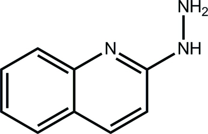
Abstract
In the title compound, C9H9N3, the 12 non-H atoms are essentially planar (r.m.s. deviation = 0.068 Å). The maximum deviation from planarity is reflected in the torsion angle between the β-N atom of the hydrazinyl residue and the quinolinyl N atom [N—N—C—N = −12.7 (3)°]; these atoms are syn. In the crystal, supramolecular layers in the bc plane are formed via N—H⋯N hydrogen bonds.
Related literature
For applications of coordination complexes of hydrazones as organic light emitting diodes and supramolecular magnetic clusters, see: Zhang et al. (2011 ▶); Petukhov et al. (2009 ▶). For background to the synthesis of hydrazones, see: Gupta et al. (2007 ▶); Anwar et al. (2011 ▶). For a related structure, see: Najib et al. (2012 ▶).
Experimental
Crystal data
C9H9N3
M r = 159.19
Monoclinic,

a = 13.7966 (9) Å
b = 3.9648 (3) Å
c = 14.0700 (8) Å
β = 97.039 (5)°
V = 763.84 (9) Å3
Z = 4
Cu Kα radiation
μ = 0.70 mm−1
T = 100 K
0.30 × 0.08 × 0.03 mm
Data collection
Agilent SuperNova Dual diffractometer with an Atlas detector
Absorption correction: multi-scan (CrysAlis PRO; Agilent, 2012 ▶) T min = 0.476, T max = 1.000
2474 measured reflections
1542 independent reflections
1169 reflections with I > 2σ(I)
R int = 0.018
Refinement
R[F 2 > 2σ(F 2)] = 0.052
wR(F 2) = 0.158
S = 1.10
1542 reflections
121 parameters
H atoms treated by a mixture of independent and constrained refinement
Δρmax = 0.32 e Å−3
Δρmin = −0.23 e Å−3
Data collection: CrysAlis PRO (Agilent, 2012 ▶); cell refinement: CrysAlis PRO; data reduction: CrysAlis PRO; program(s) used to solve structure: SHELXS97 (Sheldrick, 2008 ▶); program(s) used to refine structure: SHELXL97 (Sheldrick, 2008 ▶); molecular graphics: ORTEP-3 (Farrugia, 1997 ▶) and DIAMOND (Brandenburg, 2006 ▶); software used to prepare material for publication: publCIF (Westrip, 2010 ▶).
Supplementary Material
Crystal structure: contains datablock(s) global, I. DOI: 10.1107/S1600536812026906/su2456sup1.cif
Structure factors: contains datablock(s) I. DOI: 10.1107/S1600536812026906/su2456Isup2.hkl
Supplementary material file. DOI: 10.1107/S1600536812026906/su2456Isup3.cml
Additional supplementary materials: crystallographic information; 3D view; checkCIF report
Table 1. Hydrogen-bond geometry (Å, °).
| D—H⋯A | D—H | H⋯A | D⋯A | D—H⋯A |
|---|---|---|---|---|
| N2—H1n⋯N3i | 0.93 (3) | 2.18 (3) | 3.077 (2) | 164 (2) |
| N3—H2n⋯N1ii | 0.89 (2) | 2.31 (2) | 3.200 (2) | 175.1 (19) |
| N3—H3n⋯N2iii | 0.90 (2) | 2.58 (2) | 3.295 (2) | 136.4 (16) |
Symmetry codes: (i)  ; (ii)
  ; (iii)
  .
.
Acknowledgments
We gratefully acknowledge funding from the Brunei Research Council, and thank the Ministry of Higher Education (Malaysia) for funding structural studies through the High-Impact Research scheme (UM.C/HIR/MOHE/SC/3).
supplementary crystallographic information
Comment
Hydrazones are versatile nitrogen donor ligands which have been used extensively for making coordination complexes for a variety of applications from organic light emitting diode (OLED) materials (Zhang et al., 2011) to supramolecular magnetic clusters (Petukhov et al., 2009). These ligands are made by condensation of a carbonyl compound with an organic hydrazine or hydrazide (Anwar et al., 2011). We have previously reported the solid-state structure of the zinc(II) complex of 3,5-dimethyl-1- (2'-quinolyl)pyrazole (Najib et al., 2012). The ligand in that complex was made by the condensation of acetylacetone with the title compound (Gupta et al., 2007). Herein, the crystal and molecular structure of the title compound is described.
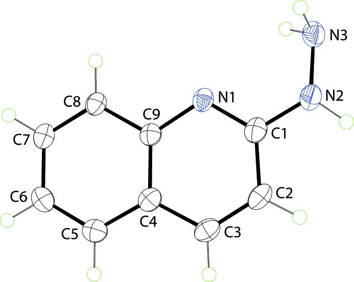
In the title compound, Fig. 1, the 12 non-hydrogen atoms are planar with a r.m.s. deviation = 0.068 Å and maximum deviations of 0.068 (2) and -0.152 (2) Å for the N1 and N3 atoms, respectively. The amine-N3 group is syn with the quinolinyl-N1 atom with the N3—N2—C1—N1 torsion angle being -12.7 (3)°.
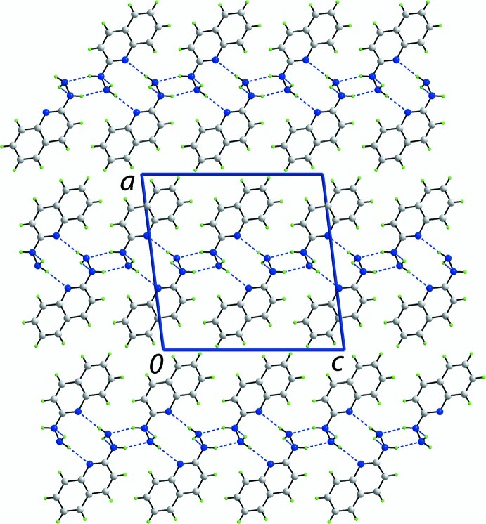
In the crystal, molecules assemble into supramolecular layers in the bc plane via N—H···N hydrogen bonds, Fig. 2 and Table 1. The secondary amine-H hydrogen bonds to the primary amine-N2 atom. One of the primary amine-H atoms forms a hydrogen bond with the quinolinyl-N atom and the other forms a weak interaction with the secondary amine-N2 atom. The layers stack along the a axis with no specific interactions between them, Fig. 3.
Experimental
The title compound was prepared by modification of a literature procedure (Gupta et al., 2007). 2-Chloroquinoline (10.06 g) and hydrazine monohydrate (64–65% N2H4) in water (10 ml) were refluxed for 2 h. The water was removed using a rotary evaporator to provide a scarlet residue which was triturated with water and filtered. This scarlet solid was recrystallized from CH2Cl2 and hexane to provide 6.48 g (66.6%) of the title compound [M.p. = 417 K]. Spectroscopic data for the title compound are given in the archived CIF.
Refinement
C-bound H-atoms were placed in calculated positions [C—H = 0.95 Å, Uiso(H) = 1.2Ueq(C)] and were included in the refinement in the riding model approximation. The N-bound H-atoms were located in a difference Fourier map and refined freely.
Figures
Fig. 1.
The molecular structure of the title molecule showing the atom-labelling scheme. The displacement ellipsoids are drawn at the 50% probability level.
Fig. 2.
A view of the supramolecular layer in the bc plane in the crystal of the title compound. The N—H···N hydrogen bonds are shown as blue dashed lines (see Table 1 for details).
Fig. 3.
A view of the unit-cell contents of the title compound in projection down the b axis. The N—H···N hydrogen bonds are shown as blue dashed lines (see Table 1 for details).
Crystal data
| C9H9N3 | F(000) = 336 |
| Mr = 159.19 | Dx = 1.384 Mg m−3 |
| Monoclinic, P21/c | Cu Kα radiation, λ = 1.54184 Å |
| Hall symbol: -P 2ybc | Cell parameters from 799 reflections |
| a = 13.7966 (9) Å | θ = 3.2–75.8° |
| b = 3.9648 (3) Å | µ = 0.70 mm−1 |
| c = 14.0700 (8) Å | T = 100 K |
| β = 97.039 (5)° | Plate, red |
| V = 763.84 (9) Å3 | 0.30 × 0.08 × 0.03 mm |
| Z = 4 |
Data collection
| Agilent SuperNova Dual diffractometer with an Atlas detector | 1542 independent reflections |
| Radiation source: SuperNova (Cu) X-ray Source | 1169 reflections with I > 2σ(I) |
| Mirror monochromator | Rint = 0.018 |
| Detector resolution: 10.4041 pixels mm-1 | θmax = 76.0°, θmin = 3.2° |
| ω scan | h = −16→17 |
| Absorption correction: multi-scan (CrysAlis PRO; Agilent, 2012) | k = −3→4 |
| Tmin = 0.476, Tmax = 1.000 | l = −17→17 |
| 2474 measured reflections |
Refinement
| Refinement on F2 | Primary atom site location: structure-invariant direct methods |
| Least-squares matrix: full | Secondary atom site location: difference Fourier map |
| R[F2 > 2σ(F2)] = 0.052 | Hydrogen site location: inferred from neighbouring sites |
| wR(F2) = 0.158 | H atoms treated by a mixture of independent and constrained refinement |
| S = 1.10 | w = 1/[σ2(Fo2) + (0.1P)2] where P = (Fo2 + 2Fc2)/3 |
| 1542 reflections | (Δ/σ)max < 0.001 |
| 121 parameters | Δρmax = 0.32 e Å−3 |
| 0 restraints | Δρmin = −0.23 e Å−3 |
Special details
| Experimental. Spectroscopic data for the title compound: IR \v/cm-1: 3282, 3188, 3042, 2954, 2926, 2854, 1621, 1529, 1462, 1404, 1377, 1307, 1146, 1116, 955, 816, 746. 1H NMR 400MHz (CDCl3) δ: 7.82 (1H, d), 7.71 (1H, d), 7.60 (1H, d), 7.54 (1H, dd), 7.23 (1H, dd), 6.75 (1 H, d), 4.0 (3H, br s). 13C NMR 100MHz (CDCl3) δ: 158.8, 147.3, 137.4, 129.7, 127.5, 126.3, 124.2, 122.8, 110.6. |
| Geometry. All e.s.d.'s (except the e.s.d. in the dihedral angle between two l.s. planes) are estimated using the full covariance matrix. The cell e.s.d.'s are taken into account individually in the estimation of e.s.d.'s in distances, angles and torsion angles; correlations between e.s.d.'s in cell parameters are only used when they are defined by crystal symmetry. An approximate (isotropic) treatment of cell e.s.d.'s is used for estimating e.s.d.'s involving l.s. planes. |
| Refinement. Refinement of F2 against ALL reflections. The weighted R-factor wR and goodness of fit S are based on F2, conventional R-factors R are based on F, with F set to zero for negative F2. The threshold expression of F2 > σ(F2) is used only for calculating R-factors(gt) etc. and is not relevant to the choice of reflections for refinement. R-factors based on F2 are statistically about twice as large as those based on F, and R- factors based on ALL data will be even larger. |
Fractional atomic coordinates and isotropic or equivalent isotropic displacement parameters (Å2)
| x | y | z | Uiso*/Ueq | ||
| N1 | 0.35068 (10) | 0.3907 (4) | 0.51371 (10) | 0.0241 (4) | |
| N2 | 0.44545 (11) | 0.4959 (4) | 0.65825 (10) | 0.0315 (4) | |
| N3 | 0.52127 (11) | 0.2769 (4) | 0.63649 (11) | 0.0305 (4) | |
| C1 | 0.35855 (13) | 0.5229 (5) | 0.60074 (12) | 0.0261 (4) | |
| C2 | 0.28104 (14) | 0.6982 (5) | 0.63892 (12) | 0.0285 (4) | |
| H2 | 0.2907 | 0.7905 | 0.7017 | 0.034* | |
| C3 | 0.19420 (13) | 0.7297 (4) | 0.58430 (13) | 0.0277 (4) | |
| H3 | 0.1422 | 0.8465 | 0.6084 | 0.033* | |
| C4 | 0.18015 (13) | 0.5882 (5) | 0.49047 (12) | 0.0250 (4) | |
| C5 | 0.09162 (13) | 0.6087 (5) | 0.42974 (13) | 0.0281 (4) | |
| H5 | 0.0375 | 0.7219 | 0.4510 | 0.034* | |
| C6 | 0.08226 (13) | 0.4670 (5) | 0.33990 (13) | 0.0287 (4) | |
| H6 | 0.0220 | 0.4808 | 0.2995 | 0.034* | |
| C7 | 0.16246 (13) | 0.3017 (5) | 0.30833 (12) | 0.0271 (4) | |
| H7 | 0.1558 | 0.2029 | 0.2464 | 0.033* | |
| C8 | 0.25074 (13) | 0.2802 (4) | 0.36567 (12) | 0.0244 (4) | |
| H8 | 0.3044 | 0.1700 | 0.3428 | 0.029* | |
| C9 | 0.26140 (12) | 0.4220 (4) | 0.45841 (11) | 0.0225 (4) | |
| H1n | 0.4430 (19) | 0.564 (7) | 0.721 (2) | 0.058 (7)* | |
| H2n | 0.5538 (16) | 0.370 (6) | 0.5919 (16) | 0.038 (6)* | |
| H3n | 0.4950 (14) | 0.090 (6) | 0.6069 (14) | 0.025 (5)* |
Atomic displacement parameters (Å2)
| U11 | U22 | U33 | U12 | U13 | U23 | |
| N1 | 0.0259 (7) | 0.0276 (8) | 0.0193 (7) | −0.0045 (6) | 0.0042 (5) | 0.0015 (5) |
| N2 | 0.0317 (8) | 0.0407 (10) | 0.0218 (7) | −0.0017 (7) | 0.0016 (6) | −0.0034 (7) |
| N3 | 0.0277 (8) | 0.0396 (10) | 0.0238 (8) | −0.0043 (7) | 0.0017 (6) | 0.0025 (6) |
| C1 | 0.0283 (8) | 0.0283 (10) | 0.0222 (8) | −0.0074 (7) | 0.0051 (6) | 0.0016 (6) |
| C2 | 0.0377 (10) | 0.0283 (9) | 0.0208 (8) | −0.0052 (8) | 0.0095 (7) | −0.0026 (7) |
| C3 | 0.0339 (9) | 0.0252 (9) | 0.0258 (9) | −0.0012 (7) | 0.0111 (7) | 0.0003 (7) |
| C4 | 0.0298 (9) | 0.0228 (9) | 0.0234 (8) | −0.0028 (7) | 0.0069 (6) | 0.0030 (6) |
| C5 | 0.0273 (9) | 0.0266 (10) | 0.0310 (9) | 0.0005 (7) | 0.0067 (7) | 0.0039 (7) |
| C6 | 0.0253 (8) | 0.0288 (10) | 0.0312 (9) | −0.0017 (7) | −0.0005 (6) | 0.0040 (7) |
| C7 | 0.0312 (9) | 0.0286 (10) | 0.0214 (8) | −0.0035 (7) | 0.0026 (7) | 0.0002 (6) |
| C8 | 0.0270 (8) | 0.0266 (9) | 0.0202 (8) | −0.0021 (7) | 0.0046 (6) | 0.0008 (6) |
| C9 | 0.0236 (8) | 0.0232 (9) | 0.0212 (8) | −0.0033 (7) | 0.0051 (6) | 0.0031 (6) |
Geometric parameters (Å, º)
| N1—C1 | 1.324 (2) | C3—H3 | 0.9500 |
| N1—C9 | 1.380 (2) | C4—C5 | 1.405 (2) |
| N2—C1 | 1.366 (2) | C4—C9 | 1.421 (2) |
| N2—N3 | 1.421 (2) | C5—C6 | 1.375 (3) |
| N2—H1n | 0.93 (3) | C5—H5 | 0.9500 |
| N3—H2n | 0.89 (2) | C6—C7 | 1.404 (2) |
| N3—H3n | 0.90 (2) | C6—H6 | 0.9500 |
| C1—C2 | 1.434 (2) | C7—C8 | 1.379 (2) |
| C2—C3 | 1.348 (3) | C7—H7 | 0.9500 |
| C2—H2 | 0.9500 | C8—C9 | 1.412 (2) |
| C3—C4 | 1.426 (2) | C8—H8 | 0.9500 |
| C1—N1—C9 | 116.89 (15) | C5—C4—C3 | 123.37 (16) |
| C1—N2—N3 | 122.42 (15) | C9—C4—C3 | 116.97 (16) |
| C1—N2—H1n | 114.1 (16) | C6—C5—C4 | 120.82 (16) |
| N3—N2—H1n | 119.8 (17) | C6—C5—H5 | 119.6 |
| N2—N3—H2n | 110.1 (15) | C4—C5—H5 | 119.6 |
| N2—N3—H3n | 109.6 (13) | C5—C6—C7 | 119.52 (16) |
| H2n—N3—H3n | 102.9 (19) | C5—C6—H6 | 120.2 |
| N1—C1—N2 | 118.89 (16) | C7—C6—H6 | 120.2 |
| N1—C1—C2 | 123.91 (16) | C8—C7—C6 | 121.16 (16) |
| N2—C1—C2 | 117.19 (15) | C8—C7—H7 | 119.4 |
| C3—C2—C1 | 118.89 (15) | C6—C7—H7 | 119.4 |
| C3—C2—H2 | 120.6 | C7—C8—C9 | 120.05 (16) |
| C1—C2—H2 | 120.6 | C7—C8—H8 | 120.0 |
| C2—C3—C4 | 120.13 (16) | C9—C8—H8 | 120.0 |
| C2—C3—H3 | 119.9 | N1—C9—C8 | 118.03 (15) |
| C4—C3—H3 | 119.9 | N1—C9—C4 | 123.20 (15) |
| C5—C4—C9 | 119.66 (15) | C8—C9—C4 | 118.77 (15) |
| C9—N1—C1—N2 | 179.57 (15) | C4—C5—C6—C7 | −0.4 (3) |
| C9—N1—C1—C2 | −1.1 (3) | C5—C6—C7—C8 | −0.3 (3) |
| N3—N2—C1—N1 | −12.7 (3) | C6—C7—C8—C9 | 0.8 (3) |
| N3—N2—C1—C2 | 167.89 (16) | C1—N1—C9—C8 | −179.37 (16) |
| N1—C1—C2—C3 | 0.7 (3) | C1—N1—C9—C4 | 0.4 (2) |
| N2—C1—C2—C3 | −179.94 (16) | C7—C8—C9—N1 | 179.17 (16) |
| C1—C2—C3—C4 | 0.4 (3) | C7—C8—C9—C4 | −0.6 (3) |
| C2—C3—C4—C5 | 179.51 (18) | C5—C4—C9—N1 | −179.88 (16) |
| C2—C3—C4—C9 | −1.0 (2) | C3—C4—C9—N1 | 0.6 (3) |
| C9—C4—C5—C6 | 0.6 (3) | C5—C4—C9—C8 | −0.1 (3) |
| C3—C4—C5—C6 | −179.92 (17) | C3—C4—C9—C8 | −179.61 (15) |
Hydrogen-bond geometry (Å, º)
| D—H···A | D—H | H···A | D···A | D—H···A |
| N2—H1n···N3i | 0.93 (3) | 2.18 (3) | 3.077 (2) | 164 (2) |
| N3—H2n···N1ii | 0.89 (2) | 2.31 (2) | 3.200 (2) | 175.1 (19) |
| N3—H3n···N2iii | 0.90 (2) | 2.58 (2) | 3.295 (2) | 136.4 (16) |
Symmetry codes: (i) −x+1, y+1/2, −z+3/2; (ii) −x+1, −y+1, −z+1; (iii) x, y−1, z.
Footnotes
Supplementary data and figures for this paper are available from the IUCr electronic archives (Reference: SU2456).
References
- Agilent (2012). CrysAlis PRO Agilent Technologies, Yarnton, England.
- Anwar, M. U., Elliott, A. S., Thompson, L. K. & Dawe, L. N. (2011). Dalton Trans. 40, 4623–4635. [DOI] [PubMed]
- Brandenburg, K. (2006). DIAMOND Crystal Impact GbR, Bonn, Germany.
- Farrugia, L. J. (1997). J. Appl. Cryst. 30, 565.
- Gupta, L. K., Bansal, U. & Chandra, S. (2007). Spectrochim. Acta Part A, 66, 972–975. [DOI] [PubMed]
- Najib, M. H. bin, Tan, A. L., Young, D. J., Ng, S. W. & Tiekink, E. R. T. (2012). Acta Cryst. E68, m571–m572. [DOI] [PMC free article] [PubMed]
- Petukhov, K., Alam, M. S., Rupp, H., Strömsdörfer, S., Müller, P., Scheurer, A., Saalfrank, R. W., Kortus, J., Postnikov, A., Ruben, M., Thompson, L. K. & Lehn, J.-M. (2009). Coord. Chem. Rev. 253, 2387–2398.
- Sheldrick, G. M. (2008). Acta Cryst. A64, 112–122. [DOI] [PubMed]
- Westrip, S. P. (2010). J. Appl. Cryst. 43, 920–925.
- Zhang, W. H., Hu, J. J., Chi, Y., Young, D. J. & Hor, T. S. A. (2011). Organometallics, 30, 2137–2143.
Associated Data
This section collects any data citations, data availability statements, or supplementary materials included in this article.
Supplementary Materials
Crystal structure: contains datablock(s) global, I. DOI: 10.1107/S1600536812026906/su2456sup1.cif
Structure factors: contains datablock(s) I. DOI: 10.1107/S1600536812026906/su2456Isup2.hkl
Supplementary material file. DOI: 10.1107/S1600536812026906/su2456Isup3.cml
Additional supplementary materials: crystallographic information; 3D view; checkCIF report


