Abstract
Background
Infected leg ulcers are major health problems resulting in morbidity and disability and are usually chronic and refractory to antimicrobial treatment.
Aims
The present study is aimed at determining the bacteria involved in leg ulcers and their resistance patterns to commonly used antibiotics as well as to determine whether Aloe Vera has antibacterial activity against multi-drug resistant organisms and promotes wound healing.
Method
A total of 30 cases with leg ulcers infected with multi-drug resistant organisms were treated with topical aloe vera gel and 30 age and sex-matched controls were treated with topical antibiotics. Culture and sensitivity was done from the wounds on alternate days and the ulcer was clinically and microbiologically assessed after 10 days. The results were compiled and statistically analysed.
Results
Cultures of the study group who were using aloe vera dressings showed no growth by the fifth day in 10 (33.3%) cases, seventh day in another 16 (53.3%) and ninth day in two of the remaining four cases (6.7%) while in two (6.7%) cases there was no decrease in the bacterial count. This means that of the 30 cases, 28 showed no growth by the end of 11 days while two cases showed no decrease in bacterial count. Growth of bacteria in study group is decreased from 100% (30 cases) to 6.7% (2 cases) by day 11 with P<0.001. Cultures of the control group did not show any decrease in the bacterial growth by day 11.
Conclusion
Aloe vera gel preparation is cheap and was effective even against multi-drug resistant organisms as compared to the routinely used topical anti-microbial agents.
Keywords: Leg ulcers, multi-drug resistant bacteria, Aloe vera
What this study adds:
Even though we are in the 21st century, leg ulcers continue to be a major cause of morbidity in India. In an effort to improve healing rates, herbal formulations have been tried. There is preliminary evidence that Aloe vera may promote wound healing. In the era of antibiotic resistance, this study offers cost-effective alternative therapy for chronic infected wounds resistant to conventional antibiotics.
Background
Infected leg ulcers are major health problems resulting in morbidity and disability. Leg ulceration affects about 1% of the middle-aged and elderly population. It commonly occurs after a minor injury in association with: chronic venous insufficiency, chronic arterial insufficiency and diabetes. There are also many less common causes of ulcers including skin cancer, systemic sclerosis, vasculitis and various skin conditions. 1
Longstanding leg ulcers are frequently colonised by micro- organisms in a biofilm. They are usually multi-drug resistant and refractory to treatment. Topical, as well as systemic antibiotics and agents have been used, solely and in combination, to eradicate the resistant infections.. Moreover, these agents have lead to the emergence and subsequent rapid overgrowth of resistant bacterial strains, drug side-effects like allergy and organ specific toxicity. In the present context where there is evolution of super bugs, it is very important to find alternatives. Various herbal products have been used in the management and treatment of wounds over the years. Many substances like tissue extracts, vitamins and minerals and a number of plant products have been reported to possess pro healing effects.2 In this context, Aloe vera, which is cheap, cost effective and easily available, would open up a reasonable solution.
Medicinal properties of Aloe vera have been recognised for a long time. 3 The antiseptic and antimicrobial agents present in Aloe vera provide the ability to attack, reduce, control, or even eliminate infections as the gel penetrates directly into the deeper layers of the skin. The analgesic property helps to be a fast and effective painkiller. The polysaccharides in Aloe vera are an important stimulus to the immune system, and also act as a catalyst for the healing properties of Aloe vera.4
Although there are many studies to prove its effectiveness in cosmetology, studies done to evaluate the efficacy of topical Aloe vera in decreasing the bacterial count and bringing about clinical improvement in chronic ulcers are few and far between. Therefore the present study is aimed at determining the bacteria involved in leg ulcers and their resistance patterns to commonly used antibiotics as well as to determine whether Aloe vera has antibacterial activity against multi-drug resistant organisms and promotes wound healing.
Method
This was a prospective, interventional study conducted over a period of three months from May to July 2011 in the Department of Microbiology of a tertiary care hospital attached to a medical college. Thirty patients between 18- 65 years attending the surgical outpatient department of our hospital with chronic leg ulcers (> 6 months) of Grade 1 (Wagner's Grading of Ulcer; Superficial ulcer) which included traumatic ulcers, diabetic ulcers, varicose ulcers and burn wounds which were all included as the study group. These patients were not showing clinical improvement with topical antibiotics and, when tested, had multi-drug resistant organisms. They had previously been treated with oral/IV antibiotics. Thirty age and sex-matched controls attending the surgery OPD with chronic leg ulcers were enrolled in the study. These were patients who had multi-drug resistant infected ulcers and were also not responding to topical antibacterial treatment. Patient selection was done randomly and written informed consent was taken from each patient to be enrolled in the study as well as for use of their photographs. Ethical clearance was obtained from the institutional ethical clearance committee. Patients with critical limb ischemia due for amputation were excluded from the study.
The Aloe vera gel used in the study was obtained by a commercial vendor who prepared the gel as follows. Fresh gel was extracted from the leaves of Spotted Aloe vera species, Aloe barbadensis. This gel was obtained by peeling the outer layer and the inner gel was pulverised and filtered and then sterilised by pasteurisation. No chemicals were added and the original composition was maintained as per our instructions. The gel was stored in sterile containers at room temperature.
The sterility of Aloe vera was tested and confirmed in the Microbiology Department by inoculating a loopful of the undiluted Aloe vera on blood agar, MacConkey agar, and Sabouraud's agar. No organism growth was observed on bacteriological media even after 48h, and cultures on Sabouraud's agar were sterile for up to three weeks.
At the initial visit, the patient was assessed through detailed history and thorough clinical examination. Peripheral neuropathy and vascular sufficiency were judged clinically through sensory, motor and trophic changes. The surface area of all wounds was examined to assess the initial size and evaluate the progress. For irregular wounds, surface area was calculated by multiplying the two largest dimensions. After debridement, material was collected with a cotton tipped sterile swab from the deeper parts of the ulcers used for bacteriological study by standard culture and sensitivity on Day 1, 3, 5, 7, 9 and 11. Subsequent to sampling for bacteriology, the wound was washed with hyper-tonic saline (3%; instead of normal saline) and the Aloe vera gel applied on ulcers in the morning. Subsequently the patients and their relatives were taught the application of gel in the afternoon and night. Colour photographs were taken before treatment and at 11th day intervals after treatment to determine clinical improvement by assessing the colour, edge, area, and granulation tissue at the base of the ulcer. The collection of specimens and treatment was being done by the same person for all the 30 cases and the assessment of ulcers was done by a general surgeon who was blinded to the treatment approach.
The specimens which were received by the Department of Microbiology were processed immediately. Gram's stained smears prepared from the pus and inoculation of the specimen was done on McConkey agar and blood agar. The plates were incubated at 37°C for 24-48 hours and any colonies obtained at the end of this period were qualitatively assessed and identified by using standard techniques.5 This was done by a microbiologist who was blinded to the case and control groups.
Antibiotic susceptibility was tested with the Kirby–Bauer disc diffusion method, according to the CLSI guidelines on Mueller Hinton agar (Hi Media, Mumbai) was prepared from a dehydrated base according to the manufacturer's instructions. The zone size around each antimicrobial disk was interpreted as sensitive or resistant according to the CLSI guidelines of 201.6 Multi-drug resistant organisms were those that are resistant to three or more anti-bacterial groups.
The results were compiled and statistically analysed.
Results
In this study, 30 patients with non-healing, infected leg ulcers were included of whom 26 (86.7%) were males and 4 (13.3%) were females. Of these, 8 (26.7%) were diabetics, 12 (40%) were post-traumatic, 6 (20%) were varicose ulcers and 4 (13.3%) were burns wounds. Thirty age and sex- matched controls were enrolled in the study that were being treated with topical antibiotics but were not responding to therapy. Of the controls, 26 (86.7%) were males, 49 (13.3%) were females. Of these, 8 (26.7%) were diabetics, 12 (40%) were post-traumatic, 6 (20%) were varicose ulcers and 4 (13.3%) were burns wounds. The culture done on Day 1 among the study group isolated Staphylococcus aureus 14 (46.7%) of which 10 (71.4%) were Methicillin Resistant Staphylococcus aureus (MRSA), Pseudomonas aeruginosa 8 (26.7%), Citrobacter koserii 4 (13.3%), Proteus vulgaris 2 (6.7%) and Enterobacter spp 2 (6.7%), among the 30 patients.
The antibiogram revealed that most of the organisms were multi-drug resistant. Staphylococcus aureus isolates were resistant to Penicillin (85.7%), Clindamycin (85.7%), Cotrimoxazole (71.4%) and Cefoxitin (57.1%). Among the Gram-negative bacilli, all strains (100%) of Pseudomonas aeruginosa were resistant to Amoxiclav, Ceftizoxime, Cotrimoxazole, Ofloxacin. All Citrobacter koserii, Proteus vulgaris and Enterobacter spp strains (100%) were resistant to Amoxicillin-Clavulanic acid, Aztreonam, Cotrimoxazole and Ofloxacin.
As shown in Table 1, subsequent cultures of the study group who were using Aloe vera dressings showed no growth by the fifth day in 10 (33.3%) cases, seventh day in another 16 (53.3%) and ninth day in two (6.7%) of the remaining four cases while in two (6.7%) cases there was no decrease in the bacterial count and there was bacterial growth in these cases on Day 11 also. All the other 28 patients showed no bacterial growth by Day 11 Growth of bacteria in study group is decreased from 100% (30 cases) to 6.7% (2 cases) in day 11 with P<0.001.
Table 1: Bacterial growth in control & study group on alternate days of sampling.
| Day 1 | Day 3 | Day 5 | Day 7 | Day 9 | Day 11 | |
|---|---|---|---|---|---|---|
| Control | 30 | 30 | 30 | 30 | 30 | 30 |
| Study group | 30 | 30 | 30 | 30 | 30 | 30 |
The culture done on Day 1 among the control group also isolated the same group of organisms which were more sensitive to the commonly used antibiotics. Subsequent cultures of the control group who were using topical antibiotics did not show any decrease in the bacterial growth on Days 3, 5, 7, 9 and 11. The same organisms with identical pattern of antibiogram were isolated.
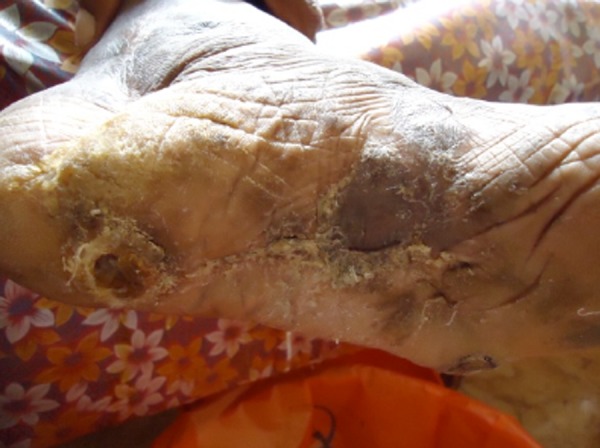
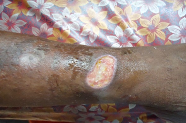
Figure 1 shows a leg ulcer with inflammation and debris and Figure 2 depicts the same ulcer after a period of 11 days during which Aloe vera was topically applied showing clinical improvement.
Figure 1. Case 03, Day 1.
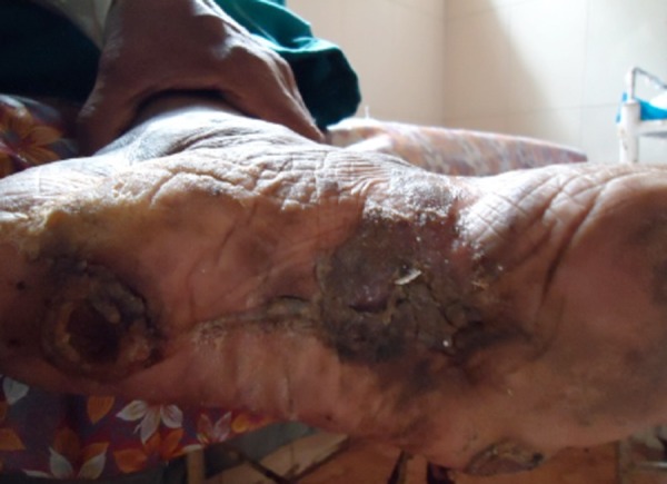
Figure 2. Case 03, Day 11 Image of leg ulcer with inflammation and debris showing clinical improvement after 10 days.
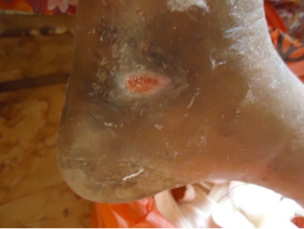
Similarly, Figure 3 shows an image of a leg ulcer with oedema and discharge without granulation tissue and Figure 4 shows clinical improvement after 11 days with healthy granulation tissue and decrease in oedema following regular topical application of Aloe vera gel.
Figure 3. Case 06, Day 1.
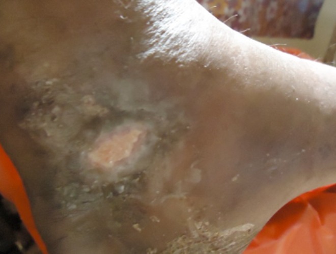
Figure 4. Case 06, Day 11 Image of leg ulcer with oedema and discharge without granulation tissue showing clinical improvement after 10 days with healthy granulation tissue and decrease in oedema.
Note: Growth of bacteria in study group is decreased from 100% (30 cases) to 6.7%(2 cases) in day 11 with P<0.001** while the growth of bacteria is remains constant in control
On the other hand, Figure 5 shows an image of a leg ulcer from the patient in the control group and there is no clinical improvement after 11 days of application of topical antibiotics and alternate day dressing as shown in Figure 6.
Figure 5. Control 11, Day 1.

Figure 6. Control 11, Day 11 Image of leg ulcer from a patient in the control group showing no clinical improvement after 10 days of application of topical antibiotics and alternate day dressing.
Discussion
The use of plant extracts, with known antimicrobial properties, can be of great significance in the treatment of various microbial infections. In the last decade, numerous studies have been conducted in different countries to prove such efficiency in a number of medicinal plants. According to the World Health Organization (WHO), medicinal plants would be the best source for obtaining a variety of drugs. Aloe vera has been known since antiquity as an effective wound dressing. The emergence of multi-drug resistant strains and the financial burden of modern dressings have revived Aloe vera as a cost-effective dressing particularly in developing countries.7
In our study, where topical Aloe vera gel was used in leg ulcers infected with multi-drug resistant organisms, it was observed that no organisms were isolated by Day 11 in all but two cases. This is similar to the study done by Zawahry et al 3 who found that Aloe vera was effective against most of the common organisms causing wound infection and Moghazy et al 8 who studied the cost effectiveness of Aloe vera dressings in the treatment of diabetic foot ulcers over a period of three months and found that complete healing was significantly achieved in 43.3% of the patients. Decrease in size and healthy granulation was significantly achieved in another 43.3% of patients. Bacterial load of all ulcers was significantly reduced after the first week of Aloe vera dressings.
In our study we also found that Aloe vera is effective even against MRSA which is similar to a study by Bashir et al.4
Further, we also noticed a considerable clinical improvement in the form of rapid diminution of inflammation, discharge and deodarisation of the wound by the end of 10 days along with the appearance of healthy granulation tissue.
Aloe vera gel is said to promote wound healing due to the presence of components like anthraquinones and hormones 9 which possess antibacterial antifungal and antiviral activities. In vitro studies have shown that Aloe vera gel showed 100% activity against gram negative isolates and 75.3% against all tested gram positive isolates. The gel possesses a 100% inhibitory effect on P. aeruginosa and Staphylococcus aureus which are known to cause skin infection especially at burns sites, wounds, pressure sores and ulcers.4
In two cases in which we isolated Enterobacter spp and Pseudomonas aeruginosa, there was no decrease in bacterial growth even on Day 11 and both these cases were diabetics with uncontrolled glucose parameters. This is similar to the study of Moghazy et al 8 who reported failure of treatment with Aloe vera in 6.7% of ulcers.
Conclusion
Aloe vera is cheap, cost-effective and easily available when compared to conventional and modern dressings and can be useful in a resource limited country. Since it is effective against multi-drug resistant organisms, it is a boon to many patients with chronic, non-healing, infected ulcers not responding to antibiotics. The simplicity of the dressing techniques which are done by the patient themselves or their relatives is an added advantage. Aloe vera does not have side effects and does not lead to drug resistance. Therefore, it can be tried in cases of infected leg ulcers thereby decreasing the emergence of multi-drug resistant “Super Bugs”. However, it may not be effective in certain patients such as those suffering from uncontrolled diabetics.
Footnotes
PEER REVIEW
Not commissioned. Externally peer reviewed.
CONFLICTS OF INTEREST
The authors declare that they have no competing interests
FUNDING
Self funded
ETHICS COMMITTEE APPROVAL
Institutional ethical committee
Please cite this paper as: Banu A, Sathyanarayana BC, Chattannavar G. Efficacy of fresh Aloe vera gel against multi- drug resistant bacteria in infected leg ulcers. AMJ 2012, 5, 6, 305-309. http//dx.doi.org/10.4066/AMJ.2012.1301.
References
- 1.DermNet NZ. Leg ulcers. Available at http://dermnetnz.org/site-age-specific/leg-ulcers.html.. Accessed on 12 July 2011. [Google Scholar]
- 2.Vishwanathan V, Kesavan R, Kavitha KV, Kumpatla S. A pilot study on the effects of a polyherbal formulation cream on diabetic foot ulcers. Indian J Med Res. 2011;134:168–173. [PMC free article] [PubMed] [Google Scholar]
- 3.Zawahry M E, Hegazy RM, Helal M. Use of aloe in treating leg ulcers and dermatoses. Int J Dermatol. 1973;12:68–73. doi: 10.1111/j.1365-4362.1973.tb00215.x. [DOI] [PubMed] [Google Scholar]
- 4.Bashir A, Saeed B, Talat Y, Jehan MN. Comparative study of antimicrobial activities of Aloe vera extracts and antibiotics against isolates from skin Infections. African Journal of Biotechnology. 2011;10(19):3835–3840. [Google Scholar]
- 5.Collee JG, Fraser AG, Marmion BP, Siminons A. 14th. Churchill Livingston: New York.: 1996. Mackie and McCartney Practical Medical Microbiology. [Google Scholar]
- 6.CLSI. Twenty First Informational Supplement. CLSI document M100-S21. Wayne, PA: Clinical and Laboratory Standards Institute;; 2011. Performance standards for Antimicrobial Susceptibility testing. [Google Scholar]
- 7.Thiruppathi S, Ramasubramanian V, Sivakumar T, Arasu TV. Antimicrobial activity of Aloe vera (L.) Burm. f. against pathogenic microorganisms. J. Biosci. Res. 2010;1(4):251–258. [Google Scholar]
- 8.Moghazy AM, Shams ME, Adly OA, Abbas AH, El- Badawy MA, Elsakka DM, Hassan SA, Abdelmohsen WS, Ali OS, Mohamed BA. The clinical and cost effectiveness of Aloe vera dressing in the treatment of diabetic foot ulcers. Diabetes Res Clin Pract. 2010;89:276–281. doi: 10.1016/j.diabres.2010.05.021. [DOI] [PubMed] [Google Scholar]
- 9.Davis RH, Leitner MG, Russo JM. Aloe vera. A natural approach for treating wounds edema and pain in diabetes. J Am Podiatr Med Assoc. 1988;78:80–88. doi: 10.7547/87507315-78-2-60. [DOI] [PubMed] [Google Scholar]


