Abstract
A 16-hour-old white foal, born to a registered quarter horse mare, was examined for signs of colic. The foal had Overo lethal white syndrome, which causes ileocolonic agangliosis. This was confirmed by DNA testing. Since there is no treatment for Overo lethal white syndrome, the foal was euthanized.
A 14-year-old, pregnant mare with American Quarter Horse Association registration papers was purchased at auction in March 2000, with the knowledge that she had been bred to a paint stallion. The mare was registered as solid chestnut with a star, strip and snip, a dark spot above her nostrils, a white spot in the right nostril, and no other white markings. The mare's sire, a registered American paint horse, was sorrel with an Overo pattern.
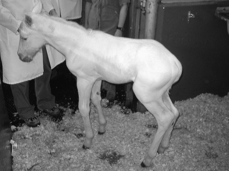
Gestation and parturition were uneventful, and the filly foal suckled spontaneously within 2 h of birth. The mare quickly accepted the foal, which was very mobile and inquisitive in the paddock. The only unusual observations were the distinctively white coat of the foal and failure to pass the meconium (Figure 1). Within 12 to 16 h of birth, the foal started to show signs of colic.
Figure 1. Overo lethal white foal born to a registered Quarter horse mare.
When examined on day 1, the foal appeared very uncomfortable, alternating between standing, lying down, and rolling on its back. It was completely white with blue irides. The pupillary light reflex and menace response were present. Oral mucous membranes and capillary refill time were normal, but the heart rate was slightly elevated (110–120 beats/min). There was a mild increase in lung sounds in the cranioventral lung fields. All joints, the umbilicus, and the rectal temperature (38°C) were normal. The most significant finding was the lack of borborygmi on ascultation of the abdomen. Feces were present deep within the rectum. The clinical findings suggested a meconium impaction or, more likely, Overo lethal white syndrome (OLWS). As supportive therapy, 120 mL of mineral oil was administered as an enema to assist with the passage of the meconium plug, and flunixin meglumine (Cronyxn; Vetrepharm, Belleville, Ontario), 150 mg, IV, was given to relieve abdominal pain. Response to the analgesia was marked and the foal became recumbent.
On day 2, the foal continued to show signs of colic. Euthanasia was recommended, and blood samples were taken from the mare and foal to confirm the diagnosis of OLWS. The foal was euthanized and a postmortem was performed. On gross examination, the foal lacked pigmentation and the abdomen was grossly distended. The lungs were mildly edematous, but the liver, spleen, kidneys, and adrenal glands appeared normal. The peritoneal cavity contained serosanguinous brown fluid, suspected to have leaked from the intestines ante- and postmortem. There was no evidence of an intestinal accident and no strictures were identified along the length of the intestine. The serosal surface of the small colon and rectum was pale. Most of the intestinal tract was gas filled, and the caudal aspect of the large intestine was distended with particulate matter. Samples (skin, liver, lung, intestine, heart, spleen, kidney, and eye) were collected into 10% buffered formalin and submitted for histopathologic examination. Histologically, no melanin was seen in the skin and there were few active hair follicles. Many follicles were devoid of hair or were in catagen phase (transition between active and resting hair growth). The liver and lungs were mildly congested. In a few areas of the lung, aspirated squamous epithelial cells and a light mononuclear cell infiltrate were seen. The colon was normal, except for the absence of ganglion cells, which was compatible with ileocolonic aganglionosis, as seen in OLWS (2).
Blood samples from the mare and foal were submitted for DNA analysis (Minnesota Veterinary Diagnostic Laboratory, College of Veterinary Medicine, University of Minnesota, St. Paul, Minnesota, USA), which confirmed that the mare was heterozygous and that the foal was homozygous for the OLW gene.
Overo lethal white syndrome occurs in newborn foals that receive a copy of the mutated OLW gene from each parent. Horses with white Overo patterning are more likely carriers of the gene than solid-colored horses (2). The mutated gene alters neural crest cell migration or survival, which affects the progenitor cells for melanocytes and intestinal ganglia. Affected foals suffer from aganglionosis of the submucosal and myenteric ganglia of the distal part of the small intestine and of the large intestine, resulting in intestinal immotility and colic (2). Phenotypically, the altered gene causes lack of skin pigmentation and white coat color. The Overo coat pattern is described as white markings on the lateral and ventral aspects of the neck and torso, whereas a pattern with more white on the dorsal cervical and lumbar regions and the legs is called tobiano (3). The Overo coat pattern is seen in the American paint horse, American miniature horse, half-Arabian, Thoroughbred, and crop-out (unregistered because of excessive white marking) quarter horse (QH).
The lethal OLWS gene is an autosomal dominant with variable expression. Heterozygotes demonstrate assorted white coat patterns, and, on very rare occasions, may be solid-colored; for example, if the dominant lethal gene is not being expressed or has spontaneously mutated. Additional studies are necessary to explain the sporadic occurrence of Overo foals from nonspotted QH parents. Two carriers of the mutated gene must be mated to produce a homozygous lethal white foal. According to Mendelian genetics, an Overo × Overo mating would be expected to produce 25% solid-colored foals, and 50% Overo foals, and 25% OLW foals (1).
Stud book records and observation of born foals show that the probability of producing an OLWS offspring is less than 25%. Possible factors contributing to this unexpectedly low frequency may include failure to report OLW foals to breed registrations, early embryonic loss of homozygote foals, or the relative proportion of carriers in the breeding population (1).
Understanding the inheritance of the lethal gene is important for economic reasons, as paints are desirable in western horse shows, and it would be an advantage to be able identify horses carrying the Overo gene (3). Inaccurate data on the risk of OLWS may deter people from using breeding stock with Overo blood lines. With accurate genetic information, breeders could avoid the psychological and economic losses associated with the Overo lethal gene by testing breeding stock for carrier status and breeding known Overos only to proven non-Overos.
As there is no treatment for OLWS, testing is essential to prevent its occurrence (1). Before DNA testing was available, carriers were identified phenotypically by the proportion of white in the coat: the more white, the greater risk of being a carrier. Although this technique identified most carriers, it was inaccurate. A DNA-based test that identifies horses that are heterozygous for the Overo lethal white gene has been developed. The allele-specific polymerase chain reaction test locates and amplifies the specific mutated site in the endothelin receptor B gene (EDNRB gene). This site has been identified in humans with Hirschsprung disease, in whom similar gastrointestinal effects from a mutation of the EDNRB gene are seen. Sequencing of the DNA of the EDNRB gene revealed a dinucleotide thymine-cytosine to adenine-guanine mutation. This results in substitution of the amino acid isoleucine for lysine in the first transmembrane domain of the EDNRB protein (termed Ile118Lys mutation) (4). The EDNRB protein is responsible for regulation of embryonic neural crest cells that develop into ganglia and melanocytes. Foals homozygous for the Ile118Lys mutation in the EDNRB gene have only 20% of the functional protein ability of control horses (5). Innervation to the intestine is impaired, causing fatal constipation. Heterozygotes commonly have the Overo coat pattern without intestinal abnormalities. In a study to determine the phenotype of heterozygotes, > 95% were Overo and < 1% were solid colored (2). Variation in the expression of the Overo pattern and inability to predict the exact genotype from the phenotype are due to augmentation of the white coloring by other genes. In order to explain the sporadic occurrence of an OLW foal from a solid-colored QH, further research is required to understand this multifactorial inheritance pattern. The association between coat color spotting, intestinal aganglionosis, and mutations of the EDNRB gene has been studied in mouse models (6,7). It is possible that the EDNRB gene in the QH spontaneously mutates or mutates at a higher rate than in rodents (2).
Proper sampling is important for DNA analysis. Blood or hair samples can be used, but there are difficulties in obtaining DNA from blood, and the blood must be unclotted, kept refrigerated, and delivered to the laboratory within 24 h. Hair samples must include the roots and 15 to 20 hairs, and can be collected from the mane or tail, and require no specific packaging (Minnesota Veterinary Diagnostic Laboratory, University of Minnesota, personal communication).
This case demonstrates the classic clinical and pathological presentation of an OLW foal with unusual parental lineage. The QH mare represents the small proportion of solid-colored heterozygotes.
Footnotes
Acknowledgements
The author thanks Dr. Peter Brouwers, Peterborough Veterinary Services, for his encouragement and guidance with this case. CVJ
Address correspondence and reprint requests to Dr. Lightbody.
Dr. Lightbody's current address is Peterborough Veterinary Services, 720 The Kingsway, Peterborough, Ontario K9J 6W6.
References
- 1.McCabe L, Griffin LD, Kinzer A, Chandler M, Beckwith JB, McCabe RB. Overo lethal white foal syndrome; equine model of aganglionic megacolon (Hirschsprung disease). Am J Med Genet 1990;36:336–340. [DOI] [PubMed]
- 2.Vrotsos PD, Santschi EM, Purdy AK, Mickelson JR. Incidence of an endothelin receptor B mutation in white patterned horses; evidence for genetic heterogeneity in the overo coat pattern. Plant Anim Genome VII Conf, San Diego, California, 1999.
- 3.Bowling AT. Dominant inheritance of overo spotting in paint horses. J Hered 1994;85:222–225. [DOI] [PubMed]
- 4.Yang GC, Croaker D, Zhang AL, Manflick P, Cartmill T, Cass D. A dinucleotide mutation in the endothelin-B receptor gene is associated with lethal white foal syndrome (LWFS); a horse variant of Hirschsprung disease. Human Mol Gene 1998;6:1047–52. [DOI] [PubMed]
- 5.James RM, Santschi EM. Role of the endothelin receptor B gene in overo coat color pattern and lethal white foal syndrome. Plant Anim Genome VII Conf, San Diego, California, 1999.
- 6.Lane PW, Liu HM. Association of megacolon with a new dominant spotting gene (Dom) in the mouse. J Hered 1984;75:435–439. [DOI] [PubMed]
- 7.Santschi EM, Purdy AK, Valberg SJ, Vrotsos PD, Kaese H, Mickelson JR. Endothelin receptor B polymorphism associated with lethal white foal syndrome in horses. Mamm Genome 1998; 4:306–309. [DOI] [PubMed]


