Summary
A 32-year-old woman hospitalized for subarachnoid hemorrhage showed rare arterial variation on the right side with anomalous origins of the vertebral artery, aberrant subclavian artery and persistent trigeminal artery. Angiography showed the right vertebral artery to originate from the right common carotid artery, the right subclavian artery to arise separately from the descending aorta, and persistent trigeminal artery on the right side.
The possible embryonic mechanism of this previously unreported variant combination is discussed.
Key words: aberrant subclavian artery, persistent trigeminal artery, vertebral artery
Introduction
Anomalous origin of the right vertebral artery from the right common carotid artery is rare 1,2, and has been reported coexisting with an aberrant right subclavian artery 3. However, there have been no previous reports of these variants in combination with persistent trigeminal artery.
Here, we describe a case of right persistent trigeminal artery associated with aberrant right subclavian artery in combination with anomalous origin of the right vertebral artery from the right common carotid artery.
Case Report
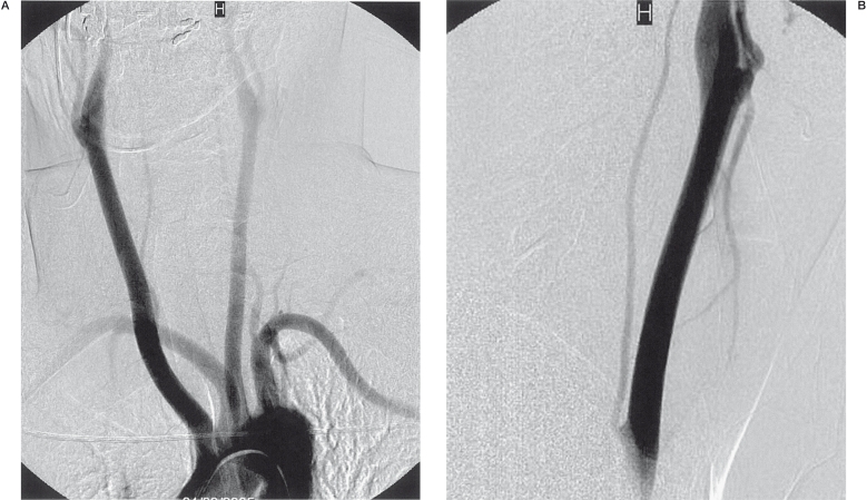
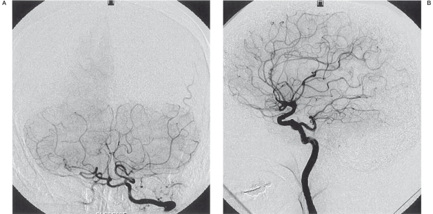
A 32-year-old woman was admitted to our hospital with sudden onset severe headache. Brain computed tomography (CT) indicated subarachnoid hemorrhage, the center of which was located anterior to the mesencephalon. Digital subtraction angiography was performed to determine the source of bleeding. Aortogram showed the right subclavian artery originating separately from the descending aorta (Figure 1A). The right vertebral artery could not be visualized on selective right subclavian artery injection. Angiography of the right common carotid artery indicated that the right vertebral artery originated from the common carotid artery (Figure 1B). The right vertebral artery entered the transverse process of the fourth cervical vertebra. Intracranial examination showed a hypoplastic basilar artery (Figure 2A) and persistent trigeminal artery located on the right (Figure 2B). The source of bleeding could not be determined despite repeated digital subtraction angiography.
Figure 1.
Arch aortogram (A) in left anterior oblique projection showing the right subclavian artery originating from the descending aorta. Selective right common carotid arteriogram (B) in lateral projection showing the right vertebral artery originating from the common carotid artery.
Figure 2.
Selective left vertebral arteriogram (A) showing the hypoplastic basilar artery. Selective right internal carotid arteriogram in lateral (B) projection showing the right persistent trigeminal artery.
Discussion
In humans, the development of vertebral arteries begins at the 7 mm embryonic stage. The vertebral arteries arise from the postcostal longitudinal anastomosis between the C1 and the C7 intercostal arteries and the cervical intercostal obliteration zone, the sixth cervical intersegmental artery persists as a trunk for the subclavian artery and the vertebral artery 4,5, and the other intersegmental arteries located between the aorta and the longitudinal anastomosis disappear by the 14- to 17-mm stage 1. The vertebral arteries normally enter the foramen of the transverse process of the cervical vertebra at the sixth level, and therefore variants of the vertebral artery are related to the persistence and/or disappearance of these intersegmental arteries.
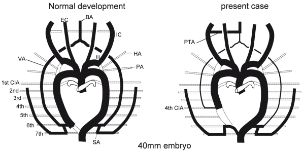
The incidence of anomalous origin of the right vertebral artery from the right common carotid is 0.18% 6. Embryologically, this is explained as due to persistence of one of the third to fifth cervical intersegmental arteries instead of the sixth 1. In this case, the right dorsal aorta distal to the persistent cervical intersegmental artery was obliterated, resulting in a right aberrant subclavian artery. In the present case, the fourth cervical intersegmental artery was considered to be persistent, while the right dorsal aorta distal to the fourth cervical intersegmental artery was obliterated (Figure 3), because the right vertebral artery enters the fourth transverse process of the cervical vertebra.
Figure 3.
Schematic representation of the embryological mechanisms underlying anatomic variation involving anomalous origins of the subclavian artery, the vertebral artery, and persistent trigeminal artery. Left, Normal embryological development. Right, Present case in which the right VA arises at the 4th CIA level, with involution of the right dorsal aorta distal to the 4th CIA. The PTA is associated with hypoplastic VAs. BA, basilar artery; CIA, cervical segmental artery; HA, hypoglossal artery; IC, internal carotid artery; PA, proatlantal artery; PTA, persistent trigeminal artery; SA, subclavian artery; VA, vertebral artery.
Primitive embryonic anastomotic vessels formed between the carotid and basilar arterial systems occasionally persist into adulthood. In early fetal development, there are four transient anastomoses between the posterior vascular plexus and the anterior carotid artery, which normally disappear by the 7-12 mm embryonic stage with the development of the vertebobasilar system. The trigeminal artery disappears at the last of these four anastomoses. Hypoplastic vertebral and basilar arteries have been documented in association with the persistent trigeminal artery 7. In the present case, the persistence of the trigeminal artery may have been due to a developmental abnormality of the vertebral artery.
The case described here represents a rare combination of the right vertebral artery originating from the right common carotid artery, aberrant right subclavian artery, and persistent right trigeminal artery. This combination of anomalies is likely due to the effect of a developmental abnormality of the vertebral artery causing the aberrant right subclavian artery and persistence of the trigeminal artery.
References
- 1.Haughton VM, Rosenbaum AE. The normal and anomalous aortic arch and brachiocephalic arteries. In: Newton TH, Potts DG, editors. Radiology of the skull and brain. St. Louis: Mosby; 1974. pp. 1145–1163 . Book 2. [Google Scholar]
- 2.Lemke AJ, Benndorf G, Liebig T, et al. Anomalous origin of the right vertebral artery: review of the literature and case report of the right vertebral artery origin distal to the left subclavian artery. Am J Neuroradiol. 1999;20:1318–1321 . [PMC free article] [PubMed] [Google Scholar]
- 3.Cairney J. The anomalous right subclavian artery considered in the light of recent findings in arterial development; with a note on two cases of an unusual relation of the innominate artery to the trachea. J Anat. 1925;59:265–296 . [PMC free article] [PubMed] [Google Scholar]
- 4.Newton TH, Mani RL. The vertebral artery. In: Newton TH, Potts DG, editors. Radiology of the skull and brain. St. Louis: Mosby; 1974. pp. 1659–1672 . [Google Scholar]
- 5.Padget DH. Designation of the embryonic intersegmental arteries in reference to the vertebral artery and subclavian stem. Anat Rec. 1954;119:349–356 . doi: 10.1002/ar.1091190306. [DOI] [PubMed] [Google Scholar]
- 6.Palmer FJ. Origin of the right vertebral artery from the right common carotid artery: angiographic demonstration of three cases. Br J Radiol. 1977;50:185–187 . doi: 10.1259/0007-1285-50-591-185. [DOI] [PubMed] [Google Scholar]
- 7.Pasco A, Papon X, Bracard S, et al. Persistent carotid-vertebrobasilar anastomoses: how and why differentiating them? J Neuroradiol. 2004;31:391–396 . doi: 10.1016/s0150-9861(04)97022-8. [DOI] [PubMed] [Google Scholar]


