Abstract
Context
Comprehensive analysis of ankle, knee, and hip kinematics and kinetics during anterior lunge performance in young adults has not been studied. In addition, the effects of adding external resistance on the kinematics and kinetics are unknown.
Objective
To determine the effects of external load on ankle, knee, and hip joint kinematics and kinetics during the anterior lunge.
Design
Crossover study.
Setting
Laboratory environment.
Patients or Other Participants
A total of 16 recreationally active, college-aged adults (8 men, 8 women).
Intervention(s)
Anterior lunges under 4 external-load conditions, 0% (control), 12.5%, 25%, and 50% of body mass.
Main Outcome Measure(s)
Ankle, knee, and hip peak flexion, net joint extensor moment impulse, and eccentric and concentric work were computed during the interval when the stepping limb was in contact with the ground. Additionally, 3 summary lunge characteristics were calculated.
Results
No significant (P > .05) load effects were noted for peak flexion angles or the lunge characteristics except for peak vertical total-body center-of-mass displacement. Trend analysis of significant condition-by-joint interactions revealed significant linear trends for all 3 joints, with the hip greater than the ankle and the ankle greater than the knee. Additionally, as the external load increased, mechanical work increased linearly at the hip and ankle but not at the knee.
Conclusions
From a kinematic perspective, the lunge involves greater motion at the knee, but from a kinetic perspective, the anterior lunge is a hip-extensor–dominant exercise. Adding external weight prompted the greatest joint kinetic increases at the hip and ankle, with little change in the knee contributions. These results can assist clinicians in deciding whether the characteristics of the anterior lunge match a patient's exercise needs during rehabilitation and performance-enhancement programs.
Key Words: resistance training, closed kinetic chain exercise, kinematics, kinetics
Key Points
Kinematically, the anterior lunge involves greater motion at the knee than at the ankle and hip, but kinetically, the exercise is hip-extensor dominant.
Increasing external loading during the exercise increased the ankle and hip contributions but had minimal effect at the knee.
Optimal exercise selection for promoting restoration, maintenance, and enhancement of functional capacities involves pairing the demands imposed by an exercise with the needs of the patient or client.1 Similarly, choosing an exercise or task to use during preexisting or residual impairment evaluations requires matching the challenges imposed by the exercise with the targeted areas of focus. Both of these applications, using an exercise for training or evaluation, require a full understanding of the mechanical demands imposed on the musculoskeletal system. Unfortunately, for many commonly used exercises, such as the anterior lunge, the specific biomechanical characteristics are largely unknown,2,3 leaving exercise selection and progression decisions to be largely based on intuition and clinical experience.
As do other closed chain exercises, the anterior lunge offers the advantages of promoting activation patterns similar to those of functional activity, especially with respect to muscle coactivation,4–6 and replicating functional movements such as gait.2 Furthermore, the anterior lunge is described as a relatively safe exercise, even for older adults,4 patients with patellofemoral pain,7 and patients after anterior cruciate ligament reconstruction,2,8 with the additional benefit of requiring minimal equipment.4 Qualitative observation of the anterior lunge reveals an exercise that largely involves the ankle, knee, and hip-extensor muscle groups as prime movers in the sagittal plane, with frontal- and transverse-plane muscle groups serving in stabilizer roles. The results of several studies3,5,9,10 using electromyography also support this observation.
Electromyography may be used to determine which individual muscles are being activated during a particular exercise. However, the complexity of the electromyography-force relationship prevents our interpreting signal amplitude as representative of muscle force production. Thus, in addition to understanding which muscles are active, quantifying joint kinematics and kinetics provides additional insight regarding the mechanical demands of an exercise. For example, joint kinematics reveal the range of motion used during an exercise, whereas joint kinetic measures, such as net joint moment impulse and angular work, provide information regarding the relative muscle group magnitudes over time and angular distance, respectively. Knowing the range of motion used with the relative muscle-group contributions reflected by impulse and work can provide clinicians with further rationale to guide exercise selection decisions.
Research examining the anterior lunge from a mechanical perspective has largely focused on knee-joint kinematics and kinetics in patients with anterior cruciate ligament5,8,12,13 or patellofemoral7 conditions. This is likely a result of the frequent use of the anterior lunge in knee exercise and rehabilitation programs. Direct comprehensive study of sagittal-plane ankle, knee, and hip-joint kinematics and kinetics has been limited to 2 investigations.4,11 The most comprehensive kinematic and kinetic study revealed the hip joint makes the greatest relative contribution to the anterior lunge, both kinematically and kinetically.4 However, because only older adults were studied, it remains unknown whether younger, physically active adults performing lunges would also demonstrate the greatest relative contributions at the hip joint. A second study11 using kinematic and kinetic measurements demonstrated that trunk position during the anterior lunge influenced the mechanical demands of the ankle, knee, and hip joints. Specifically, completing anterior lunges with a flexed-trunk position increased the ankle and hip-extensor net joint moment impulses (NJMIs), whereas using an extended-trunk position significantly increased the knee NJMI. Although the investigation involved a small sample of young, healthy adults, no between-joints statistical comparisons were conducted; thus, the question about the relative mechanical demands imposed on the ankle, knee, and hip during the anterior lunge in young adults remains.
In addition to the kinematic and kinetic characteristics, many other pertinent features of the anterior lunge remain unknown. Similar to any exercise, once an individual adapts to the imposed demands, the challenge of the exercise is often augmented by adding or increasing external resistance. Specific to the anterior lunge, does adding external resistance increase the mechanical demands equally, in proportion to the magnitude of the load across the ankle, knee, and hip joints? Thus, the aims of our study were to confirm that the anterior lunge is a hip-extensor–dominant exercise in young, healthy adults and to determine the effects of external loads on sagittal-plane ankle-, knee-, and hip-joint kinematics and kinetics during the anterior lunge. We hypothesized that in our sample of young, healthy adults, the hip joint would make the greatest relative contribution and that ankle, knee, and hip kinetics would increase equally as the external loads were increased, but kinematics would not change. In addition, because external loads might influence how the lunges were performed, thereby potentially confounding interpretation of the kinematic and kinetic changes, a third aim was to examine the effect of external loads on 3 performance summary variables: stepping-limb contact time, peak vertical total-body center-of-mass displacement (TBCM), and peak anterior TBCM displacement. We hypothesized that external loads would have no effect on these performance summary variables.
METHODS
Participants
A total of 16 recreationally active, college-aged volunteers (8 men, 8 women, age = 20.4 ± 1.2 years, height = 1.70 ± 0.08 m, mass = 70.6 ± 11.2 kg) participated in the investigation. All were healthy at the time of testing, which was operationally defined as no history of previous ankle, knee, hip, or back musculoskeletal conditions that could influence their ability to perform the anterior lunge. Additionally, they were screened for recent head injuries and any preexisting visual, vestibular, or balance disorders by being asked about any previously diagnosed conditions using a comprehensive health and medical history questionnaire. Recreationally active was operationally defined as participation in some form of physical activity for 20 minutes at least 3 times per week. Volunteers were given a verbal description of the study procedures, the opportunity to ask questions, and the option to decline to participate; those who were willing to continue were asked to read and sign a consent form approved by the Institutional Review Board for the Protection of Human Research Participants, which also approved the study.
Experimental Design
Participants were required to attend a 45-minute testing session. During that session, they completed 6 anterior lunges under 4 external-load conditions (0% [control], 12.5%, 25%, and 50% of body mass) while kinematic and ground reaction force data were collected. To assist in controlling for multiple exposure and fatigue effects, each participant was randomly assigned an external-load condition order.
Anterior Lunge Procedures
For the anterior lunge, the participant was barefoot and used the dominant limb as the stepping limb. The dominant limb was operationally defined as the preferred limb for kicking a ball. Lunge stepping distance was standardized to 70% of dominant-leg length, measured from the greater trochanter to the lateral malleolus, by placing tape strips at the starting point and target step distance. Participants were instructed to complete each repetition by stepping forward with the dominant limb and then lowering the body as much as comfortably possible. Once they reached the lowest position, they were instructed to immediately push backward through the dominant limb and return to the full-standing starting position. They were also instructed to maintain an erect torso, perpendicular to the horizontal, during the entire lunge. Participants attempted to complete the lowering phase of each repetition within 2 seconds; an acoustic metronome set to 60 beats per minute was used to assist with pacing. During the 0% load (control) condition, participants held 1 oak dowel (0.32-m long, 0.03-m diameter, 0.11-kg mass) in each hand with arms at their sides and the forearms in neutral position (palms facing body). For the external-load conditions, we could not use standard iron dumbbells due to the electromagnetic tracking system that collected kinematic data. Instead, 2 disks filled with sand and concrete were attached to the ends of each dowel to produce a dumbbell-like apparatus that equaled half (within 1.5 kg) the target external loads (12.5%, 25%, and 50% of body mass) and was held using a similar grip to that in the 0% condition. Several familiarization trials (3 to 6 trials) were allowed for each external-load condition before data collection so that the participants could become comfortable with the movements and loads. A 10-minute rest period followed. Data-collection trials under each external-load condition were completed in 2 sets of 3 continuous repetitions.
Data Collection
An extended-range, 9-sensor electromagnetic tracking system (MotionStar, Ascension Technology Corporation, Inc, Burlington, VT) with all the hardware settings in the default mode collected 3-dimensional kinematic data (100 Hz) using the MotionMonitor acquisition software package (version 8.4; Innovative Sports Training, Inc, Chicago, IL). The manufacturer indicated that this system measures positional data accurately to 0.5°, with a resolution of 0.1° at a distance of 1.52 m. Sensors were attached to the C7 posterior spinous process, sacrum, and both feet, shanks, and thighs using double-sided tape and elastic tape after the familiarization trials were completed. During setup, the ankle- and knee-joint centers were calculated by locating midpoints between contralateral points at each respective joint with the ninth electromagnetic sensor attached to a customized calibrated stylus. The hip-joint center was established using a series of 8 points along a circumduction cycle for each hip to estimate the apex of femoral motion.14 The participant's height and mass were also recorded for the anthropometric calculations required for locating each segment's center of mass using the Dempster parameters as reported by Winter.15 Ground reaction force data under the stance and stepping limbs were collected (100 Hz) using 2 nonconducting force plates (model BP400600NC 2000; Advanced Mechanical Technology, Inc, Watertown, MA) synchronized with the electromagnetic system.
Data Reduction
The 3-dimensional ankle, knee, and hip-joint angles and net joint moments and the segment center-of-mass 3-dimensional linear position data (feet, shanks, thighs, pelvis, trunk) were calculated using the MotionMonitor software. These data and the ground reaction force data were exported as text files for further processing using MATLAB-based scripts (The MathWorks, Inc, Natick, MA). First, all data were low-pass filtered with a zero-phase lag Butterworth filter (10-Hz cutoff). The instantaneous TBCM position was determined for each trial using the segment center of mass and anthropometric data. The beginning and end of a trial were operationally defined as occurring when vertical TBCM velocity exceeded −0.15 m/s and 0.15 m/s, respectively. Three of the 6 trials under each condition were selected for analysis using a based graphic user interface display of the vertical TBCM trajectory. Criteria for selection included achievement of similar vertical TBCM displacement (± 0.01 m) and repetition time across the 3 trials.
For both the ankle and hip kinematic and kinetic data, the polarity was reversed so that extension and net joint extensor moments would be positive, thereby matching the knee. The interval of interest for kinematic and kinetic variables was the duration of time the stepping limb was in contact with the second anteriorly located force plate (ground contact > 10 N, ground off < 10 N) . Four dependent variables at each joint (ankle, knee, hip) from the kinematic and kinetic data were determined: peak flexion angles, NJMI, eccentric work, and concentric work. The peak flexion angles were expressed relative to each participant's standing (double-leg) calibration position. Net joint flexor-extensor moments were normalized to body mass, with impulses calculated as the integrated magnitude of the net joint moment curve. To calculate eccentric and concentric work, net joint power was first calculated as the product of angular velocity (radians) and net joint moment (normalized to body mass). Eccentric and concentric work was calculated as the integrated magnitude of the absolute net joint power curve. For eccentric work only, the descent phase was considered (stepping-limb ground contact to minimal vertical TBCM position), whereas for concentric work only, the ascent phase was considered (minimal vertical TBCM position to stepping-limb ground off).
Finally, to examine differences in lunge performance among the 4 external-load conditions, stepping-limb contact time and peak anterior TBCM displacement were also determined for each selected trial. The averages of these 3 characteristics were calculated across the 3 trials.
Data Analysis
For each dependent variable, the average across the 3 trials within each of the 4 external-load conditions was calculated and used for statistical analysis. The α level for all statistical analyses was set at .05. Separate repeated-measures analyses of variance (ANOVAs) were used to compare the 3 lunge characteristic variables (stepping-limb contact time, peak vertical TBCM displacement, and peak anterior TBCM displacement) among the 4 external-load conditions. Two-factor repeated-measures ANOVAs (condition by joint) were used for statistical comparison of the peak flexion angles and NJMI. A 3-factor repeated-measures ANOVA (condition by joint by phase) was used for statistical comparison of mechanical work, with phase having 2 levels (eccentric and concentric). Because eccentric work has negative polarity, the absolute value of eccentric work was entered into the model so that comparisons between eccentric and concentric magnitudes could be conducted. In all analyses, a Greenhouse-Geisser correction factor was applied when sphericity was indicated. When statistical significance was evident for the 4 lunge characteristics, Bonferroni post hoc tests were used. For NJMI and mechanical work, because the levels of external load are ordinal (ordered), polynomial trend analyses were conducted to examine significant weight-condition effects. Coefficients for unequal intervals were calculated according to procedures outlined by Grandage.16 By conducting trend analyses, we could determine changes in NJMI and mechanical work across the 4 external-load conditions. Identifying a significant linear trend would suggest that NJMI/work increased linearly with increased external loading, whereas a concurrent significant linear and quadratic trend would suggest that NJMI/work increased to a certain point and then plateaued. In addition to the trend analyses, Bonferroni post hoc comparisons between joints within each level of condition were conducted when significant condition-by-joint-interactions were revealed. Finally, for mechanical work, simple main-effects post hoc analyses were conducted to examine significant phase-by-joint interactions using Bonferroni adjusted P values (.05/9 comparisons = .0056).
RESULTS
Peak Flexion Angles
Descriptive statistics for the peak flexion angles are presented in Table 1. The addition of external weight did not change the peak flexion angles attained during the anterior lunge as evidenced by the condition-by-joint interaction (F2.4,35.9 = 0.416, P = .698, η2p = 0.03) and main effect for condition (F3,45 = 1.86, P = .163, η2p = 0.11). A main effect for joint was noted (F1.4,20.5 = 470 , P < .001, η2p = 0.97). Post hoc analysis revealed greater peak knee flexion than peak ankle (P < .001, d = 10.63) and hip flexion (P = .001, d = 1.0) and greater peak hip flexion than peak ankle flexion (P < .001, d = 4.8).
Table 1.
Peak Flexion Angles for the Ankle, Knee, and Hip Across the 4 Weight Conditions During the Anterior Lunge (Mean ± SD)

Net Joint Moment Impulses
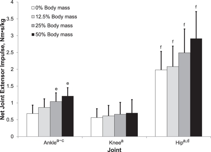
A significant condition-by-joint interaction (F3,45.6 = 15.80, P < .001, η2p = 0.51) was revealed for NJMI magnitude (Figure 1). As weight increased, NJMI for the ankle (F1,15 = 152.95, P < .001, η2 = 0.91), knee (F1,15 = 5.65, P = .032, η2 = 0.27), and hip (F1,15 = 72.59, P < .001, η2 = 0.83) demonstrated significant linear trends. The linear trend for the hip was greater than for the ankle (F1,15 = 37.63, P < .001, η2 = 0.72 ) and knee (F1,15 = 18.65, P = .001, η2 = 0.55), and the linear trend for the ankle was greater than for the knee (F1,15 = 49.28, P < .001, η2 = 0.77). A significant quadratic trend for the ankle was seen (F1,15 = 8.82, P = .010, η2 = 0.37). No other significant trends were revealed. Post hoc comparisons among joints within each weight condition demonstrated that hip NJMI was greater than ankle and knee NJMI (P ≤ .001, d = 1.88 to 2.77). The NJMI at the ankle was greater than at the knee for the 25% (P = .014, d = 0.83) and 50% (P = .003, d = 1.0) weight conditions.
Figure 1.

Ankle, knee, and hip net joint extensor impulse across the 4 weight conditions during the anterior lunge. aSignificant linear trend (P < .05). bSignificant quadratic trend (P < .05). cLinear trend greater than at the knee (P < .05). dLinear trend greater than at the ankle and knee (P < .05). eGreater than at the knee for the same weight condition (P < .05). fGreater than at the ankle and knee for the same weight condition (P < .05).
Concentric and Eccentric Work
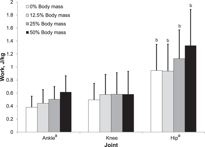
Significant condition-by-joint (F6,90 = 5.24, P < .001, η2p = 0.259) and phase-by-joint (F6,90 = 22.15, P < .001, η2p = 0.596) interactions were revealed. As weight increased (Figure 2), mechanical work increased linearly at the ankle (F1,15 = 39.56, P < .001, η2 = 0.72) and hip (F1,15 = 32.45, P < .001, η2 = 0.68) but not at the knee (F1,15 = 2.18, P = .160, η2 = 0.13). No other higher-order trends were revealed. The linear trend for the hip was not different than at the ankle (F1,15 = 4.47, P = .052, η2 = 0.23). Post hoc comparisons among joints within each weight condition showed that hip work was greater than ankle and knee work (P ≤ .046, d = 0.68 to 1.47). No differences were observed between ankle and knee work across any of the weight conditions.
Figure 2.

Ankle, knee, and hip work across the 4 weight conditions during the anterior lunge. aSignificant linear trend (P < .05). bGreater than at the ankle and knee for the same weight condition (P < .05).
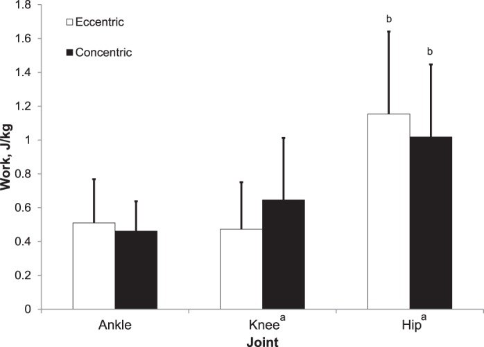
Simple main-effects post hoc analyses of the phase-by-joint interaction (Figure 3) revealed that eccentric work was greater than concentric work at the knee (P = .001, d = 0.98), whereas eccentric work was less than concentric work at the hip (P < .001, d = 1.3). No difference in work between the phases existed for the ankle (P = .144, d = 0.39). Comparing joints within each of the phases showed that eccentric and concentric hip work were greater at the ankle (eccentric: P = .001, d = 1.2; concentric: P < .001, d = 1.2) and knee (eccentric: P < .001, d = 1.4; concentric: P = .033, d = 0.73). No differences in eccentric (P = .713, d = 0.09) or concentric (P = .278, d = 0.45) work between the ankle and knee were observed.
Figure 3.

Eccentric and concentric work, collapsed across loading condition, for the ankle, knee, and hip during the anterior lunge. aDifference between phases (P < .05). bSignificantly greater than at the ankle and knee for the same phase.
Lunge Characteristics
Descriptive statistics for the lunge characteristics are presented in Table 2. Except for peak vertical TBCM displacement (F3,45 = 9.10, P < .001, η2p = 0.378), the additional weight had no effect on stepping-limb contact time (F2.1,45 = 2.97, P = .064, η2p = 0.165), and peak anterior TBCM displacement (F3,45 = 2.07, P = .117, η2p = 0.122). Post hoc analysis of the peak vertical TBCM displacements demonstrated less displacement between the 50% condition and the 0% (P =.001, d = 1.26) and 12.5% (P = .015, d = 0.91) conditions.
Table 2.
Anterior Lunge Characteristics Across the 4 Weight Conditions (Mean ± SD)

DISCUSSION
The results of this investigation support our hypothesis that the anterior lunge is a hip-extensor–dominant exercise in young, healthy adults. Part of our second hypothesis, that external load would not change the kinematics, was supported by the data. In contrast to the kinetic portion of our second hypothesis, increasing the external load during an anterior lunge did not increase kinetics of the ankle, knee, and hip joints equally. For NJMI, as the external load increased, the greatest increase was at the hip, followed by the ankle and then the knee. Although no difference was evident in the linear trends between the hip and ankle for work, both joints demonstrated linear increases, compared with no significant trends for the knee. These results confirm that the anterior lunge is a hip-dominant exercise and that increasing the external load affects the hip, and secondarily the ankle, to a greater extent than the knee.
Consistent with the findings of previous researchers,4,11,17 the kinematic results identify the knee as reaching a larger peak flexion angle than the hip or ankle. This result was not unexpected because the knee has greater available range of motion than the hip and ankle. No differences in peak flexion were demonstrated across the 3 joints related to external loading; however, peak TBCM vertical displacement demonstrated a slight but statistically significant difference between the 50% condition and the 0% and 12.5% conditions. Peak TBCM vertical displacement represents the cumulative effects of ankle, knee, and hip flexion to reach the lowest point of the eccentric phase compared with the full upright standing position. Specifically, as a result of the heavier load, participants did not lower themselves to the same depth (2 cm less) under the 50% condition compared with the 0% and 12.5% conditions.
Although neither Flanagan et al4 nor Farrokhi et al11 conducted statistical comparisons of ankle, knee, and hip-joint kinetics, their descriptive statistics and graphical data displays also show the anterior lunge to be a hip-extensor–dominant exercise. The peak net joint moment, NJMI, and mechanical energy expenditure (sum of absolute eccentric and concentric work) at the hip were all greater than at the knee and ankle in older adults performing the anterior lunge.4 Specifically, the hip provided 53% of the total support impulse, compared with 26% and 21% provided by the knee and ankle joints, respectively.4 Calculating the relative contribution of the hip, knee, and ankle joints to the total NJMI from the data provided by Farrokhi et al11 reveals 48% for the hip, 31% for the knee, and 21% for the ankle. During the 0% (control) condition in our study, which is most similar to the 2 previous studies, relative contributions to total impulse were 62% by the hip, 17% by the knee, and 21% by the ankle. Although the exact percentages differ slightly between the current and the 2 previous studies, all support greater kinetic contributions to the anterior lunge by the hip than either the knee or ankle.
To date, most research on the anterior lunge has been conducted using only body weight as the external load. Ebben et al3 compared electromyography during anterior lunges at participants' 6-repetition maximum, but they did not compare performance with any other external weight, nor did they consider any muscles other than the hamstrings and quadriceps. Thus, a major purpose of our investigation was to determine the effects of increasing the external load on the kinematics and relative kinetic contributions of the ankle, knee, and hip joints. The external-load protocol (0%, 12.5%, 25%, and 50% of body mass) was selected to provide a full range of external loads when performing anterior lunges with dumbbells. Typically, a person performing anterior lunges with loads greater than 50% of body mass uses a weighted barbell because holding heavier-weight dumbbells at one's side would begin to reach the limit of grip strength. Unexpected in our results was the observation that the relative kinetic contributions did not remain the same as external load was increased. For NJMI, based on the linear trends being different among the joints (hip > ankle > knee), we concluded that as external load increased, the contributions of the hip and ankle increased, with little change in the knee NJMI. The detection of both linear and quadratic trends in the ankle NJMI suggests that the ankle may have reached a point of maximum contribution; however, further research with heavier external loads is needed to confirm this idea. For mechanical work, based on the trend analyses, no increase in the contribution of the knee was detected, but increased contributions were revealed for the ankle and hip. Given the lack of differences in stepping-limb contact time and the range of motion (peak flexion) among external-loading conditions, the effects of the external load in increasing NJMI and mechanical work may be attributed to higher net joint moments.
Although external load had no effect on the peak hip-, knee-, or ankle-flexion angles, small changes in upper extremity or trunk kinematics might explain the shift in relative ankle, knee, and hip contributions. For example, as the external load increased, participants might have changed the way in which they held the dumbbells. Moving the dumbbells to a more anterior position would likely prompt effects similar to those Farrokhi et al11 detected with respect to anterior positioning of the trunk. Specifically, in the same way that a flexed trunk shifts the TBCM anteriorly, moving the dumbbells anteriorly would also shift more mass anteriorly, thereby potentially prompting increased hip and ankle contributions. Future research is recommended to examine whether upper extremity and trunk positioning changes with increased mass and explains the relative contribution changes we noted.
It is important to recognize that joint kinetic measures, such as the NJMI and work measures used in the current investigation and previous investigations,1,4,11 reflect the net result of all structures, both passive and active, acting about a joint. These measures do not estimate the activities of individual muscles or muscle groups but rather describe the relative agonist-antagonist contributions acting on the ankle, knee, and hip joints to produce the anterior lunge.15,18 When agonist-antagonist coactivation occurs, as between the quadriceps and hamstrings, the actual muscle moments produced by each muscle group are likely underestimated.15,18 Electromyography can identify the activation of individual muscles; however, the relationship between electromyographic amplitude and active muscle force production during dynamic movements is not always linear, nor does it take into account the forces contributed by inactive muscles as a result of passive lengthening. Thus, combining the results of joint kinematic and kinetic measures with electromyography provides more insight into the anterior lunge than these measurement techniques can offer in isolation. We did not use electromyography because several groups2,5,6,9,10 that examined muscle activation during the anterior lunge provided consistent electromyographic results that could be used to interpret our joint kinematic and kinetic data. Furthermore, adding electromyographic measurement to the current study would have required participants to accommodate the addition of electrodes and their associated cables, as well as increased each participant's time during setup and calibration.
Both the current and previous studies examining joint kinetics have largely considered dependent variables such as NJMI and mechanical energy expenditure, which are computed across the entire repetition. Exceptions are peak joint extensor moments4,11 and peak joint extensor power4,11; the locations of these values do not appear to have been taken into account. Considering these measures within specific parts of the lunge may be relevant for future researchers because the contributions of various muscles and their resulting effects about a joint likely change throughout the exercise, secondary to factors such as the length-tension relationship and passive and active insufficiency. Furthermore, during a lunge, as a result of the specific muscle contributions changing, similar changes may occur in the relative contributions of the ankle, knee, and hip to the overall support moment through particular phases of the exercise. For example, as the TBCM reaches its lowest point toward the end of the eccentric phase, the gluteus maximus and vasti muscles are elongated, whereas the rectus femoris and biarticular portions of the hamstrings muscles experience little change in length because the knee and hip are flexed. Further examination of these factors will advance our understanding of the anterior lunge.
In interpreting the results, several factors need to be recognized. First, our methods included providing stepping-distance targets, cues to keep the trunk vertical, and a metronome to help with pacing. Anticipating the effect of the increasing external load on how the exercise is performed, we included 3 lunge characteristic variables. This allowed us to account for changes in overall lunge performance as viable alternative explanations for changes in the joint kinematics and kinetics detected under the external-load conditions. Except for peak vertical TBCM displacement, the lack of potent changes in lunge characteristics supports our conclusions that changes in external load prompted the documented joint kinetic changes. It is also noteworthy that despite an attempt to standardize pacing, our participants completed the lunge at a slightly faster pace than targeted, although no differences were seen among the 4 load conditions. Future research is recommended to consider the effects of standardizing step length and pacing on lunge performance because these are not usually controlled in applied settings. We also increased the external load during the lunges by having the participants hold dumbbells. Whether similar results would be attained with the use of a barbell is unknown and therefore also recommended for future investigation. Finally, in order to limit the potential confounding effects of different shoes on lunge performance, participants performed the lunges unshod. The extent to which shoes may influence the kinematic and kinetic results attained is unknown and should be recognized as a deviation from the normal method of performing anterior lunges.
Although in terms of absolute values, knee flexion was greatest, followed by hip flexion and ankle dorsiflexion, during the anterior lunge, the percentage of flexion relative to the normal available range of motion for each joint was slightly greater for the hip (approximately 69%) and knee (approximately 70%) compared with the ankle (approximately 60%). Therefore, if the anterior lunge is used in rehabilitation, the patient's available hip, knee, and ankle range of motion and specific soft tissue healing limitations need to be considered when deciding whether the anterior lunge is an appropriate exercise.
Moreover, many clinicians and fitness professionals use the anterior lunge with the primary intent of strengthening the quadriceps. However, as this study illustrated, based on the NJMI and mechanical work results, the anterior lunge appears to be a hip-extensor–dominant exercise with nearly equal relative ankle and knee contributions at lower external loads. This finding does not suggest that the quadriceps are not active contributors to the anterior lunge movement but rather that the hip extensors provide a greater contribution to the total support moment. As expected, given that the anterior lunge is performed with greater external loads, the contributions of all 3 joints based on the NJMI progressively increased. The NJMI increase was not equal across the joints but was greatest at the hip, followed by the ankle and then the knee. Additionally, as external loads increased, mechanical work increased for the hip and ankle but did not change for the knee. These findings were unexpected, but they have implications for exercise and rehabilitation programs. If the knee extensors are the target muscle group, practitioners may want to select a different exercise. Furthermore, hip-extension impairments may limit the external weight progression and resultant knee-extensor activation.
Of the 3 anterior lunge characteristics we considered (stepping-limb contact time, peak vertical TBCM displacement, and peak anterior TBCM displacement), only vertical TBCM displacement demonstrated changes with increased external load. Practitioners may need to monitor the depth of the anterior lunge and provide cues when the exercise is performed with heavier external loads to ensure that clients and patients perform through the full range of motion.
CONCLUSIONS
The biomechanical characteristics of many exercises used in sports medicine, both from the rehabilitation and strength-training perspectives, need continued examination and documentation. From a kinematic perspective, the anterior lunge involves significantly greater motion at the knee, but from a kinetic perspective, the exercise is hip-extensor dominant. External loading prompted increases in hip and ankle contributions but had minimal effect at the knee. These results can assist clinicians in deciding whether the characteristics of the anterior lunge match a patient's exercise needs during rehabilitation and performance-enhancement programs.
REFERENCES
- 1.Flanagan S, Salem GJ, Wang MY, Sanker SE, Greendale GA. Squatting exercises in older adults: kinematic and kinetic comparisons. Med Sci Sports Exerc. 2003;35(4):635–643. doi: 10.1249/01.MSS.0000058364.47973.06. [DOI] [PMC free article] [PubMed] [Google Scholar]
- 2.Crill MT, Kolba CP, Chleboun GS. Using lunge measurements for baseline fitness testing. J Sport Rehabil. 2004;13(1):44–53. [Google Scholar]
- 3.Ebben WP, Feldmann CR, Dayne A, Mitsche D, Alexander P, Knetzger KJ. Muscle activation during lower body resistance training. Int J Sports Med. 2009;30(1):1–8. doi: 10.1055/s-2008-1038785. [DOI] [PubMed] [Google Scholar]
- 4.Flanagan SP, Wang MY, Greendale GA, Azen SP, Salem GJ. Biomechanical attributes of lunging activities for older adults. J Strength Cond Res. 2004;18(3):599–605. doi: 10.1519/1533-4287(2004)18<599:BAOLAF>2.0.CO;2. [DOI] [PMC free article] [PubMed] [Google Scholar]
- 5.Hefzy MS, al Khazim M, Harrison L. Co-activation of the hamstrings and quadriceps during the lunge exercise. Biomed Sci Instrum. 1997;33:360–365. [PubMed] [Google Scholar]
- 6.Pincivero DM, Aldworth C, Dickerson T, Petry C, Shultz T. Quadriceps-hamstring EMG activity during functional, closed kinetic chain exercise to fatigue. Eur J Appl Physiol. 2000;81(6):504–509. doi: 10.1007/s004210050075. [DOI] [PubMed] [Google Scholar]
- 7.Escamilla RF, Zheng N, Macleod TD, et al. Patellofemoral joint force and stress between a short- and long-step forward lunge. J Orthop Sports Phys Ther. 2008;38(11):681–690. doi: 10.2519/jospt.2008.2694. [DOI] [PubMed] [Google Scholar]
- 8.Alkjaer T, Simonsen EB, Magnusson SP, Aagaard H, Dyhre-Poulsen P. Differences in the movement pattern of a forward lunge in two types of anterior cruciate ligament deficient patients: copers and non-copers. Clin Biomech (Bristol, Avon) 2002;17(8):586–593. doi: 10.1016/s0268-0033(02)00098-0. [DOI] [PubMed] [Google Scholar]
- 9.Boudreau SN, Dwyer MK, Mattacola CG, Lattermann C, Uhl TL, McKeon JM. Hip-muscle activation during the lunge, single-leg squat, and step-up-and-over exercises. J Sport Rehabil. 2009;18(1):91–103. doi: 10.1123/jsr.18.1.91. [DOI] [PubMed] [Google Scholar]
- 10.Ekstrom RA, Donatelli RA, Carp KC. Electromyographic analysis of core trunk, hip, and thigh muscles during 9 rehabilitation exercises. J Orthop Sports Phys Ther. 2007;37(12):754–762. doi: 10.2519/jospt.2007.2471. [DOI] [PubMed] [Google Scholar]
- 11.Farrokhi S, Pollard CD, Souza RB, Chen YJ, Reischl S, Powers CM. Trunk position influences the kinematics, kinetics, and muscle activity of the lead lower extremity during the forward lunge exercise. J Orthop Sports Phys Ther. 2008;38(7):403–409. doi: 10.2519/jospt.2008.2634. [DOI] [PubMed] [Google Scholar]
- 12.Heijne A, Fleming BC, Renstrom PA, Peura GD, Beynnon BD, Werner S. Strain on the anterior cruciate ligament during closed kinetic chain exercises. Med Sci Sports Exerc. 2004;36(6):935–941. doi: 10.1249/01.mss.0000128185.55587.a3. [DOI] [PubMed] [Google Scholar]
- 13.Stuart MJ, Meglan DA, Lutz GE, Growney ES, An KN. Comparison of intersegmental tibiofemoral joint forces and muscle activity during various closed kinetic chain exercises. Am J Sports Med. 1996;24(6):792–799. doi: 10.1177/036354659602400615. [DOI] [PubMed] [Google Scholar]
- 14.Leardini A, Cappozzo A, Catani F, et al. Validation of a functional method for the estimation of hip joint centre location. J Biomech. 1999;32(1):99–103. doi: 10.1016/s0021-9290(98)00148-1. [DOI] [PubMed] [Google Scholar]
- 15.Winter DA. Biomechanics and Motor Control of Human Movement. 3rd ed. Hoboken, NJ: John Wiley & Sons Inc; 2005. [Google Scholar]
- 16.Grandage A. Query: orthogonal coefficients for unequal intervals. Biometrics. 1958;14(2):287–289. [Google Scholar]
- 17.Dwyer MK, Boudreau SN, Mattacola CG, Uhl TL, Latterman C. Comparision of lower extremity kinematics and hip muscle activation during rehabilitation tasks between sexes. J Athl Train. 2010;45(2):181–190. doi: 10.4085/1062-6050-45.2.181. [DOI] [PMC free article] [PubMed] [Google Scholar]
- 18.Whittlesey SN, Robertson DGE. Two-dimensional inverse dynamics. In: Robertson G, Caldwell G, Hamill J, Kamen G, Whittlesey S, editors. Research Methods in Biomechanics. Champaign, IL: Human Kinetics; 2004. In. eds. [Google Scholar]


