Abstract
Human senescence patterns—late onset of mortality increase, slow mortality acceleration, and exceptional longevity—are often described as unique in the animal world. Using an individual-based data set from longitudinal studies of wild populations of seven primate species, we show that contrary to assumptions of human uniqueness, human senescence falls within the primate continuum of aging; the tendency for males to have shorter life spans and higher age-specific mortality than females throughout much of adulthood is a common feature in many, but not all, primates; and the aging profiles of primate species do not reflect phylogenetic position. These findings suggest that mortality patterns in primates are shaped by local selective forces rather than phylogenetic history.
Humans are thought to age more slowly than other mammalian taxa [(1), but see (2)] on the basis of their low early-adult mortality, slow mortality acceleration, and long life span. However, it is not known if these human features are unique or are shared with other primates (3, 4). The rapid increase in human life expectancy in the 20th century (5) has increased the proportion of individuals in older age classes (6), raising questions about the flexibility of human aging patterns and the limits of the human life span [e.g., (7–9)]. These questions necessitate a deeper understanding of natural aging patterns in other primates, which represent our closest living relatives (10).
Nonhuman primates, like humans, are cognitively and socially complex and behaviorally flexible. However, their long lives and the challenges of continuous, long-term observation make longitudinal demographic data on nonhuman primates uncommon, especially for wild populations [(11); see also (12)]. We compiled rare data sets from seven species that span the Primate Order [one Indriid (a Madagascan prosimian), two New World monkeys, two Old World monkeys, and two great apes] and carried out a comparative demographic analysis of mortality. Our analyses used data from 226 observation-years of births and deaths on more than 2800 individually recognized male and female primates (13, 14).
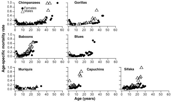
We produced species-specific mortality tables for each sex and computed actuarial estimates of age-specific survival and mortality for each of the primate populations (15). Analysis of mortality rates revealed the expected pattern for mammals: high infant mortality, followed by a period of low mortality during the juvenile stage, and an extended period of increasing age-specific mortality during mid to late life (Fig. 1). We focused on mid- to late-life demography and modeled initial mortality rate at the start of adulthood for each species, defined in Table 1, through the last age interval for which we had census data. For humans, we used published male and female age-at-death data, from age 15 through 100 years, from the U.S. Department of Health and Human Services life tables (16) and repeated the analyses with a second, independent life table for humans (17), which confirmed our findings.
Fig. 1.
Age-specific mortality at age x, ux, for each study species, illustrating high infant mortality, low juvenile mortality, and mortality increasing with age over the adult life span. No sex-specific first-year mortality estimates are available for sifaka because individuals were not sexed and individually identified until their first birthday. For blue monkey males and both sexes of capuchins and muriquis, mortality estimates extend only through age 6, 20, and 32 years, respectively; in each case, this is much less than the suspected full life span, making it difficult to estimate the shape of the mortality curve at the end of life.
Table 1.
Summary of study populations. Details about and references for study sites are in (15).
| Common name | Species | Family | Country | Avg. annual rainfall (mm)* | Life-style | Start year † | Sample size
|
Adult age‡
|
Predominant dispersing sex | Mean age of first dispersal (years) | ||
|---|---|---|---|---|---|---|---|---|---|---|---|---|
| M | F | M | F | |||||||||
| Sifaka | Propithecus verreauxi | Indriidae | Madagascar | 578 | Arboreal | 1984 | 291 | 219 | 5–6 | 6–7 | M | 4–5 |
| Northern Muriqui | Brachyteles hypoxanthus | Atelidae | Brazil | 1180 | Arboreal | 1983 | 192 | 212 | 6–7 | 8–9 | F | 6–7 |
| Capuchin | Cebus capucinus | Cebidae | Costa Rica | 1736 | Arboreal | 1983 | 98 | 58 | 6–7 | 6–7 | M¶ | 4–5 |
| Yellow baboon | Papio cynocephalus | Cercopithecidae | Kenya | 347 | Semi-terrestrial | 1971 | 489 | 437 | 7–8 | 5–6 | M | 7–8 |
| Blue monkey | Cercopithecus mitis | Cercopithecidae | Kenya | 1962 | Arboreal | 1979 | 128 | 194 | 8–9 | 7–8 | M | 7–8 |
| Chimpanzee | Pan troglodytes | Hominidae | Tanzania | 1330 | Semi-terrestrial | 1963 | 122 | 144 | 14–15 | 14–15 | F | 12–13 |
| Gorilla | Gorilla beringei | Hominidae | Rwanda | 1358 | Terrestrial | 1967 | 128 | 120 | 15–16 | 9–10 | M# F |
15–16 7–8 |
Average annual rainfall for each study, representative of the study years. Rainfall data for gorillas were collected by the Rwandan Government Meteorological Office at a location several kilometers from the field site and at a lower elevation. Rainfall data for other studies were collected at the study site.
Year study was established. Latest census date for all populations in these analyses was December 2008.
Mean age class at which adulthood is attained for each sex. Male onset of adult stage was defined as mean age of likely first reproduction (using physical criteria such as copulation with ejaculation, behavioral criteria such as the onset of mate guarding behavior, or genetically confirmed paternity). Female onset of adult stage is defined as the mean age of first live birth.
Twelve percent of female capuchins disperse. The average age interval of dispersing capuchin females is 6 to 7 years.
Both sexes disperse in gorillas.
Understanding flexibility and constraints in the expression and evolution of aging requires a careful analysis of key aging metrics (1, 18, 19). We used a maximum-likelihood framework for estimating two metrics that, together, describe the pattern of senescence for a population: the initial adult mortality rate (IMR, the risk of death at onset of adulthood) and the rate of aging (RoA, the rate of increase in the age-specific mortalities with advancing adult age). These aging metrics are often best estimated by fitting the Gompertz model of increasing failure time. We thus tested among competing models for accelerating risk of death with advancing age on the basis of the Gompertz family of models in program WinModest (20) model fitting as described in (21). Our tests included a standard two-parameter Gompertz model and the Gompertz-Makeham and Logistic models. In all but 2 of the 13 species and sex comparisons we examined, the standard two-parameter Gompertz model yielded the best fit to the nonhuman primate data. In the other two cases (sifaka females and capuchin males), the Gompertz-Makeham model was recommended, but because of particular features of those two data sets (see table S1), we proceeded with the standard Gompertz model for males and females of all species. Our model was of the form ux = IMR × e(RoA)x, where ux is the age-specific mortality, i.e., instantaneous mortality probability, at age x (results in Tables 2 and 3 for females and males, respectively).
Table 2.
Gompertz estimates of female mortality parameters and life-span summary statistics. Adult age interval is the age interval containing the mean age of first live birth; IMR (= Gompertz a) is the Gompertz estimate of instantaneous mortality rate at the first adult age interval [with its 95% confidence interval (CI)]; RoA (= Gompertz b) is the adult rate of aging estimated with Gompertz acceleration (with its 95% CI); MRDT is the mortality rate doubling time during adulthood; Oldest age reached is the age class of the oldest observed individual; Median age is the 50% survival age with its range.
| Species | Adult age interval (years) | IMR (/year) | 95% CI | RoA | 95% CI | MRDT (no. of years) | Oldest age reached (years)
|
Median age | [Range] | |
|---|---|---|---|---|---|---|---|---|---|---|
| Estimated | Known* | |||||||||
| Sifaka | 6–7 | 0.0278 | [0.019, 0.0410] | 0.0991 | [0.072, 0.136] | 7.0 | 31–32 | 23–24 | 10 | [9, 12] |
| Muriqui | 8–9 | 0.00170 | [0.00042, 0.00685] | 0.129 | [0.0722, 0.230] | 5.4 | 40–41 | 26–27 | 25 | [18, 33] |
| Capuchin | 6–7 | 0.0415 | [0.0150, 0.114] | 0.165 | [0.055, 0.494] | 4.2 | 26–27 | 19–20† | 11 | [10, 13] |
| Baboon | 5–6 | 0.0285 | [0.020, 0.040] | 0.123 | [0.0926, 0.165] | 5.6 | 27–28 | 27–28 | 8 | [7, 9] |
| Blue monkey | 7–8 | 0.00723 | [0.00367, 0.0143] | 0.160 | [0.123, 0.209] | 4.3 | 33–34 | 26–27 | 18 | [17, 22] |
| Chimpanzee | 14–15 | 0.00774 | [0.0038, 0.0156] | 0.0992 | [0.070, 0.140] | 7.0 | 53–54 | 38–39 | 16 | [10, 25] |
| Gorilla | 9–10 | 0.00028 | [0.00004, 0.00214] | 0.211 | [0.148, 0.300] | 3.3 | 43–44 | 38–39 | 33 | [31, 35] |
| Human‡ | 0.00009 | [0.00008, 0.00009] | 0.0961 | [0.0956, 0.0967] | 7.2 | 100+ | 100+ | 83.5 | ||
Oldest individual with known date of birth.
Truncated at 18–19 for mortality analysis because of relatively smaller sample sizes of deaths and transitions in later age classes.
Data from (16), modeled beginning at age interval 15–16 years through 99–100 years.
Table 3.
Gompertz estimates of male mortality and life-span summary statistics. Adult age interval is the mean age class of likely first reproduction. IMR (= Gompertz a) is the instantaneous mortality at adulthood (with its 95%CI); RoA (= Gompertz b) is the rate of aging estimated with Gompertz acceleration (with its 95% CI); MRDT is the mortality rate doubling time during adulthood; Oldest age reached is the age class of the oldest observed individual; Median age is the 50% survival age with its range.
| Species | Adult age interval (years) | IMR (/year) | 95% CI | RoA | 95% CI | MRDT (no. of years) | Oldest age reached (years)
|
Median age | [Range] | |
|---|---|---|---|---|---|---|---|---|---|---|
| Estimated | Known* | |||||||||
| † Sifaka | 5–6 | 0.0201 | [0.0140 0.0290] | 0.186 | [0.155, 0.222] | 3.73 | 26–27 | 19–20 | 12 | [12, 13] |
| Muriqui | 6–7 | 0.00187 | [0.00044, 0.00784] | 0.148 | [0.0820, 0.266] | 4.70 | 33–34 | 26–27 | 24 | [22, 27] |
| Capuchin†‡ | 6–7 | 0.010 | [0.0027, 0.036] | 0.294 | [0.159, 0.542] | 2.36 | 24–25¶ | 12–13 | 4 | [3, 5] |
| Baboon†‡ | 7–8 | 0.0371 | [0.0266, 0.0517] | 0.213 | [0.177, 0.256] | 3.26 | 24–25 | 22–23 | 10 | [9, 11] |
| Blue monkey | 8–9 | No est. | No est. | 19–20 | 19–20 | No est. | ||||
| Chimpanzee | 14–15 | 0.00787 | [0.00346, 0.0179] | 0.137 | [0.0980, 0.190] | 5.07 | 43–44 | 40–41 | 11 | [9, 14] |
| Gorilla | 15–16 | 0.00594 | [0.00139, 0.0254] | 0.182 | [0.106, 0.313] | 3.81 | 38–39 | 34–35 | 23 | [22, 29] |
| Human|| | 0.00024 | [0.00023, 0.00025] | 0.086 | [0.0854, 0.0863] | 8.07 | 100+ | 100+ | 79.5 | ||
Oldest individual with known date of birth.
Distribution of deaths imputed from onset of adulthood with age structure. See methods in the Supporting Online Material (SOM).
Age structure corrected for population growth. See methods in the SOM.
Truncated at 18–19 for mortality analysis because of relatively smaller samples sizes of deaths and transitions in later age classes.
Data from (16), modeled beginning at age interval 15–16 years through 99–100 years.
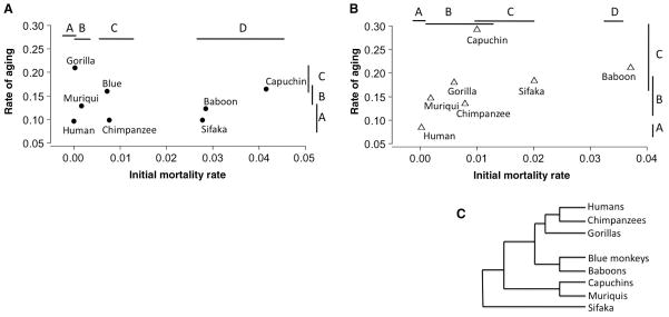
We found significantly positive values for RoA in all study species, indicating that mortality rate increased with advancing age [Tables 2 and 3 and fig. S1; see also (22, 23)]. Notably, humans fell along a continuum with the other primate species for both IMR and RoA (Fig. 2). Furthermore, in neither females nor males did we find evidence of a negative correlation between IMR and RoA, which would be indicative of a trade-off between these two parameters (Fig. 2). Instead, our data suggest that they can evolve independently. Humans had low values for both parameters, which explains their exceptional longevity.
Fig. 2.
IMR versus RoA for (A) females and (B) males. Phylogenetic relationships among species are shown in (C). Letters over bars denote statistically significant groupings. [Female IMR: human, gorilla (A) ≤ gorilla, muriqui (B) < blue monkey, chimpanzee (C) < sifaka, baboon, capuchin (D); female RoA: human, chimpanzee, sifaka, baboon, muriqui (A) ≤ muriqui, blue monkey, capuchin (B) ≤ blue monkey, capuchin, gorilla (C); male IMR: human (A) < muriqui, gorilla, chimpanzee, capuchin (B) ≤ capuchin, sifaka (C) < baboon (D); male RoA: human (A) < chimpanzee, muriqui, gorilla, sifaka (B) ≤ muriqui, gorilla, sifaka, baboon, capuchin (C).] See table S2 for tests of pairwise comparisons of IMR and RoA.
For females, we identified four distinct groups of IMR across the eight species (Fig. 2A). All species comparisons were computed on the basis of χ2 tests of pairwise comparisons of the log-likelihood ratio of models with unique versus identical Gompertz parameters (table S2). We identified three significant groups for RoA (Fig. 2A). The coefficient of variation among species for female IMR was 111%, much greater than that for RoA, which was 30%; females of these primate species exhibited a wide range of IMR values, whereas RoA was less variable (equality of variance test: F7,7 = 6.44, P = 0.02). Moreover, all combinations of high and low IMR with high and low RoA were found in the females of the seven nonhuman species. For example, female chimpanzees were characterized by both low IMR and low RoA, whereas female sifaka exhibited high IMR but relatively low RoA. In contrast, female gorillas had low IMR and high RoA, while female capuchins exhibited both high IMR and high RoA. The RoA for human females was statistically indistinguishable from that of the four other slowly aging female primates (Fig. 2A and table S2). Human females had one of the two lowest IMRs (statistically indistinguishable from gorilla; Fig. 2A and table S2), but this trait is arguably more reflective of environmental plasticity than is RoA (24). This similarity between humans and non-human primates indicates that aging in humans is not evolutionarily divergent from that in other primate species [see also (1)]. This similarity is particularly noteworthy given that our human-nonhuman comparison was a conservative one, in that it used data from modern human populations rather than hunter-foragers or historical populations [which might resemble wild nonhuman primates more than modern humans do (23, 25)].
Among males, the coefficient of variation for IMR was 107%, much greater than the coefficient of variation in RoA, which was 40% (equality of variance F6,6 = 26.0, P = 0.001). Males and females showed similar variation in IMR, but males showed greater variation than females in RoA. Males exhibited fewer combinations of IMR and RoA than females: Baboon, sifaka, and capuchin males were characterized by high IMR and high RoA, whereas gorilla, muriqui, and chimpanzee males had intermediate IMR and intermediate RoA. Like females, males exhibited four significant groupings of IMR and three significant groupings of RoA (Fig. 2B and table S2). RoA in human males, unlike in human females, was significantly lower than the next closest value, that of chimpanzees, and the IMR for human males was relatively even lower (Fig. 2B).
Males of monogamous animal species tend to age at rates similar to those of females, whereas males of polygynous species exhibit increased aging rates relative to females (26, 27). All of the nonhuman primate species studied here are polygynous (or more accurately polygynandrous, as multiple mating is exhibited by females as well as males). Further, six of the seven experience relatively intense male-male competition for access to mates [see (28) for genus-level data on Cebus, Cercopithecus, Gorilla, Papio, and Pan; (29) for data on Propithecus]. The exception is the muriqui, a sexually monomorphic species in which male-male competition for access to females appears to be absent (30). In the species with relatively intense male-male competition for mates, males and females showed significant differences in either IMR or RoA, and male life span was shorter than female life span (baboons, sifaka, gorillas, chimpanzees, and capuchins; we lacked mortality data for male blue monkeys; see Fig. 1 and table S3). In contrast, male and female muriquis were indistinguishable in their IMRs, RoAs, and life spans (table S3). This male-female similarity in muriqui aging patterns, combined with the observation of multiple mating by both sexes in all of our study species, suggests that the male-male competitive environment, not just multiple mating by males, may be a key factor driving faster aging in males in polygynandrous species [see also (26)].
If demographic patterns of aging were evolutionarily constrained, we would expect closely related species of primates to exhibit similar aging patterns. Instead, the species rankings of IMR and RoA in males and females showed no relationship to phylogeny (Fig. 2C and fig. S1). This implies that the study species have not been constrained phylogenetically to high or low aging rates, and have the flexibility to respond to evolutionary forces at the species level or potentially even the local population level. Furthermore, within-species comparisons of baboons (31), chimpanzees (23, 32), and humans (23, 25) all support the view that both IMR and RoA can vary substantially among populations within a species. Notably, in all three species, populations existing in more demanding habitats, without benefit of modern medical intervention (e.g., hunter-forager humans and wild as opposed to captive primates), exhibit higher IMR and, for both chimpanzees and humans, higher RoA. That is, aging appears to be both evolutionarily labile and phenotypically plastic. The slowing of aging-related disease under dietary restriction (33) is further evidence of the flexibility of aging rates in primates.
We examined our data for the existence of mortality plateaus (34), a subject of much recent interest in the aging literature, but none of the age-specific mortality relationships in our non-human primate analyses demonstrated the type of leveling off that has been shown in human and fly data sets [e.g., (35)]. Whether additional long-term data from natural primate populations will demonstrate a generalized mortality deceleration in old age remains an open question that should motivate future comparative analyses of aging in other natural populations.
Supplementary Material
Acknowledgments
The National Evolutionary Synthesis Center (NESCent) and the National Center for Ecological Analysis and Synthesis (NCEAS) jointly supported the Primate Life Histories Working Group. H. Lapp and X. Liu at NESCent provided expert assistance in designing and implementing the Primate Life Histories Database (PLHD). J. Moorad commented on the manuscript. The governments of Brazil, Costa Rica, Kenya, Madagascar, Rwanda, and Tanzania provided permission for our field studies, and all research complied with guidelines in the host countries. For study-specific acknowledgments and Institutional Animal Care and Use Committee compliance, see http://demo.plhdb.org. The nonhuman primate data used in these analyses are available in the Dryad database (http://dx.doi.org/10.5061/dryad.8682).
Footnotes
References and Notes
- 1.Finch CE, Pike MC, Witten M. Science. 1990;249:902. doi: 10.1126/science.2392680. [DOI] [PubMed] [Google Scholar]
- 2.Ricklefs RE. Proc Natl Acad Sci USA. 2010;107:10314. doi: 10.1073/pnas.1005862107. [DOI] [PMC free article] [PubMed] [Google Scholar]
- 3.Finch C. Longevity, Senescence and the Genome. Univ. of Chicago Press; Chicago: 1990. [Google Scholar]
- 4.Sacher GA. In: Handbook of the Biology of Aging. Finch CE, Hayflick L, editors. Van Nostrand Reinhold; New York: 1977. pp. 582–638. [Google Scholar]
- 5.Sanderson WC, Scherbov S. Science. 2010;329:1287. doi: 10.1126/science.1193647. [DOI] [PubMed] [Google Scholar]
- 6.Lutz W, Sanderson W, Scherbov S. Nature. 2008;451:716. doi: 10.1038/nature06516. [DOI] [PubMed] [Google Scholar]
- 7.Gavrilova NS, et al. Hum Biol. 1998;70:799. [PubMed] [Google Scholar]
- 8.Evert J, Lawler E, Bogan H, Perls T. J Gerontol A Biol Sci Med Sci. 2003;58:232. doi: 10.1093/gerona/58.3.m232. [DOI] [PubMed] [Google Scholar]
- 9.Westendorp RGJ, Kirkwood TBL. Nature. 1998;396:743. doi: 10.1038/25519. [DOI] [PubMed] [Google Scholar]
- 10.Kirkwood TBL. Nature. 2008;451:644. doi: 10.1038/451644a. [DOI] [PubMed] [Google Scholar]
- 11.Brunet-Rossinni AK, Austad SN. In: Handbook of the Biology of Aging. Masoro EJ, Austad SN, editors. Elsevier; Amsterdam: 2006. pp. 243–266. [Google Scholar]
- 12.Clutton-Brock T, Sheldon BC. Science. 2010;327:1207. doi: 10.1126/science.1187796. [DOI] [PubMed] [Google Scholar]
- 13.Morris WF, et al. Am Nat. 2011;177:E14. doi: 10.1086/657443. [DOI] [PMC free article] [PubMed] [Google Scholar]
- 14.Strier KB, et al. Methods Ecol Evol. 2010;1:199. doi: 10.1111/j.2041-210X.2010.00023.x. [DOI] [PMC free article] [PubMed] [Google Scholar]
- 15.Supporting material is provided on Science Online.
- 16.Arias E. National Vital Statistics Reports. Vol. 56. National Center for Health Statistics; Hyattsville, MD: 2007. United States life tables, 2004. [PubMed] [Google Scholar]
- 17.Anderson RN. National Vital Statistics Reports. Vol. 47. National Center for Health Statistics; Hyattsville, MD: 1999. United States life tables, 1997. [PubMed] [Google Scholar]
- 18.Wachter KW. Popul Dev Rev. 2003;29(suppl):270. [PMC free article] [PubMed] [Google Scholar]
- 19.Wachter KW, Finch CE. Between Zeus and the Salmon: The Biodemography of Longevity. National Academy Press; Washington, DC: 1997. [PubMed] [Google Scholar]
- 20.Pletcher SD. J Evol Biol. 1999;12:430. [Google Scholar]
- 21.Bronikowski AM, Promislow DEL. Trends Ecol Evol. 2005;20:271. doi: 10.1016/j.tree.2005.03.011. [DOI] [PubMed] [Google Scholar]
- 22.Gage TB. Annu Rev Anthropol. 1998;27:197. doi: 10.1146/annurev.anthro.27.1.197. [DOI] [PubMed] [Google Scholar]
- 23.Hawkes K, Smith KR, Robson SL. Am J Hum Biol. 2009;21:578. doi: 10.1002/ajhb.20890. [DOI] [PMC free article] [PubMed] [Google Scholar]
- 24.Promislow DEL, Tatar M, Khazaeli AA, Curtsinger JW. Genetics. 1996;143:839. doi: 10.1093/genetics/143.2.839. [DOI] [PMC free article] [PubMed] [Google Scholar]
- 25.Gurven M, Kaplan H. Popul Dev Rev. 2007;33:321. [Google Scholar]
- 26.Clutton-Brock TH, Isvaran K. Proc Biol Sci. 2007;274:3097. doi: 10.1098/rspb.2007.1138. [DOI] [PMC free article] [PubMed] [Google Scholar]
- 27.Allman J, Rosin A, Kumar R, Hasenstaub A. Proc Natl Acad Sci USA. 1998;95:6866. doi: 10.1073/pnas.95.12.6866. [DOI] [PMC free article] [PubMed] [Google Scholar]
- 28.Mitani JC, Gros-Louis J, Richard AF. Am Nat. 1996;147:966. [Google Scholar]
- 29.Lawler RR, Richard AF, Riley MA. J Hum Evol. 2005;48:259. doi: 10.1016/j.jhevol.2004.11.005. [DOI] [PubMed] [Google Scholar]
- 30.Strier KB. Behaviour. 1994;130:151. [Google Scholar]
- 31.Bronikowski AM, et al. Proc Natl Acad Sci USA. 2002;99:9591. doi: 10.1073/pnas.142675599. [DOI] [PMC free article] [PubMed] [Google Scholar]
- 32.Hill K, et al. J Hum Evol. 2001;40:437. doi: 10.1006/jhev.2001.0469. [DOI] [PubMed] [Google Scholar]
- 33.Colman RJ, et al. Science. 2009;325:201. doi: 10.1126/science.1173635. [DOI] [PMC free article] [PubMed] [Google Scholar]
- 34.Vaupel JW, et al. Science. 1998;280:855. doi: 10.1126/science.280.5365.855. [DOI] [PubMed] [Google Scholar]
- 35.Rau R, Soroko E, Jasilionis D, Vaupel JW. Popul Dev Rev. 2008;34:747. [Google Scholar]
Associated Data
This section collects any data citations, data availability statements, or supplementary materials included in this article.


