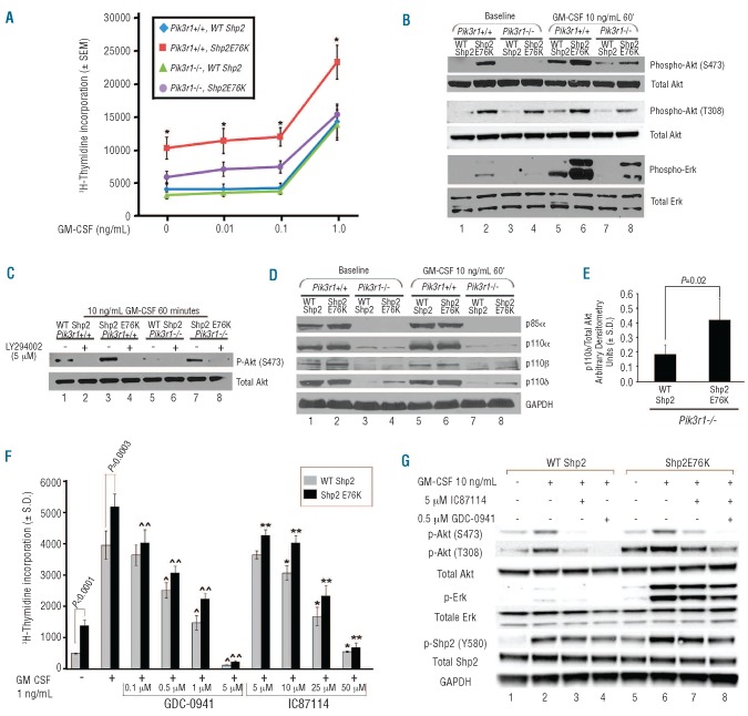
Figure 2.
Ablation of p85α, p55α, and p50α and inhibition with PI3K catalytic isoform-specific inhibitors normalizes gain-of-function Shp2-induced GM-CSF hypersensitivity. (A) Day 14.5 WT or Pik3r1−/− fetal liver cells were transduced with WT Shp2 or Shp2 E76K and subjected to [3H]-thymidine incorporation assays; 3 independent experiments combined with n=3 replicates per experiment, *P<0.01 for Pik3r1+/+, Shp2 E76K (red) versus Pik3r1−/−, Shp2 E76K (purple) at each concentration of GM-CSF; statistics performed using Prentice’s rank sum test for replicated block data.19 (B) Immunoblots demonstrating reduced phospho-Akt and phospho-Erk in Shp2 E76K-expressing Pik3r1−/− cells compared to Pik3r1+/+ cells, experiment repeated on 3 independent occasions. (C) Immunoblots demonstrating LY294002-mediated inhibition of Akt activation in Shp2 E76K-expressing Pik3r1−/− cells, experiment repeated on 2 independent occasions. (D) Immunoblots examining p110α, p110β, and p110δ levels in WT Shp2- and Shp2 E76K-expressing Pik3r1+/+ and Pik3r1−/− cells. (E) Quantitation using den-sitometry of immunoblot analyses comparing p110δ levels in WT Shp2- and Shp2 E76K-expressing Pik3r1−/− cells normalized to total Akt expression; n=5, P=0.02, statistics performed using unpaired, two-tailed students’ t test. (F) Proliferation of WT Shp2- and Shp2 E76K-transduced bone marrow LDMNCs in response to GM-CSF 1 ng/mL in the presence of increasing concentrations of GDC-0941 or of IC87114 (p110δ-specific); representative of 2 independent experiments, n=4–5, ^P=0.001, ^P<0.0001, and ^P<0.0001 comparing WT Shp2-expressing cells in the absence to the presence of GDC-0941 0.5 μM, 1 μM, and 5 μM, respectively; ^^P=0.002, ^^P<0.0001, ^^P<0.0001, and ^^P<0.0001 comparing Shp2 E76K-expressing cells in the absence to the presence of GDC-0941 0.1 μM, 0.5 μM, 1 μM, and 5 μM, respectively; *P=0.005, *P<0.0001, and *P<0.0001 comparing WT Shp2-expressing cells in the absence to the presence of IC87114 10 μM, 25 μM, and 50 μM, respectively; **P=0.002, **P=0.0005, **P<0.0001, and **P<0.0001 comparing Shp2 E76K-expressing cells in the absence to the presence of IC87114 5 μM, 10 μM, 25 μM, and 50 μM, respectively; statistics performed using unpaired, two-tailed students’ t-test. (G) Immunoblots demonstrating reduced phospho-Akt, phospho-Erk, and phospho-Shp2 in Shp2 E76K-expressing cells treated with 0.5 μM GDC-0941 or 5 μM IC87114, experiment repeated on 2 independent occasions.

