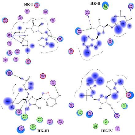
Figure 6.

Molecular docking of ATP showing binding mode variations in the kinase domains of HKs. Arrow marks indicate hydrogen bonding between ATP and kinase domain residues and blue shaded region indicates the solvent contacts made by ATP.

Molecular docking of ATP showing binding mode variations in the kinase domains of HKs. Arrow marks indicate hydrogen bonding between ATP and kinase domain residues and blue shaded region indicates the solvent contacts made by ATP.