Figure 5.
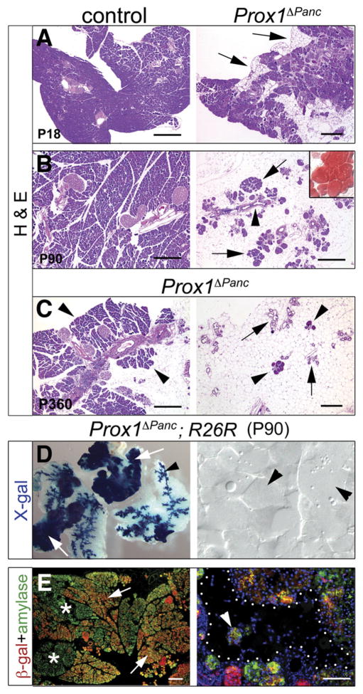
Loss of acinar tissue and lipomatosis in Prox1-deficient pancreata. (A) Prox1ΔPanc pancreata experience gradual loss of acinar tissue (arrows) starting at approximately P15–P18. (B) By P90, an extensive mass of adipocytes (oil red O+; inset) appears, engulfing some ducts (arrowhead) and a few small acini (arrows) in the mutant pancreas. Similar features are also observed in the pancreas of older Prox1ΔPanc mice (C, right), although these animals always retain a few intact acinar lobes (C, arrowheads, left). Lineage tracing results of P90 Prox1ΔPanc;R26R pancreata stained with X-gal (D) or with anti–β-gal and anti-amylase antibodies (E, asterisks show acini devoid of β-gal) support that both the remnants of acinar tissue (arrows, left) and the ducts (arrowhead, left) are derived from cells in which Prox1 was deleted. In contrast, X-gal staining (D, right, arrowheads) or immunohistochemistry results (E, right, dotted area; arrowhead shows an acinus) rule out that the adipocyte infiltrates of Prox1-deficient pancreata originate from pancreatic epithelial cells. Scale bar = 50 μm (A–C), 100 μm (E, right), and 200 μm (E, left).

