Abstract
Background
Patients with COPD use a higher proportion of their peak aerobic capacity during the performance of domestic activities of daily life (ADLs) compared to healthy peers, accompanied by a higher degree of task-related symptoms. To date, the influence of body mass index (BMI) on the task-related metabolic demands remains unknown in patients with COPD. Therefore, the aim of our study was to determine the effects of BMI on metabolic load during the performance of 5 consecutive domestic ADLs in patients with COPD.
Methodology
Ninety-four COPD patients and 20 healhty peers performed 5 consecutive, self-paced domestic ADLs putting on socks, shoes and vest; folding 8 towels; putting away groceries; washing up 4 dishes, cups and saucers; and sweeping the floor for 4 min. Task-related oxygen uptake and ventilation were assessed using a mobile oxycon, while Borg scores were used to assess task-related dyspnea and fatigue.
Principal Findings
1. Relative task-related oxygen uptake after the performance of domestic ADLs was increased in patients with COPD compared to healthy elderly, whereas absolute oxygen uptake is similar between groups; 2. Relative oxygen uptake and oxygen uptake per kilogram fat-free mass were comparable between BMI groups; and 3. Borg symptom scores for dyspnea en fatigue were comparable between BMI groups.
Conclusion
Patients with COPD in different BMI groups perform self-paced domestic ADLs at the same relative metabolic load, accompanied by comparable Borg symptom scores for dyspnea and fatigue.
Introduction
Chronic obstructive pulmonary disease (COPD) is not only characterised by pathological changes of the respiratory system, but has also significant systemic consequences like involuntary weight loss and skeletal muscle dysfunction [1], [2]. These systemic changes adversely affect patients exercise performance, quality of life and prognosis [2].
Involuntary weight loss and muscle wasting have traditionally been an active field of interest in patients with COPD, but overweight and obesity are increasingly common [1], [3]. An overweight or obese body mass index (BMI, kg/m2) is strongly associated with a decreased compliance of the respiratory system, a restrictive lung function, increased airway resistance, and increased work of breathing [1]. This can result in a worsening of dyspnea, increased physical limitations and decreased quality of life [1]. On the other hand, being overweight or obese has been associated with less osteoporosis [4] and a decreased mortality among patients with severe COPD; a phenomenon commonly referred to as the ‘obesity paradox’ [1], [2].
Functional exercise performance and physical activity levels have been shown to be reduced in patients with COPD compared to healthy subjects, even in the earliest stage of the disease [3], [5]–[9]. Moreover, overweight and obese COPD patients have a significantly shorter six minute walk distance compared to normal and low BMI patients [10], [11]. However, walk-work (the product of six minute walk distance in meters and body weight in kilogram) is similar between obese en non-obese COPD patients [10]. This suggests that the reduced functional exercise performance reflects, at least in part, the increased work burden of overweight and obesity. Indeed, obesity has been associated with reduced physical activity levels in patients with COPD, irrespective of the degree of lung function impairment [12].
Patients with COPD use a higher proportion of their peak aerobic capacity during the performance of simple domestic ADLs compared to healthy peers, accompanied by a higher degree of task-related dyspnea and fatigue [7], [13]–[16]. These studies did not study the possible effects of BMI on task-related oxygen uptake and symptoms in patients with COPD. Therefore, the influence of BMI on the task-related metabolic demands remains unknown in patients with COPD. Nevertheless, earlier was already shown that oxygen uptake and the ventilatory response are dependent on the corporal size. So, it seems reasonable to hypothesize that patients with low or normal BMI have a lower task-related oxygen uptake and ventilation during domestic activities in daily life compared to overweight and obese COPD patients. This is probably accompanied by lower Borg symptom scores, due to lower metabolic and ventilatory requirements during weight-bearing activities of daily life [10], [11]. Therefore, the aim of our study was to determine the effects of BMI on metabolic load during the performance of 5 consecutive domestic activities of daily life in patients with COPD.
Methods
One hundred COPD patients and 25 healthy peers volunteered to participate in the present study, which was approved by the institutional review board of the Maastricht University Medical Centre (MEC08-3-032). Patients were recruited at CIRO + in Horn (the Netherlands) before the start of a comprehensive pulmonary rehabilitation program [17] and had not participated in clinical studies before [7]. Healthy volunteers were recruited through posters and among healthy subjects who had participated in previous trials. At enrolment, written informed consent was obtained from potential participants and study eligibility was determined.
Patients were clinically stable (no exacerbation in past 4 weeks) and were on various (non-) pulmonary drug therapies (Table S1). None of the healthy subjects used physician-prescribed drugs. Exclusion criteria were the use of long-term oxygen therapy, neuromuscular co-morbidities, a cardiac pacemaker and/or an implantable cardioverter defibrillator and the disability to perform the domestic ADLs were included in the study. All tests were in accordance with the World Medical Association declaration of Helsinki.
Clinical Phenotyping
General demographics, co-morbidities, post-bronchodilator pulmonary function, dyspnea, body composition and peak aerobic capacity were assessed as described previously [14], [18]. Co-existing morbidities were scored using the Charlson Comorbidity Index and the Charlson Age Comorbidity Index, as described before [7], [19], [20]. The scores were based on patients’ self-reported co-morbidities and data from medical records.
Domestic ADLs
Participants performed 5 domestic ADLs in the kitchen of the Dept. of Occupational Therapy at CIRO + as described before [7]: putting on 2 socks (sitting in chair), 2 shoes (sitting in chair) and a vest (standing, ADL1); folding 8 towels (standing, ADL2); putting away groceries (e.g., 6 cans of beans of 400 grams each) in a cupboard (standing and walking, ADL3); washing up 4 dishes, 4 cups and 4 saucers (standing, ADL4); and sweeping the floor for 4 minutes (standing and walking, ADL5). These ADLs have all be identified as problematic valued life activities in patients with COPD [9], [21]. Participants were asked to perform ADLs at their own pace; and all ADLs were performed consecutively and, unlike to our previous study [7], without four minute rest intervals between ADLs.
Online breath-by-breath calculations of oxygen uptake and ventilation were recorded using a mobile oxycon (Oxycon Mobile, CareFusion, San Diego, CA, USA) which provides reliable measurements of oxygen uptake and ventilation. A mobile oxycon has been used before in patients with moderate to very severe COPD and in patients with chronic heart failure without adverse events [7], [15], [22]–[24].
Heart rate was monitored using a Polar belt. Moreover, all participants were asked to score the degree of dyspnea and fatigue at the beginning ADL1 and at the end of all ADLs using a modified Borg symptom score ranging from 0 (no symptoms) to 10 points (worst symptoms). For unknown reasons a download of the data failed in 6 patients and 5 healthy subjects. Therefore, the final group comprised 94 COPD patients and 20 healthy subjects.
A priori, relative task-related oxygen uptake during ADLs (ie, task-related oxygen uptake expressed as a proportion of the pre-determined peak aerobic capacity) was chosen as the primary outcome. Task-related oxygen uptake (mL/min) per kilogram fat-free mass (FFM, using whole-body dual-energy x-ray absorptiometry (DEXA, GE Medical Systems Lunar Prodigy, Madison, USA)), task-related ventilation (expressed as a proportion of pre-determined peak ventilation), time to accomplish the first four ADLs (ADL5 was always set at 4 min), and Borg symptom scores for dyspnea and fatigue at the end of each ADL were chosen as secondary outcomes.
Statistics
Data are presented as mean and standard deviation, unless noted otherwise. Student’s t-test was used to determine differences between patients with COPD and healthy subjects. Patients were divided according to BMI: low BMI: <21 kg/m2; normal BMI: 21 kg/m2 to 25 kg/m2; overweight BMI: >25 to 30 kg/m2; or obese BMI: >30 kg/m2. Differences between BMI groups were compared using analysis of variance (ANOVA) and Bonferroni post hoc comparisons. Chi squared (with Cramer’s V measure for strength of association) was used to assess the differences in gender distribution between groups. A priori, the level of significance was set at ≤0.05. Data were analyzed with SPSS, version 19.0.
Results
Characteristics
Subject characteristics are listed in table 1. On average, patients had mild to very severe COPD, a normal BMI and a poor exercise capacity. Patients had a significantly lower BMI and FFM compared to healthy subjects. Moreover, patients had higher scores on the (age-adjusted) Charlson Comorbidity Index. Most common co-morbidities were cardiovascular diseases. Furthermore, 19.6% of the COPD patients had a history of orthopaedic problems. Present comorbidities had no significant effect on exercise tolerance.
Table 1. Characteristics.
| Healthy subjects (n = 20) | COPD patients (n = 94) | |
| Men (%) | 60 | 61 |
| Age (years) | 62.1 (5.6) | 60.4 (9.3) |
| FEV1 (L) | 3.34 (0.61) | 1.42 (0.57)† |
| FEV1 (% predicted) | 119.4 (24.2) | 51.2 (19.1)† |
| FEV1/FVC (%) | 76.8 (4.6) | 42.5 (13.0)† |
| TLCO (%) | – | 56.7 (19.8) |
| PaO2 (kPa)* | – | 9.75 (1.24) |
| PaCO2 (kPa) | – | 5.20 (0.66) |
| SaO2 (%) | – | 95.2 (2.1) |
| GOLD stage I/II/III/IV (n) | – | 5/36/37/16 |
| MRC grade 1/2/3/4/5 (n) | – | 7/28/35/12/12 |
| BODE score (points) | – | 2.88 (1.89) |
| Body weight (kg)** | 79.2 (12.3) | 69.8 (17.3)† |
| Height (cm) | 171.1 (7.7) | 168.4 (9.7) |
| Body mass index (kg/m2) | 27.0 (3.0) | 24.5 (5.1)† |
| FFM (kg) | 55.6 (9.9) | 45.5 (9.1)† |
| FFMI (kg/m2) | 18.9 (2.3) | 16.0 (2.3)† |
| Women <15 kg/m2 (%) | 25.0 | 62.2 |
| Men <16 kg/m2 (%) | 0.0 | 45.5 |
| Charlson CMI (points) | 0.0 | 2.0 (1.4)† |
| Charlson CMI 2 (points) | 1.7 (0.7) | 3.7 (2.0)† |
| ≥1 co-morbidities (%) | 0.0 | 56.1† |
| Peak VO2 (mL/min) | 2155 (770) | 1202 (360)† |
| Peak VO2 (mL/min/kg BW) | 27.5 (10.2) | 17.5 (4.4)† |
| Peak VO2 (mL/min/kg FFM) | 38.8 (12.2) | 26.4 (6.0)† |
| Peak VE (L) | 87.3 (28.2) | 47.4 (14.6)† |
| Peak VE (% MVV) | 65.4 (17.1) | 89.8 (26.1)† |
| Peak HR (bpm) | 154 (18.9) | 132 (20.0)† |
| Peak HR (% max HR) | 97.5 (10.7) | 83.0 (11.5)† |
| Borg dyspnoea (points) | 5.5 (2.2) | 6.6 (2.1)† |
| Borg fatigue (points) | 5.7 (2.2) | 5.9 (2.2) |
Results are presented as mean (standard deviation). FEV1 = forced expiratory volume in the first second; L = litre; FVC = forced vital capacity; kg = kilogram; kg/m2 = kilogram per squared meters; Charlson CMI = Charlson co-morbidity index [19]; Charlson CMI 2 = Charlson age comorbidity index [20]; 6MWD = six-minute walking distance; VO2 = oxygen uptake; mL = millilitre; min = minute; BW = body weight; FFM = fat free mass; VE = minute ventilation; MVV = maximum voluntary ventilation; HR = heart rate; bpm = beats per minute. *1 kPa = 7.5 mm Hg; **1 kilogram = 2.2046 pounds; †p≤0.05 vs. healthy subjects.
Patients with COPD had a significantly lower peak aerobic capacity (mL/min), peak heart rate (beats/min), and peak ventilation (L) than healthy subjects. Peak Borg dyspnea scores of patients were significantly higher than those of healthy subjects, whereas peak Borg fatigue scores were comparable between groups (Table 1).
Characteristics of the patients with COPD per BMI group are listed in table 2. Results showed no significant differences in lung function between BMI groups; while, obviously, there were significant differences in body composition. Peak aerobic capacity (mL/min) was significantly lower in low BMI patients compared to those with an overweight and obese BMI. Peak ventilation, peak heart rate, and peak Borg symptom scores were comparable between BMI groups (Table 2).
Table 2. Baseline characteristics by BMI categories.
| <21 kg/m2 (n = 24) | 21–25 kg/m2 (n = 31) | 25–30 kg/m2 (n = 26) | >30 kg/m2 (n = 13) | |
| Men (%) | 50 | 45 | 77*+ | 85*+ |
| Age (years) | 57.2 (7.8) | 59.5 (9.1) | 66.3 (8.7)†+ | 56.8 (8.8)# |
| FEV1 (L) | 1.25 (0.61) | 1.37 (0.52) | 1.54 (0.64) | 1.59 (0.44) |
| FEV1 (% predicted) | 44.8 (19.9) | 51.7 (18.3) | 56.9 (20.1) | 50.5 (15.1) |
| FEV1/FVC (%) | 39.2 (14.7) | 41.0 (11.1) | 44.3 (13.4) | 48.5 (12.0) |
| TLCO (%) | 53.3 (18.6) | 54.4 (19.3) | 57.3 (20.4) | 61.1 (19.0) |
| PaO2 (kPa)* | 9.79 (1.41) | 9.65 (1.05) | 9.77 (1.17) | 9.85 (1.56) |
| PaCO2 (kPa) | 5.33 (0.92) | 5.14 (0.56) | 5.11 (0.53) | 5.34 (0.62) |
| SaO2 (%) | 94.7 (3.2) | 95.4 (1.6) | 95.4 (1.5) | 95.3 (1.8) |
| GOLD stage I/II/III/IV (n) | 1/6/6/11 | 1/14/13/3 | 2/11/11/2 | 1/5/6/1 |
| MRC grade 1/2/3/4/5 (n) | 3/5/9/4/3 | 2/10/11/5/3 | 1/9/10/3/3 | 1/4/6/1/1 |
| BODE score (points) | 4.2 (2.0) | 2.4 (1.4)† | 2.4 (2.0)† | 2.5 (1.5)† |
| Body weight (kg)** | 52.6 (7.9) | 64.9 (7.7)† | 76.7 (11.0)†+ | 99.7 (7.8)†+# |
| Height (cm) | 167.0 (9.2) | 167.7 (9.7) | 168.5 (10.5) | 172.4 (8.6) |
| Body mass index (kg/m2) | 18.8 (1.7) | 23.0 (1.0)† | 26.9 (1.4)†+ | 33.7 (3.6)†+# |
| FFM (kg) | 39.0 (5.4) | 42.9 (5.7) | 48.5 (8.8)†+ | 59.3 (6.7)†+# |
| FFMI (kg/m2) | 14.0 (1.2) | 15.2 (1.3)† | 17.0 (1.6)†+ | 20.0 (1.5)†+# |
| Women <15 kg/m2 (%) | 75.0 | 76.5 | 16.7 | 0.0 |
| Men <16 kg/m2 (%) | 91.7 | 71.4 | 20.0 | 0.0 |
| Charlson CMI (points) | 1.7 (0.9) | 1.9 (1.1) | 2.5 (1.8) | 2.1 (1.9) |
| Charlson CMI 2 (points) | 3.0 (1.5) | 3.4 (1.8) | 4.8 (2.2)† | 3.4 (2.6) |
| ≥1 co-morbidities (%) | 57.6 | 55.6 | 56.7 | 53 |
| 6MWD (m) | 487 (111) | 500 (96) | 483 (114) | 485 (108) |
| Peak VO2 (mL/min) | 1009 (306) | 1172 (330) | 1290 (343)† | 1458 (374)† |
| Peak VO2 (mL/min/kg BW) | 19.2 (4.9) | 18.0 (4.3) | 16.8 (3.6) | 14.6 (3.7)† |
| Peak VO2 (mL/min/kg FFM) | 25.7 (6.1) | 27.3 (6.5) | 26.7 (5.2) | 24.7 (6.1) |
| Peak VE (L) | 42.6 (11.2) | 45.7 (15.6) | 50.6 (15.3) | 53.8 (14.1) |
| Peak VE (% MVV) | 96.9 (31.3) | 83.5 (17.5) | 91.5 (29.0) | 88.0 (25.4) |
| Peak HR (bpm) | 130 (17.8) | 137 (19.8) | 130 (19.4) | 131 (25.5) |
| Peak HR (% max HR) | 80.1 (10.8) | 85.2 (10.3) | 84.7 (12.5) | 79.8 (12.6) |
| Borg dyspnoea (points) | 6.5 (2.5) | 7.2 (2.0) | 6.2 (1.7) | 6.2 (2.4) |
| Borg fatigue (points) | 5.6 (2.5) | 6.4 (2.6) | 5.6 (1.6) | 5.6 (2.0) |
Results are presented as mean (standard deviation). FEV1 = forced expiratory volume in the first second; L = litre; FVC = forced vital capacity; kg = kilogram; kg/m2 = kilogram per squared meters; Charlson CMI = Charlson co-morbidity index [19]; Charlson CMI 2 = Charlson age comorbidity index [20]; 6MWD = six-minute walking distance; VO2 = oxygen uptake; mL = millilitre; min = minute; BW = body weight; FFM = fat free mass; VE = minute ventilation; MVV = maximum voluntary ventilation; HR = heart rate; bpm = beats per minute. *1 kPa = 7.5 mm Hg; **1 kilogram = 2.2046 pounds; †p<0.05 vs. <21 kg/m2 +p<0.05 vs. 21–25 kg/m2 #p<0.05 vs. 25–30 kg/m2.
Domestic ADLs
Patients with COPD versus healthy subjects
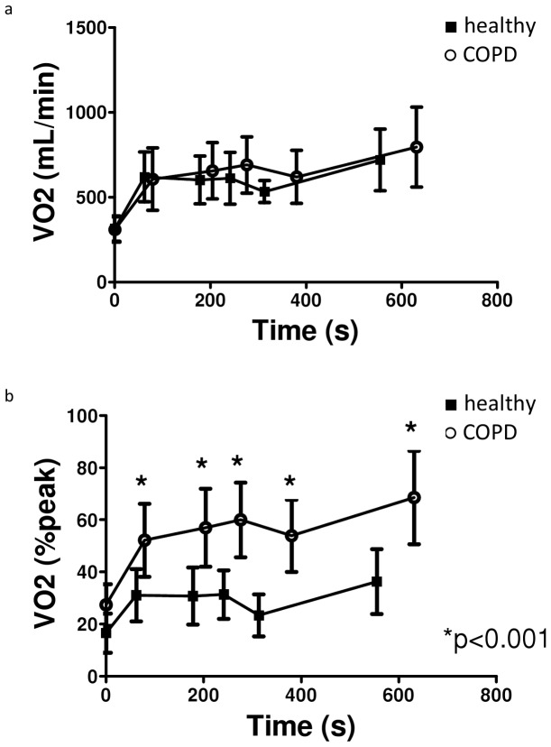
Basal values before performance of ADL-protocol as well as absolute task-related oxygen uptake (mL/min) at the end of the 5 ADLs were similar between COPD patients and healthy peers (Figure 1a). Consequently, patients performed the domestic ADLs at a significantly higher proportion of their peak aerobic capacity compared to healthy subjects: mean difference (95%CI) ADL1∶21.1 (14.6–27.6)%; ADL2∶26.2 (19.2–33.2)%; ADL3∶28.5 (21.8–35.3)%; ADL4∶30.4 (24.1–36.8)% and ADL5∶32.4 (24.0–40.7)%; all p<0.001 (Figure 1b). See Text S1.
Figure 1. Task-related oxygen uptake in patients with COPD and healthy controls.
a. Absolute task-related oxygen uptake (mL/min). b. Relative task-related oxygen uptake (%peakVO2).
COPD patients stratified by body mass index
All patients were able to complete ADL1 to ADL4 without stops, with no significant differences in time to complete the domestic tasks between BMI groups (figure S2). Thirty patients with COPD needed 20 to 145 s rest before they were able to start ADL5, while seven patients were not able to complete ADL5 due to exertional dyspnea. No differences were found between BMI groups. Moreover, there are no significant difference in task-related metabolic load between patients who did rest before start of ADL 5 and patients who did not.
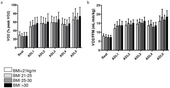
Task-related oxygen uptake expressed as a percentage of peak aerobic capacity was comparable between BMI groups at rest and after performance of all ADLs (Figure 2a). Moreover, oxygen uptake per kilogram FFM was also comparable between BMI groups for all ADLs (Figure 2b).
Figure 2. Task-related oxygen uptake in patients with COPD.
a. Relative task-related oxygen uptake (%peakVO2). b. Task-related oxygen uptake per kilogram FFM (mL/min/kg FFM).
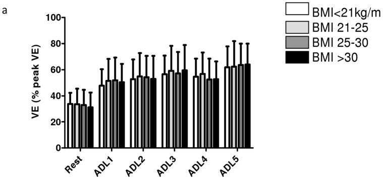
Relative task-related ventilation (ventilation expressed as a proportion of the pre-determined peak ventilation) was comparable between BMI groups at rest and after performance for all ADLs (Figure 3a).
Figure 3. Task-related ventilation in patients with COPD.
a. Relative task-related ventilation (%peakVE).
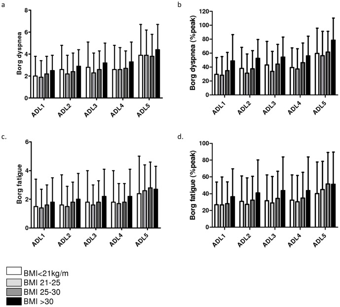
Although Borg scores for dyspnea appeared lower in low BMI patients compared to obese COPD patients, these differences were non-significant (Figure 4a). Moreover, relative Borg dyspnea scores (expressed as percentage of peak Borg scores during CPET) were comparable between BMI groups (Figure 4b). Borg scores for fatigue (absolute en relative) were also comparable between BMI groups (Figure 4c and 4d).
Figure 4. Borg symptom scores after performance of domestic ADLs.
a. absolute Borg dyspnea scores. b. relative Borg dyspnea scores (%peak). c. absolute Borg fatigue scores. d. relative Borg fatigue scores (%peak).
Discussion
Our results demonstrate differences in relative task-related oxygen uptake after the performance of five consecutive domestic ADLs between patients with COPD and healthy peers, whereas absolute oxygen uptake is similar between groups. Moreover, the present study is the first to demonstrate that relative oxygen uptake, oxygen uptake per kilogram FFM and relative ventilation during the performance of domestic ADLs were comparable between COPD patients with a low, normal, overweight or obese BMI. Moreover, Borg symptom scores for dyspnea en fatigue were comparable between BMI groups.
Previously was already reported that relative task-related oxygen uptake after the performance of domestic ADLs was increased in patients with COPD compared to healthy elderly [7], [13], [15], [16]. Our results show similar results between patients with COPD and healthy subjects during five consecutive ADLs. This may explain why patients with COPD experience more limitations in their daily life [9].
An increased BMI is associated with an increased work of breathing, increased breathing resistive load and worse exertional dyspnea [1]. On the other hand, obesity is associated with decreased resting and dynamic lung volumes [25], [26], which would positively affect exercise performance and perceived dyspnea. Indeed, overweight and obese patients with COPD did not experience greater dyspnea and exercise limitation during cycle ergometry compared to normal weight patients with comparable degree of airflow limitation [10], [27]. We also found no significant differences in dyspnea after performance of domestic ADLs between obese and non-obese COPD patients (Figure 4).
Previous studies showed that underweight COPD patients, particularly those with an abnormal low FFM, have an impaired peripheral muscle function and a poor exercise capacity [28]. Therefore, underweight patients may experience more limitations during the performance of domestic ADLs compared to normal weight COPD patients. In the presents study, underweight COPD patients had a significantly lower absolute oxygen uptake and ventilation during the performance of domestic ADLs compared to overweight or obese patients. This may indicate that obese and overweight patients experience more functional impairments in their daily life, probably caused by increases in the energetic cost of moving heavier limbs during activities and increased work of breathing [1], [25], [29], [30]. However, differences in task-related oxygen uptake and ventilation between BMI groups disappeared after correction for body weight or fat-free mass. These findings are corroborated by an a posteriori analysis of a previous study in which task-related oxygen uptake was assessed during five separate ADLs in 97 patients with COPD [7] (see Text S1 for details).
Patients with COPD use a higher proportion of their peak aerobic capacity and peak ventilation to perform domestic ADLs as compared to healthy subjects, accompanied by higher task-related Borg dyspnea scores [7], [13], [15], [16]. Moreover, task-related dynamic hyperinflation will contribute to a worsening of the task-related dyspnea in patients with COPD [22], Because of the reduced static and dynamic hyperinflation in overweight and obese COPD patients it was conceivable that these patients would experience less dyspnea during the performance of ADL compared to normal and underweight COPD patients. However, in the present study non-significant differences in task-related dyspnea were found after stratification for BMI (figure 4). This indicates that the burden of obesity restricts patients with COPD during the performance of ADL and offsets the potential beneficial influence of lower resting lung volumes.
Several methodological limitations need to be addressed. Due to the portable metabolic system we were not able to measure patients with long-term oxygen therapy, who may even experience more problematic ADLs compared to those without long-term oxygen therapy [31]. Moreover, we did not measure dynamic hyperinflation. Nevertheless, dynamic hyperinflation generally occurs during the performance of ADLs in patients with COPD [22], [32].
Fifty-six percent of the COPD patients reported ≥1 co-morbidities, which may have influenced the performance of ADLs. However, proportion of patients with co-morbidities was comparable between BMI groups and we did not found differences in task-related oxygen uptake, ventilation and dyspnea between COPD patients with and without co-morbidities (see Text S1 for details). This strengthens the validity of the current findings.
Stratification for BMI resulted in an unequal distribution over BMI groups. Indeed, the number of obese COPD patients seems rather low. However, the percentage of obese COPD patients in our study is comparable with the prevalence of obesity in a large primary care population of patients with COPD [33]. Moreover, the BMI groups were not comparable at baseline. However, earlier was already shown that there are no gender- or age-related differences in the metabolic demands during the performance of domestic ADLs (Vaes AW, Wouters EF, Franssen FM, Uszko-Lencer NH, Stakenborg KH, Westra M, et al. Task-Related Oxygen Uptake During Domestic Activities of Daily Life in Patients With COPD and Healthy Elderly Subjects. Chest. 2011;140(4)). Furthermore, there are no significant differences in age between patients with an obese, normal and low BMI. Therefore, the highest absolute task-related oxygen uptake and ventilation in the obese COPD group can not be explained by age-related differences.
In our study we focused on BMI, but we did not have information on fat distribution. It would be interesting to investigate whether central versus peripheral obesity exert similarly influence on the physiological response during performance of domestic ADLs.
Finally, the current findings need to be interpreted in the light of the number of comparisons that were made in the present study [34]. Nonetheless, multiple findings in the same direction, rather than a single statistically significant result, suggest that these are not due to chance alone.
To conclude, despite the additional mechanical load of obesity, relative task-related oxygen uptake and ventilation, oxygen uptake per kilogram FFM and Borg symptom scores for dyspnea and fatigue were comparable between patients with low, normal, overweight and obese BMI. Therefore, patients in different BMI groups experience similar functional limitations in the performance of domestic ADLs.
Supporting Information
Results of patients with COPD and healthy elderly subjects after performance of 5 simple domestic activities of daily life.
(TIF)
Time to accomplish ADLs.
(TIF)
Results after performance of simple domestic activities of daily life in patients with COPD after stratification for BMI.
(TIF)
Pulmonary and non-pulmonary drugs.
(DOCX)
Effects of Body Mass Index on Task-Related Oxygen Uptake and Dyspnea During Activities of Daily Life in COPD.
(DOC)
Acknowledgments
The authors want to acknowledge the patients with COPD and the healthy subjects to volunteer to participate in the present study. Moreover, the authors are grateful to Lucie Fransen, Marco Akkermans, Jos Peeters, Annie van de Kruijs, and Irma Timmermans for planning all tests and/or gathering the data.
Footnotes
Competing Interests: The authors have declared that no competing interests exist.
Funding: This work was supported by Foundation ‘De Weijerhorst’. The funders had no role in study design, data collection and analysis, decision to publish, or preparation of the manuscript.
References
- 1.Franssen FM, O’Donnell DE, Goossens GH, Blaak EE, Schols AM. Obesity and the lung: 5. Obesity and COPD. Thorax. 2008;63:1110–1117. doi: 10.1136/thx.2007.086827. [DOI] [PubMed] [Google Scholar]
- 2.Schols AM, Slangen J, Volovics L, Wouters EF. Weight loss is a reversible factor in the prognosis of chronic obstructive pulmonary disease. Am J Respir Crit Care Med. 1998;157:1791–1797. doi: 10.1164/ajrccm.157.6.9705017. [DOI] [PubMed] [Google Scholar]
- 3.Monteiro F, Camillo CA, Vitorasso R, Sant’anna T, Hernandes NA, et al. Obesity and Physical Activity in the Daily Life of Patients with COPD. Lung. 2012. [DOI] [PubMed]
- 4.Graat-Verboom L, Wouters EF, Smeenk FW, van den Borne BE, Lunde R, et al. Current status of research on osteoporosis in COPD: a systematic review. Eur Respir J. 2009;34:209–218. doi: 10.1183/09031936.50130408. [DOI] [PubMed] [Google Scholar]
- 5.Spruit MA, Watkins ML, Edwards LD, Vestbo J, Calverley PM, et al. Determinants of poor 6-min walking distance in patients with COPD: the ECLIPSE cohort. Respir Med. 2010;104:849–857. doi: 10.1016/j.rmed.2009.12.007. [DOI] [PubMed] [Google Scholar]
- 6.Pitta F, Troosters T, Spruit MA, Probst VS, Decramer M, et al. Characteristics of physical activities in daily life in chronic obstructive pulmonary disease. Am J Respir Crit Care Med. 2005;171:972–977. doi: 10.1164/rccm.200407-855OC. [DOI] [PubMed] [Google Scholar]
- 7.Vaes AW, Wouters EF, Franssen FM, Uszko-Lencer NH, Stakenborg KH, et al. Task-Related Oxygen Uptake During Domestic Activities of Daily Life in Patients With COPD and Healthy Elderly Subjects. Chest. 2011;140:970–979. doi: 10.1378/chest.10-3005. [DOI] [PubMed] [Google Scholar]
- 8.Watz H, Waschki B, Meyer T, Magnussen H. Physical activity in patients with COPD. Eur Respir J. 2009;33:262–272. doi: 10.1183/09031936.00024608. [DOI] [PubMed] [Google Scholar]
- 9.Annegarn J, Meijer K, Passos VL, Stute K, Wiechert J, et al. Problematic Activities of Daily Life are Weakly Associated With Clinical Characteristics in COPD. J Am Med Dir Assoc. 2011. [DOI] [PubMed]
- 10.Bautista J, Ehsan M, Normandin E, Zuwallack R, Lahiri B. Physiologic responses during the six minute walk test in obese and non-obese COPD patients. Respir Med. 2011;105:1189–1194. doi: 10.1016/j.rmed.2011.02.019. [DOI] [PubMed] [Google Scholar]
- 11.Sava F, Laviolette L, Bernard S, Breton MJ, Bourbeau J, et al. The impact of obesity on walking and cycling performance and response to pulmonary rehabilitation in COPD. BMC Pulm Med. 2010;10:55. doi: 10.1186/1471-2466-10-55. [DOI] [PMC free article] [PubMed] [Google Scholar]
- 12.Watz H, Waschki B, Kirsten A, Muller KC, Kretschmar G, et al. The metabolic syndrome in patients with chronic bronchitis and COPD: frequency and associated consequences for systemic inflammation and physical inactivity. Chest. 2009;136:1039–1046. doi: 10.1378/chest.09-0393. [DOI] [PubMed] [Google Scholar]
- 13.Jeng C, Chang W, Wai PM, Chou CL. Comparison of oxygen consumption in performing daily activities between patients with chronic obstructive pulmonary disease and a healthy population. Heart Lung. 2003;32:121–130. doi: 10.1067/mhl.2003.20. [DOI] [PubMed] [Google Scholar]
- 14.Spruit MA, Pennings HJ, Janssen PP, Does JD, Scroyen S, et al. Extra-pulmonary features in COPD patients entering rehabilitation after stratification for MRC dyspnea grade. Respir Med. 2007;101:2454–2463. doi: 10.1016/j.rmed.2007.07.003. [DOI] [PubMed] [Google Scholar]
- 15.Lahaije A, van Helvoort H, Dekhuijzen P, Heijdra Y. Physiologic limitations during daily life activities in COPD patients. Respir Med. 2010;104:1152–1159. doi: 10.1016/j.rmed.2010.02.011. [DOI] [PubMed] [Google Scholar]
- 16.Velloso M, Stella SG, Cendon S, Silva AC, Jardim JR. Metabolic and ventilatory parameters of four activities of daily living accomplished with arms in COPD patients. Chest. 2003;123:1047–1053. doi: 10.1378/chest.123.4.1047. [DOI] [PubMed] [Google Scholar]
- 17.Spruit MA, Vanderhoven-Augustin I, Janssen PP, Wouters EF. Integration of pulmonary rehabilitation in COPD. Lancet. 2008;371:12–13. doi: 10.1016/S0140-6736(08)60048-3. [DOI] [PubMed] [Google Scholar]
- 18.Spruit MA, Franssen FME, Rutten EPA, Wagers SS, Wouters EFM, et al. Age-graded reduction in quadriceps muscle strength and peak aerobic capacity in COPD. Rev Bras Fisioter In press. 2011. [DOI] [PubMed]
- 19.Charlson ME, Pompei P, Ales KL, MacKenzie CR. A new method of classifying prognostic comorbidity in longitudinal studies: development and validation. J Chronic Dis. 1987;40:373–383. doi: 10.1016/0021-9681(87)90171-8. [DOI] [PubMed] [Google Scholar]
- 20.Charlson M, Szatrowski TP, Peterson J, Gold J. Validation of a combined comorbidity index. J Clin Epidemiol. 1994;47:1245–1251. doi: 10.1016/0895-4356(94)90129-5. [DOI] [PubMed] [Google Scholar]
- 21.Katz P, Chen H, Omachi TA, Gregorich SE, Julian L, et al. The Role of Physical Inactivity in Increasing Disability Among Older Adults With Obstructive Airway Disease. J Cardiopulm Rehabil Prev. 2010. [DOI] [PMC free article] [PubMed]
- 22.Hannink JD, van Helvoort HA, Dekhuijzen PN, Heijdra YF. Dynamic hyperinflation during daily activities: does COPD global initiative for chronic obstructive lung disease stage matter? Chest. 2010;137:1116–1121. doi: 10.1378/chest.09-1847. [DOI] [PubMed] [Google Scholar]
- 23.Spruit MA, Wouters EF, Eterman RM, Meijer K, Wagers SS, et al. Task-related oxygen uptake and symptoms during activities of daily life in CHF patients and healthy subjects. Eur J Appl Physiol. 2011;111:1679–1686. doi: 10.1007/s00421-010-1794-y. [DOI] [PMC free article] [PubMed] [Google Scholar]
- 24.Sillen MJ, Janssen PP, Akkermans MA, Wouters EF, Spruit MA. The metabolic response during resistance training and neuromuscular electrical stimulation (NMES) in patients with COPD, a pilot study. Respir Med. 2008;102:786–789. doi: 10.1016/j.rmed.2008.01.013. [DOI] [PubMed] [Google Scholar]
- 25.Ofir D, Laveneziana P, Webb KA, O’Donnell DE. Ventilatory and perceptual responses to cycle exercise in obese women. J Appl Physiol. 2007;102:2217–2226. doi: 10.1152/japplphysiol.00898.2006. [DOI] [PubMed] [Google Scholar]
- 26.Ora J, Laveneziana P, Wadell K, Preston M, Webb KA, et al. Effect of obesity on respiratory mechanics during rest and exercise in COPD. J Appl Physiol. 2011;111:10–19. doi: 10.1152/japplphysiol.01131.2010. [DOI] [PubMed] [Google Scholar]
- 27.Laviolette L, Sava F, O’Donnell DE, Webb KA, Hamilton AL, et al. Effect of obesity on constant workrate exercise in hyperinflated men with COPD. BMC Pulm Med. 2010;10:33. doi: 10.1186/1471-2466-10-33. [DOI] [PMC free article] [PubMed] [Google Scholar]
- 28.Baarends EM, Schols AM, Mostert R, Wouters EF. Peak exercise response in relation to tissue depletion in patients with chronic obstructive pulmonary disease. Eur Respir J. 1997;10:2807–2813. doi: 10.1183/09031936.97.10122807. [DOI] [PubMed] [Google Scholar]
- 29.Cecere LM, Littman AJ, Slatore CG, Udris EM, Bryson CL, et al. Obesity and COPD: Associated Symptoms, Health-related Quality of Life, and Medication Use. COPD. 2011. [DOI] [PMC free article] [PubMed]
- 30.Guenette JA, Jensen D, O’Donnell DE. Respiratory function and the obesity paradox. Curr Opin Clin Nutr Metab Care. 2010;13:618–624. doi: 10.1097/MCO.0b013e32833e3453. [DOI] [PubMed] [Google Scholar]
- 31.Sandland CJ, Morgan MD, Singh SJ. Patterns of domestic activity and ambulatory oxygen usage in COPD. Chest. 2008;134:753–760. doi: 10.1378/chest.07-1848. [DOI] [PubMed] [Google Scholar]
- 32.O’Donnell DE, Revill SM, Webb KA. Dynamic hyperinflation and exercise intolerance in chronic obstructive pulmonary disease. Am J Respir Crit Care Med. 2001;164:770–777. doi: 10.1164/ajrccm.164.5.2012122. [DOI] [PubMed] [Google Scholar]
- 33.Steuten LM, Creutzberg EC, Vrijhoef HJ, Wouters EF. COPD as a multicomponent disease: inventory of dyspnoea, underweight, obesity and fat free mass depletion in primary care. Prim Care Respir J. 2006;15:84–91. doi: 10.1016/j.pcrj.2005.09.001. [DOI] [PMC free article] [PubMed] [Google Scholar]
- 34.Perneger TV. What’s wrong with Bonferroni adjustments. BMJ. 1998;316:1236–1238. doi: 10.1136/bmj.316.7139.1236. [DOI] [PMC free article] [PubMed] [Google Scholar]
Associated Data
This section collects any data citations, data availability statements, or supplementary materials included in this article.
Supplementary Materials
Results of patients with COPD and healthy elderly subjects after performance of 5 simple domestic activities of daily life.
(TIF)
Time to accomplish ADLs.
(TIF)
Results after performance of simple domestic activities of daily life in patients with COPD after stratification for BMI.
(TIF)
Pulmonary and non-pulmonary drugs.
(DOCX)
Effects of Body Mass Index on Task-Related Oxygen Uptake and Dyspnea During Activities of Daily Life in COPD.
(DOC)


