

Case presentation
A 4-year-old, neutered male domestic shorthair was presented to a local veterinary clinic with a complaint of recent lethargy after a fight with another cat. The physical examination revealed a mild fever (40.0°C), 2 bite wounds localized in the lumbar region, and acute back pain near the left coxofemoral joint. The bladder was distended but painless. According to the owner, the animal had no history or clinical signs of lower urinary tract disease. The cat was hospitalized for 2 d; it received oral antibiotics and was then released because of significant clinical improvement. Eight days after the initiation of treatment, the cat became lethargic and occasional vomiting was reported by the owner. At that time, examination by the veterinarian revealed no significant findings, except a distended nonpainful bladder. Results from routine blood analyses (complete blood cell (CBC) count and biochemical panel) were within reference ranges. A urine sample was collected by cystocentesis to determine the cause of the enlarged bladder. The urine was submitted to the clinical pathology diagnostic service of the Faculty of Veterinary Medicine, University of Montreal, in Saint-Hyacinthe.
Urologic studies
The urine was clear and pale yellow with a specific gravity of 1.038. Results from a chemical analysis, using a reagent strip (Chemstrip 9; Roche Diagnostics, Laval, Quebec), indicated a pH of 6.0 and a mild amount of blood and proteins (0.3g/L); they were negative for glucose, ketone bodies, and bilirubin. Microscopic examination of the urinary sediment revealed a mild hematuria (20–50 erythrocytes/hpf, 400X), a low leukocyte count (0–3 leukocytes/hpf), and a large number of amorphous crystals. Several large oval structures were also seen at low magnification (Figure 1).
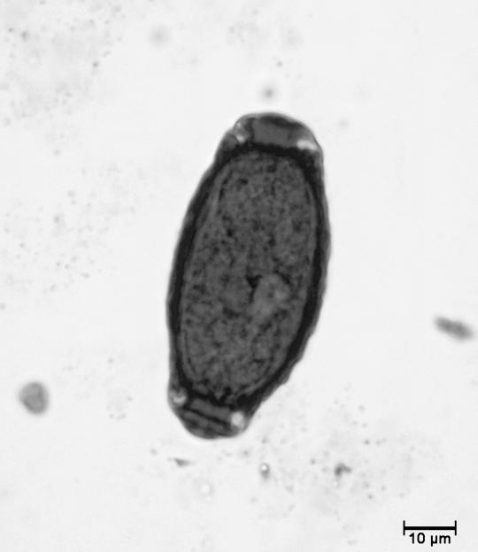
Figure 1. Photomicrograph of the urinary sediment showing the presence of a dark staining oval structure measuring approximately 25 by 60 μm (100X; Sedi-Stain, Becton Dickinson, Sparks, Maryland, USA ).
Description and interpretation
The oval structures seen in the urinary sediment were identified as Capillaria sp. eggs. Nematodes of the genus Capillaria comprise a very wide group of parasites found in all classes of vertebrates (1). The adult worms are typically attached to epithelial surfaces and, in dogs and cats, they have been associated with the intestinal and urinary tract, as well as the bronchi (1). Capillaria plica and C. feliscati are parasites rarely found in the bladder of dogs and cats, respectively (1). They are widely distributed and can be found in numerous domestic and wild carnivores (1,2).
Adult C. plica are embedded in the bladder epithelium and, occasionally, within the ureter or the renal pelvis, where they can cause mild inflammatory reaction and submucosal edema (3). Capillaria feliscati are essentially free on the surface of the bladder mucosa (4). Adult bladder worms are small threadlike parasites. Adult females measure 30 to 60 mm, males 13 to 30 mm long (2). Eggs are oval and colorless with a thick capsule and typical bipolar plugs (Figure 1). Their sizes range from 22 to 32 μm in width by 50 to 68 μm in length (3).
The life cycle of Capillaria bladder worms is not completely understood. Eggs released in the urine are not immediately infectious (1). In order to hatch and become first-stage larva, eggs must be ingested by an earthworm, which serves as a secondary host (1,2,3). The definitive host becomes infected after ingestion of earthworms or contaminated materials containing larva, likely accidentally ingested during grooming. Larva reside in the intestinal wall for a short period of time until they migrate to the bladder. Eggs usually appear in the urine 2 mo after the ingestion of the first-stage larva (2). In 3 infected dogs isolated in cages for 84 d, the daily number of eggs in the urine diminished until the eggs were undetectable (3).
Dogs and cats with urinary capillariasis usually do not show clinical signs (2). When heavily infested, they may exhibit signs of urinary tract disease: pollakiuria, dysuria, and inappropriate micturition (1,2,3). In foxes, retarded growth has also been reported (1). Urinalysis may reveal mild proteinuria, microscopic hematuria, and the presence of an increased number of transitional epithelial cells (1,3,4). The diagnosis is made by finding Capillaria eggs in the urinary sediment (2).
Infestations are usually self-limiting, but in presence of clinical signs, treatment may be necessary. One to several doses of 50mg/kg of fenbendazole, PO, have been suggested for the treatment of capillariasis in the dog (6). However, persistence of C. plica eggs in the urine of 1 dog treated with several doses of fenbendazole has been reported; this dog was then treated successfully with a single dose of 0.2 mg/kg ivermectin injected subcutaneously (SC) (7). The case reported here has been treated successfully with a single SC injection of ivermectin (0.2mg/kg). Two weeks after treatment, the microscopic examination of urinary sediment did not reveal the presence of Capillaria sp. eggs.
Cases of Capillaria bladder worms in dogs and cats are rarely reported, since most infected animals show no clinical signs, presumably because of a low parasite burden. The prevalence of Capillaria sp. in dogs and cats is, therefore, difficult to estimate. In Canada, the prevalence of capillariasis in cats is unknown but assumed to be low. To our knowledge, this is the first case reported in the province of Québec. The clinical signs reported here were not related to the Capillaria infection and the presence of eggs in the urine was an incidental finding.
Footnotes
Address correspondence and reprint requests to Dr. Christian Bédard; e-mail: christian.bedard@umontreal.ca.
References
- 1.Bowman DD. Georgis' Parasitology for Veterinarians. 7th ed. Philadelphia: WB Saunders, 1999:219–221.
- 2.Osborne CA, Delmar RF. Canine and Feline Nephrology and Urology. Baltimore: Williams & Wilkins, 1995:919–920.
- 3.Senior DF, Solomon GB, Goldschmidt MH, Joyce T, Bovee KC. Capillaria plica infection in dogs. J Am Vet Med Assoc 1980;176:901–905. [PubMed]
- 4.Wilson-Hanson S, Prescott CW. Capillaria in the bladder of the domestic cat. Aust Vet J 1982;59:190–191. [DOI] [PubMed]
- 5.Harris LT. Feline bladderworm. Vet Med Small Anim Clin 1981;76:844. [PubMed]
- 6.Gillepsie D. Successful treatment of canine Capillaria plica cystitis. Vet Med Small Anim Clin 1983;78:681–682.
- 7.Kirkpatrick CE, Nelson GR. Ivermectin treatment of urinary capillariasis in a dog. J Am Vet Med Assoc 1987;191:701–702. [PubMed]