Abstract
Granuloma faciale (GF) is a rare benign inflammatory dermatosis that usually develops as a solitary brownish-red plaque on the face. It clinically mimics and is often misinterpreted as, sarcoidosis, lupus erythematosus, lupus vulgaris, lymphoma or basal cell carcinoma.
Dermoscopy, which is valuable for evaluation and differentiation between malignant and benign skin tumors, allows better visualization of dermal vascular structures and color variations. In this context, it might serve as an adjuvant diagnostic tool in the differentiation of inflammatory disorders, too. In the current manuscript, we present the dermoscopic features observed in a lesion of GF and discuss them in correlation with the underlying histopathological alterations.
Keywords: dermoscopy, granuloma faciale
Case
Granuloma faciale (GF) is a rare benign chronic inflammatory dermatosis that usually develops as a solitary brownish-red infiltrated plaque on the face. It clinically mimics, and is often misinterpreted as, sarcoidosis, lupus erythematosus, lupus vulgaris, lymphoma or basal cell carcinoma.[1]
Dermoscopy allows better visualization of dermal vascular structures and color variations. Therefore, in recent years it is gaining appreciation in the era of inflammatory skin diseases.[2]
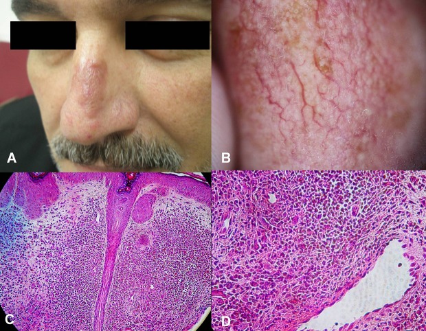
A 45-year-old man was referred to our clinic with a 5-month history of a solitary, stable in size, asymptomatic plaque on the nasal ridge. The patient did not report development of similar lesions in the past and his medical history was otherwise unremarkable. Clinical examination revealed a 1.5x2.5 cm infiltrated brownish-red plaque affecting the nasal ridge [Fig. 1A]. Prominent telangiectasias could easily be identified all over the surface of the lesion. Based on clinical findings, differential diagnosis included sarcoidosis, lupus vulgaris and basal cell carcinoma. Dermoscopically, the lesion exhibited dilated follicular openings and linear, slightly arborizing vessels in a parallel arrangement, crossing all over the surface of the plaque. Background color was red and scattered aggregations of brown dots/globules were observed [Fig. 1B]. Histopathology showed dilation of infundibula, dense admixed inflammatory cell infiltrate, with numerous eosinophils, sparing the dermal papillae (Grenz zone), and dilated venules throughout the upper half of the reticular dermis. Focal micro- haemorrhage and haemosiderin deposition was also observed [Fig. 1C,D].
Figure 1.
(A) Brownish-red plaque over the nasal ridge with telangiectasias and dilated follicular openings. (B) Arborizing vessels in a parallel arrangement, in the background of red color with scattered aggregations of brown dots/globules. (C) Dilation of infundibula, and mixed inflammation sparing the dermal papillae. (D) Numerous eosinophils and focal haemosiderin deposition close to a dilated dermal venule.
The dermoscopic features observed in our case of GF were in direct correlation to the underlying histopathologic alterations. Linear, slightly arborizing vessels in a parallel arrangement represented the predominant feature and are well known as one of the commonest vascular change taking place in GF in terms of histopathology. Dilated follicular openings have also been described as a common feature that may be useful in differentiating GF from BCC, sarcoidosis and cutaneous lymphoma.[3] The brown dots/globules, detected under dermoscopy, could possibly correspond to hemosiderin deposition which is known to be part of the pathophysiological process of GF.[3]
Taking under consideration, that evidence concerning the dermoscopic patterns of the clinical mimickers of GF, with the exception of BCC, is limited, applicability of dermoscopy in differentiating among these entities remains uncertain.
Arborizing vessels are considered the hallmark of BCC.[4] Similar vessels were observed under dermoscopy in our case of GF. However, compared to the typical arborizing vessels of BCC, in GF vessels appeared to be more prominent in terms of size and number, running in a parallel arrangement, while branching into smaller vessels and capillaries was less evident.
Sarcoidosis is dermoscopically characterized by translucent yellow to orange globular-like or structureless areas, possibly corresponding to the well defined sarcoid granulomas.[5] The latter finding is absent in GF.
Finally, dilated follicular openings that have already been proposed to enhance the differentiation of GF from its clinical simulators,4 are much more evident under dermoscopy.
In conclusion, dermoscopic findings in our case of GF were in direct correlation to the underlying histopathologic changes. In this context, dermoscopy could represent a valuable diagnostic tool that enhances clinical diagnosis. Our observations may motivate further research in the field of dermoscopy of inflammatory diseases such as GF.
References
- Thiyanaratnam J, Doherty SD, Krishnan B, Hsu S. Granuloma faciale: Case report and review. Dermatol Online J. 2009;15:3. [PubMed] [Google Scholar]
- Lallas A, Kyrgidis A, Tzellos TG, Apalla Z, Karakyriou E, Karatolias A, Lefaki I, Sotiriou E, Ioannides D, Argenziano G, Zalaudek I. Accuracy of dermoscopic criteria for the diagnosis of psoriasis, dermatitis, lichen planus and pityriasis rosea. Br J Dermatol. 2012;166:1198–1205. doi: 10.1111/j.1365-2133.2012.10868.x. [DOI] [PubMed] [Google Scholar]
- Ortonne N, Wechsler J, Bagot M, Grosshans E, Cribier B. Granuloma faciale: a clinicopathologic study of 66 patients. J Am Acad Dermatol. 2005;53:1002–1009. doi: 10.1016/j.jaad.2005.08.021. [DOI] [PubMed] [Google Scholar]
- Altamura D, Menzies SW, Argenziano G, Zalaudek I, Soyer HP, Sera F, Avramidis M, DeAmbrosis K, Fargnoli MC, Peris K. Dermatoscopy of basal cell carcinoma: Morphologic variability of global and local features and accuracy of diagnosis. J Am Acad Dermatol. 2010;62:67–75. doi: 10.1016/j.jaad.2009.05.035. [DOI] [PubMed] [Google Scholar]
- Pellicano R, Tiodorovic-Zivkovic D, Gourhant JY, Catricalà C, Ferrara G, Caldarola G, Argenziano G, Zalaudek I. Dermoscopy of cutaneous sarcoidosis. Dermatology. 2010;221:51–54. doi: 10.1159/000284584. [DOI] [PubMed] [Google Scholar]


