Abstract
Background:
For patients with newly diagnosed high-grade gliomas (HGG), the current standard-of-care treatment involves surgical resection, followed by concomitant temozolomide (TMZ) and external beam radiation therapy (XRT), and subsequent TMZ chemotherapy. For patients with recurrent HGG, there is no standard of care. Stereotactic radiosurgery (SRS) is used to deliver focused, relatively large doses of radiation to a small, precisely defined target. Treatment is usually delivered in a single fraction, but may be delivered in up to five fractions. The role of SRS in the management of patients with HGG is not well established.
Methods:
The PubMed database was searched with combinations of relevant MESH headings and limits. Case reports and/or small case series were excluded. Attention was focused on overall median survival as an objective measure, and data were examined separately for newly diagnosed and recurrent HGG.
Results:
With respect to newly diagnosed HGG, there is strong evidence that addition of an SRS boost prior to standard XRT provides no survival benefit. However, recent retrospective evidence suggests a possible survival benefit when SRS is performed after XRT. With respect to recurrent HGG, there is suggestion that SRS may confer a survival benefit but with potentially higher complication rates. Newer studies are investigating the combination of SRS with targeted molecular agents. Controlled prospective clinical trials using advanced imaging techniques are necessary for a complete assessment.
Conclusions:
SRS has the potential to provide a survival benefit for patients with HGG. Further research is clearly warranted to define its role in the management of newly diagnosed and recurrent HGG.
Keywords: Glioma, high-grade, newly diagnosed, recurrent, stereotactic radiosurgery
INTRODUCTION
Gliomas are primary malignant brain tumors that arise from glial cells, namely astrocytes and oligodendrocytes. The World Health Organization (WHO) has classified gliomas into four grades of ascending malignancy.[39] According to this classification, Grade III and Grade IV are the most aggressive and termed high-grade gliomas (HGG). Glioblastoma is a Grade IV glioma representing one of the most malignant and, at the same time, most common types of glioma.[39] The current standard of care is to treat glioblastoma patients with surgical resection, followed by temozolomide (TMZ) concomitant with external beam radiation (XRT), and then subsequently with additional TMZ cycles, according to the Stupp protocol.[68] Despite this treatment, patients have a median survival of 14.6 months and an overall survival of 27% at 2 years, that drops to under 10% at 5 years.[68] Analysis of treatment failure patterns has revealed that up to 80% of recurrences occurred within 2 cm of the tumor margins.[76] This was the basis for inclusion of a margin from the residual tumor and resection cavity, typically of 2–3 cm, when radiation treatment portals were designed. More recent data have demonstrated that now the majority of treatment failures are within the irradiated field, with the distribution affected by other factors, such as methylation status of the O6-methylguanine-DNA-methyltransferase promoter.[48] This pattern would suggest that a focal radiation delivery technique that intensifies the dose to a specific area with minimal toxicity to the surrounding areas could be beneficial in reducing treatment failures.
Stereotactic radiosurgery (SRS) refers to a technique of highly focused radiation delivery based on the use of stereotactic image guidance. Originally developed by the Swedish neurosurgeon Lars Leksell in the 1950s, SRS delivers high doses of radiation to a precisely defined target area with minimal toxicity outside the target area because of a steep dose gradient. Several systems are in use for SRS.[2,37] The first was the Gamma Knife (GK), developed by Leksell and based on the simultaneous delivery of gamma rays generated by the nuclear decay of multiple cobalt-60 sources converging on the target. Subsequent systems used linear particle accelerators (LINAC) where X-rays are generated from electron acceleration into a high-density material and then converge on the target. The latest evolution of LINAC-based systems includes the Novalis, with a multi-leaf collimator, and the Cyberknife (CBK), based on the concept of a compact LINAC mounted on a robotic arm. Specific technology aside, SRS is in widespread clinical use for a variety of intracranial pathologies. Although originally conceived as a single-fraction treatment, SRS may be divided in up to five fractions.[5] Delivery of stereotactic radiation in more than five fractions is considered stereotactic radiation therapy (SRT). In single-fraction SRS, the maximum tolerated doses range from a high of 24 Gy with target diameters less than 2 cm to a low of 15 Gy for target diameters ranging from 3 to 4 cm, which is generally considered to be the upper limit of target size.[60] The concept of fractionation allows larger target volumes to be safely irradiated and potentially higher doses delivered, while still maintaining the fundamental principles of SRS.[64]
The overall aim of this work was to review the existing literature on SRS for the treatment of HGG and provide insight into its current status. Since newly diagnosed and recurrent HGG represent distinct therapeutic challenges, consideration of SRS as a treatment modality for HGG will be examined separately for these two areas. An illustrative case is also presented.
MATERIALS AND METHODS
The PubMed database was searched using the following MESH headings and combinations: “radiosurgery,” “glioma,” high-grade glioma,” “glioblastoma,” “anaplastic astrocytoma.” Limits were set to the language “English” and species “Human” for broad inclusion of articles. Clinical case reports or small series where HGG did not constitute a majority of cases were excluded, as were the studies focusing on brainstem gliomas. Studies using the term SRT were included only if they met the definition of SRS.[5] For the articles that included both newly diagnosed and recurrent HGG, the data for each category was abstracted and presented in the designated category. Particular attention was given to median overall survival as an objective measure.
RESULTS AND DISCUSSION
Newly diagnosed high-grade gliomas
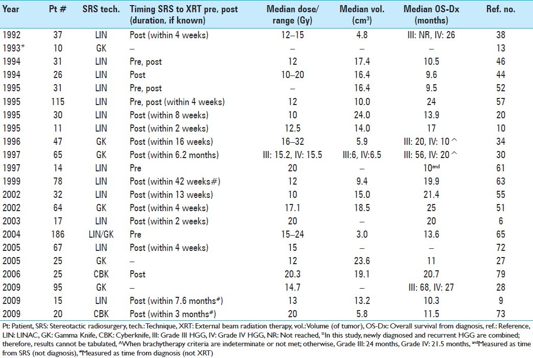
Over 20 clinical studies were identified in the literature[6,9,10,13,20,27,28,30,34,38,44,46,51,52,54,55,57,61,63,65,72,73,79] and are summarized chronologically in Table 1. Radiation Therapy Oncology Group (RTOG) 93-05 is the only randomized controlled trial (RCT) on this topic. This study tested the benefits of administering SRS before XRT with carmustine (bis-chloroethylnitrosourea or BCNU) in patients with glioblastoma.[65] A total of 186 patients were included: 97 were randomized to receive XRT and BCNU, while 89 were randomized to receive SRS 1 week prior to XRT and BCNU. Relevant eligibility criteria included age greater than 18 years, histopathologically proven diagnosis of supratentorial glioblastoma with no prior chemotherapy or radiation, tumor size less than 4 cm, and Karnofsky score greater than 60 with a life expectancy greater than 3 months. Exclusion criteria included histopathology of atypical and/or anaplastic astrocytoma and gross total resection (GTR) with no visible residual. The tumor dose delivered was volume dependent, ranging from 15 to 24 Gy, according to established maximum safely tolerated doses.[60] Of the patients in the SRS + XRT arm, 18% had unacceptable deviations from protocol, but were nonetheless included in the study. RTOG 93-05 found no significant difference in median overall survival or patterns of failure in patients with glioblastoma with the addition of SRS. Although this study provides Level I evidence against the use of SRS prior to XRT with BCNU, several important issues have been raised with respect to the applicability of these findings,[3,31,40,74] including timing of SRS (before or after XRT), type of chemotherapy (BCNU vs. TMZ), and extent of surgical resection. These limitations are reviewed and discussed below.
Table 1.
Studies of stereotactic radiosurgery as adjunct treatment for newly diagnosed high-grade gliomas

The RTOG 93-05 randomized study used SRS prior to XRT. As shown in Table 1, all the studies finding a favorable median overall survival ≥20 months performed SRS after XRT in the majority of patients, suggesting that this paradigm is more likely to have a favorable outcome. From a radiobiological perspective, the timing of SRS after XRT appears to be more advantageous based on the concepts of fractionation and cancer cell repopulation[25,29] such that high-dose radiation delivery within 4 weeks of the end of fractionated XRT should more effectively address the residual, but actively repopulating cancer cells. Although there are no radiobiological studies readily available addressing this, it is an extrapolation of basic radiobiological concepts that warrants examination. RTOG 93-05 authors explained that their choice of SRS timing was, in part, to avoid selection bias and the exclusion of patients from SRS because of progression during XRT.[74] Thoughtful subsequent analyses examined the potential selection basis inherent in the RTOG 93-05 SRS eligibility criteria.[3,40] Consistent with concerns of the RTOG 93-05 authors, one analysis did find that 41.4% of patients initially eligible for SRS could become ineligible following SRS; however, there was no significant difference in median overall survival or progression-free survival between the groups.[3] Another study applied RTOG 93-05 SRS criteria to RTOG 90-06 patients (where SRS was not used) and using recursive partitioning analysis (RPA) found that RTOG 93-05 SRS eligibility was not in and of itself associated with a survival benefit.[40] A second major issue involves the type of chemotherapeutic agent used, i.e. BCNU, rather than the currently used TMZ.[74] TMZ, unlike BCNU, may prevent radiation-induced glioma invasiveness in experimental models at clinically relevant radiation doses.[77,78] It is therefore possible that the combination of TMZ with SRS could have yielded more favorable results. Finally, there is potential selection bias by the exclusion of patients who underwent GTR. Recent evidence-based Level II data show a favorable association of aggressive surgical resection and increased survival in glioblastoma patients.[56,66,67] Therefore, the lack of inclusion of GTR patients suggests a selection bias toward patients with poor prognosis. Two studies[46,63] with roughly comparable rates of patients with GTR (~10%) and biopsies (~30–40%), however, had markedly different median survival times (10.5 months vs. 19.9 months), suggesting other factors to be relevant. One could be timing of SRS with respect to XRT. In the study with a median overall survival of 19.9 months, SRS was done after XRT,[63] while in the other one with a median survival of 10.5 months, half of the patients underwent SRS before XRT.[46] A more recent study[73] using the CBK specifically examined, via regression analysis, the relationship between RPA class, extent of resection, and survival time, and found the extent of resection to be statistically significant (P < 0.008) compared to RPA class (P = 0.07). Interestingly, in this study, 50% of patients with newly diagnosed HGG underwent GTR, but their median survival was only 11.5 months from diagnosis.[73] Although SRS was performed after XRT, it was done at a median time of 3 months after XRT, a period of time potentially too long.
Among the remainder of the non-RCT, several studies suggest a possible survival benefit in the use of SRS in the initial management of newly diagnosed HGG. The largest of these pooled 115 patients from three separate institutions.[57] The majority of these (100/115 patients, 87%) underwent SRS within 2–4 weeks of XRT. A minority (13/115 patients, 11%) underwent SRS before XRT. Overall median survival was 96 weeks or 24 months, comparing favorably with historical controls.[68] One of the centers in the study did not exclude patients based on Karnofsky or age, thereby yielding a wide range of Karnofsky scores. Multivariate analysis of various factors identified the Karnofsky score as the only significant predictor of outcome with median survival of 106 weeks for a Karnofsky score ≥70% compared to 38 weeks for a Karnofsky score <70%. Disease progression was seen in 59% of patients (68/115). However, only 29% (33/115) required re-operation for either tumor progression (25/33 patients) or radiation necrosis (8/33 patients). Complications were noted in 16% of patients (19/115), consisting mostly of radiation necrosis (17/19 patients, 90%). One patient had a transient hemiparesis and another one developed blurry vision with hydrocephalus requiring shunt placement. Overall, this study showed a favorable risk to benefit profile in favor of SRS, although no control group was included beside literature comparison. Two subsequent non-RCT studies improved on this aspect by including historical controls from their respective institutions.[51,55] These studies also used SRS at similar doses and at comparable median times after XRT (5 weeks[55] and 6 weeks,[51] respectively). The median overall survival rates were also comparable (21.4 months vs. 11.6 months for SRS vs. control group in one study,[55] and 25 months vs. 13 months for SRS vs. control group in the other).[51] SRS was identified as a significant predictor of outcome in both studies,[51,55] while one study also found Karnofsky and age as additional factors.[51] The re-operation rate was not significantly different between the SRS and control groups, but tended to be higher in the SRS group compared to controls (5/15 patients vs. 3/17 patients).[55] No acute grade 3 or 4 toxicities were reported in one study[51] and this was corroborated in a subsequent study.[9]
The potential for a favorable survival benefit of SRS as an adjunct therapeutic modality for newly diagnosed in HGG is not without contention. Several non-RCT studies listed in Table 1 have median overall survival times of less than 14.6 months,[9,20,27,44,46,52,61,65,73] consistent with a lack of benefit from SRS when compared to the median survival from the Stupp trial.[68] Among these studies, however, three had a preponderance of patients who received SRS prior to XRT,[46,52,61] which has been established by RTOG 93-05 to provide no benefit. Two studies give no information on the actual timing between SRS and XRT,[27,44] while two others list time from SRS to diagnosis rather than to XRT,[9,73] again preventing determination of the timing of SRS with respect to XRT that may affect results. In a study where SRS was given after XRT with a median interval of 4 weeks,[20] the median overall survival was 13.9 months, with 33% of patients displaying tumor progression by 7 months after SRS, determined by histopathologic analysis following craniotomy. No significant acute or late toxicities were observed and there were no documented occurrences of symptomatic radiation necrosis. These results suggest that addition of SRS may have little risk, but may also provide little benefit. One potentially confounding factor in this study, however, is the somewhat larger tumor volume (and even larger median treatment volume) which can adversely impact SRS survival outcome, as previously noted.[34]
Recurrent high-grade gliomas
The management of recurrent HGG is particularly challenging. While the role for surgery in newly diagnosed HGG is well established, the role for re-operation remains to be defined. One of the earliest studies found median postoperative survivals of 88 and 36 weeks, respectively, for Grade III and Grade IV HGG after re-operation.[26] However, complication rates were high: 5.7% morbidity and 4.3% mortality.[26] A subsequent study confirmed improvement in median survival for those taken to the operating room (36 weeks vs. 23 weeks), but acknowledged a selection bias.[4] The latest study included 20 patients with recurrent Grade IV HGG treated surgically; of these, 9 did not receive any other adjunct therapy postoperatively, while 11 underwent both SRS and chemotherapy.[42] Median survival for patients in this study[42] undergoing re-operation with adjunct postoperative therapy was in keeping with previous data,[26] but those patients who underwent surgery not followed by postoperative adjuvant therapy appeared to fare even worse than those patients managed with adjuvant therapy alone.[26] Surgical morbidity and mortality were reported to be 15% and 5%, respectively.[42] The high surgical risk coupled with minimal beneficial impact on survival suggest a potential key role for SRS in the management of recurrent gliomas.
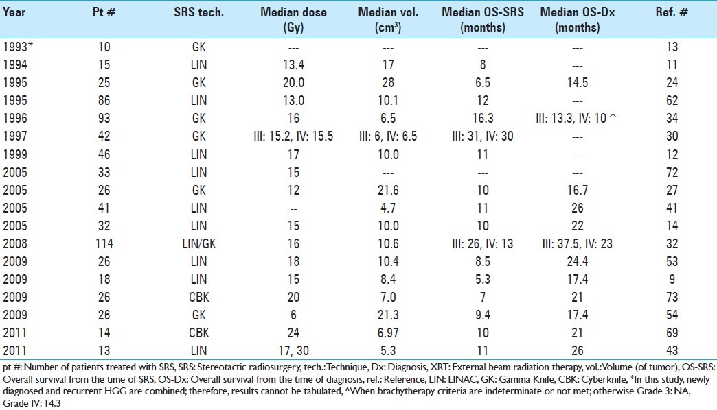
Table 2 lists studies in chronological order where SRS alone was added to the treatment regimen.[9,11–14,18,24,27,30,32,34,41,43,53,54,62,69,72,73] There are no RCT available on the use of SRS in the management of recurrent HGG. The largest study examining the impact of adding SRS as salvage treatment for recurrent HGG is a prospective cohort study with 114 patients.[32] The improvement in survival of Grade IV patients with SRS (23 months vs. 12 months) was statistically significant, while the difference in survival for Grade III HGG was not (37.5 months vs. 26 months). Univariate analysis of several factors in this study included lower pathologic grade and smaller tumor volume (<10 cm3) as significant prognostic factors. Consideration of these two factors is important when evaluating and comparing survival data. Two studies[34,62] with relatively large numbers of patients serve to illustrate this. One study includes 86 Grade IV patients with a median tumor volume of 10.1 cm3 and a median overall survival from SRS of 10.2 months.[62] The second study, with 93 patients (29% Grade III and 71% Grade IV), was notable for a median tumor volume of 6.5 cm3 and a median overall survival from SRS of 16.3 months.[34] The improved survival observed in this latter study could be expected based on both a higher proportion of lower grade patients and a lower median tumor volume. The relative weight of these two factors, however, is uncertain, but tumor grade may impact overall survival to a greater extent. Corroborating this, two studies[12,32] that included both Grade III and IV patients found an almost twofold difference in overall median survival according to grade. Furthermore, two other studies[43,62] that restricted inclusion to Grade IV, but with different median tumor volumes, were notable for a less than 10% difference in overall median survival despite an almost twofold difference in median tumor volume.
Table 2.
Studies of stereotactic radiosurgery as adjunct treatment for recurrent high-grade gliomas

The identification of prognostic factors varies between studies. In one study, multivariate analysis demonstrated tumor grade as well as Karnofsky score to be the only statistically significant factors.[12] A different study identified smaller tumor volume, younger age, and unifocality, in addition to Karnofsky score and lower tumor grade, as all significant by multivariate analysis.[34]
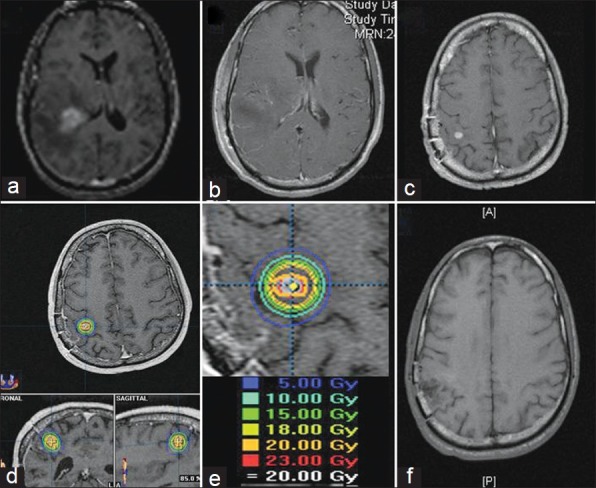
Others found neither Karnofsky score, age, nor tumor volume to be significant.[32,43] The difference may be due, in part, to the definition and/or adherence to eligibility criteria; an increase in the homogeneity of a specific factor will lower the likelihood of that factor being found significant. Alternatively, a particularly wide or narrow distribution in outcome may also obscure identification of prognostic significance. In our clinical practice, we advocate SRS for patients with recurrent HGG only for those with a small tumor volume (<3 cm in maximum longitudinal diameter). A case example is shown in Figure 1.
Figure 1.

The patient was 47 years old who presented with headache and dysphasia. Brain MRI before surgery (a) shows a periventricular contrast-enhancing mass with surrounding edema. Postoperative MRI (b) shows gross total resection and pathology confirmed glioblastoma. The patient underwent XRT and concomitant TMZ. Two months after adjuvant therapy, follow-up MRI (c) shows a small recurrent nodule outside the tumor cavity. This was targeted with SRS. Isodose lines around the lesion (d) treated with 20 Gy at the 85% isodose line (e). Follow-up MRI (f) shows radiographic control up to 19 months later
Given the extremely poor survival times in recurrent HGG, even small gains in survival with SRS may be meaningful. However, this must be balanced by awareness of potential complications and impact on quality of life. Early adverse side effects typically involve headache, nausea and/or vomiting which may be medically managed, while late complications typically involve radiation necrosis. There are some that have reported no acute or late complications from SRS treatment.[14,54] However, others have reported an incidence of radiation necrosis as high as 30%.[12,27] While mild cases may be managed by serial imaging and steroids, more severe cases may require surgical intervention. The incidence of returning to the operating room has been reported to be as high as 20%.[24,42] Interestingly, despite similar craniotomy rates, histopathologic findings may vary. In one study, histopathologic analysis of seven patients requiring re-operation demonstrated four tumor recurrences, two radiation necrosis, and one mixed specimen.[24] In a different study, in 10 patients undergoing repeat craniotomy, 2 patients had tumor recurrence, 2 radiation necrosis, and 6 mixed pathology.[12] There was no comparison with a control group in these studies, and the use of historical controls is particularly difficult with respect to selection bias.
A potentially promising concept is the combination of SRS with targeted molecular therapy,[15,19,35,52,58] as listed chronological order in Table 3. One of the first studies combined SRS with marimastat, a matrix metalloproteinase inhibitor (MMPI).[35] The rationale for using this agent is based on evidence that MMPIs can restrict tumor invasion and block neovascularization. A total of 26 patients (14 Grade IV and 12 Grade III) were enrolled and data compared to historical controls within the same institution. Results demonstrated relatively small survival advantages that were statistically significant for Grade III patients (17 months with SRS + marimastat vs. 14.8 months with SRS alone) but not for Grade IV (10.5 months with SRS + marimastat vs. 9.5 months with SRS alone) patients. The next two studies[19,58] combined SRS with a small molecule inhibitor of the epidermal growth factor receptor (EGFR), which plays a fundamental role in cancer cell proliferation and growth.[47] The EGFR inhibitors, gefitinib and vandetanib, also have potential radiosensitizing effects.[7,16] The focus of these Phase I clinical trials was safety, not evaluation of treatment effects. However, data from these studies show overall median survival ranging from 6 to 10 months, roughly comparable to survival times with SRS alone. The most recent studies[15,52] have used bevacizumab, an antibody to the vascular endothelial growth factor (VEGF), a key player in tumor angiogenesis.[59] Bevacizumab was chosen based on early clinical studies that demonstrated encouraging results with respect to progression-free survival and time to progression,[17,75] and previous experience with fractionated SRT.[23] While both studies[15,52] evaluated the safety profile of this combined therapeutic approach, one[52] also evaluated efficacy in patients with Grade IV and found a significant improvement in both overall and progression-free survival when compared to a matched cohort which underwent SRS without bevacizumab. Median overall survival from SRS was 18 months,[52] which is higher than that reported by the large majority of studies listed in Table 2 evaluating SRS without targeted molecular therapy for recurrent HGG. The incidence of mild to moderate toxicity (grades 1 and 2) in the cohort of patients receiving bevacizumab in addition to SRS was comparable to that seen in patients receiving SRS alone and was around 30%. The incidence of severe toxicity (grades 3 and 4) was also comparable, approximately 4% in both groups. Acute complications, including worsening of neurologic symptoms, headache, and fatigue, developed in approximately half the patients in both arms and typically responded to steroids. With respect to late complications, the incidence of radionecrosis was lower in the cohort that received bevacizumab compared to the group that did not (5% vs. 19%). This is in keeping with data showing that bevacizumab may decrease radiation necrosis by decreasing capillary leakage and associated brain edema.[22]
Table 3.
Studies of stereotactic radiosurgery + molecular targeting agent as adjunct treatment for recurrent high-grade gliomas

Finally, as already been pointed out in discussion of SRS for the management of newly diagnosed HGG,[3,70] it should be highlighted that the limitations of conventional imaging techniques may hamper the interpretation and potential success of SRS-based treatment. The impact of appropriate imaging may be even more important for recurrent HGG, where the distinction between tumor recurrence and radiation necrosis is crucial for determining therapeutic approaches and evaluating outcomes. A review of the accuracy of current imaging modalities demonstrates that despite progress, further research is needed to firmly establish imaging methodology to accurately and reliably distinguish between glioma recurrence and radiation necrosis.[1] Incorporation of advanced imaging techniques, such as perfusion, permeability, diffusion, and functional magnetic resonance imaging (MRI), magnetic resonance spectroscopy (MRS), and metabolically based positron emission tomography, may improve both prognostication and tumor delineation.[33,36,49,50,71] Additionally, as cellular and molecular imaging techniques evolve in the experimental setting,[21] they may enable future visualization of glioma stem cells which could have important therapeutic implications for SRS, given their role in treatment resistance.[8]
CONCLUSIONS
For newly diagnosed HGG, there is strong evidence that addition of an SRS boost prior to standard XRT provides no survival benefit. However, evidence from numerous studies suggests a possible survival benefit when SRS is performed after XRT in a timely fashion and on a well-selected patient population. A randomized controlled clinical trial evaluating the impact of SRS after XRT in patients with newly diagnosed HGG is warranted to fully define its role in therapeutic management.
For recurrent HGG, there is suggestion that SRS may potentially confer survival benefit. However, it may be limited to tumors of small volume. Complication rates may be higher, but may be justified given the particularly poor prognosis of patients with recurrent HGG. Newer studies have also provided preliminary promising data from the combination of SRS with targeted molecular agents. Controlled clinical trials are necessary to corroborate the potential role of SRS in recurrent HGG.
Incorporation of advanced imaging techniques, as well as evolving cellular and molecular strategies, grounded in fundamental radiobiological principles, may improve the overall efficacy and evaluation of SRS-based treatments. The poor survival statistics for newly diagnosed HGG patients, and even more so for patients with recurrent HGG, mandate continued investigation of SRS as a potential therapeutic modality.
Publication of this manuscript has been made possible by an educational grant from
 BRAINLAB
Footnotes
Disclaimer: The authors of this paper have received no outside funding, and have nothing to disclose.
Available FREE in open access from: http://www.surgicalneurologyint.com/text.asp?2012/3/3/118/95423
Contributor Information
Emanuela Binello, Email: emanuela.binello@mssm.edu.
Sheryl Green, Email: sheryl.green@mountsinai.org.
Isabelle M. Germano, Email: isabelle.germano@mountsinai.org.
REFERENCES
- 1.Alexiou GA, Tsiouris S, Kyritsis AP, Voulgaris S, Argyropoulou MI, Fotopoulos AD. Glioma recurrence versus radiation necrosis: Accuracy of current imaging modalities. J Neurooncol. 2009;95:1–11. doi: 10.1007/s11060-009-9897-1. [DOI] [PubMed] [Google Scholar]
- 2.Andrews DA, Bednarz G, Evans JJ, Downes B. A review of 3 current radiosurgery systems. Surg Neurol. 2006;66:559–64. doi: 10.1016/j.surneu.2006.08.002. [DOI] [PubMed] [Google Scholar]
- 3.Anker CJ, Hymas RV, Hazard LJ, Boucher KM, Jensen RL, Shrieve DC. Stereotactic radiosurgery eligibility and selection bias in the treatment of glioblastoma multiforme. J Neurooncol. 2010;98:253–63. doi: 10.1007/s11060-010-0176-y. [DOI] [PubMed] [Google Scholar]
- 4.Barker F, Chang S, Gutin P, Malec M, Mcdermott M, Prados M, et al. Survival and functional status after resection of recurrent glioblastoma multiforme. Neurosurgery. 1998;42:709–20. doi: 10.1097/00006123-199804000-00013. [DOI] [PubMed] [Google Scholar]
- 5.Barnett GH, Linskey ME, Adler JR, Cozzens JW, Friedman WA, Heilburn MP, et al. Stereotactic radiosurgery-an organized neurosurgery-sanctioned definition. J Neurosurg. 2007;106:1–5. doi: 10.3171/jns.2007.106.1.1. [DOI] [PubMed] [Google Scholar]
- 6.Baumert BG, Lutterbach J, Bernays R, Davis JB, Heppner FL. Fractionated stereotactic radiotherapy boost after post-operative radiotherapy in patients with high-grade gliomas. Radiother Oncol. 2003;67:183–90. doi: 10.1016/s0167-8140(02)00386-9. [DOI] [PubMed] [Google Scholar]
- 7.Bianco C, Tortora G, Bianco R, Caputo R, Veneziani BM, Caputo R, et al. Enhancement of antitumor activity of ionizing radiation by combined treatment with the selective epidermal growth factor receptor-tyrosine kinase inhibitor ZD1839 (Iressa) Clin Cancer Res. 2002;8:3250–8. [PubMed] [Google Scholar]
- 8.Binello E, Germano IM. Targeting glioma stem cells: A novel framework for brain tumors. Cancer Sci. 2011;102:1958–66. doi: 10.1111/j.1349-7006.2011.02064.x. [DOI] [PMC free article] [PubMed] [Google Scholar]
- 9.Biswas T, Okunieff P, Schell MC, Smudzin T, Pilcher W, Bakos RS, et al. Stereotactic radiosurgery for glioblastoma: Retrospective analysis. Radiat Oncol. 2009;4:11. doi: 10.1186/1748-717X-4-11. [DOI] [PMC free article] [PubMed] [Google Scholar]
- 10.Buatti JM, Friedman WA, Bova FJ, Mendenhall WM. Linac radiosurgery for high-grade gliomas: The University of Florida experience. Int J Radiat Oncol Biol Phys. 1995;32:205–10. doi: 10.1016/0360-3016(94)00498-A. [DOI] [PubMed] [Google Scholar]
- 11.Chamberlain MC, Barba D, Kormanik P, Shea WM. Stereotactic radiosurgery for recurrent gliomas. Cancer. 1994;74:1342–7. doi: 10.1002/1097-0142(19940815)74:4<1342::aid-cncr2820740426>3.0.co;2-y. [DOI] [PubMed] [Google Scholar]
- 12.Cho KH, Hall WA, Gerbi BJ, Higgins PD, McGuire WA, Clark HB. Single dose versus fractionated stereotactic radiotherapy for recurrent high-grade glioma. Int J Radiat Oncol Biol Phys. 1999;45:1133–41. doi: 10.1016/s0360-3016(99)00336-3. [DOI] [PubMed] [Google Scholar]
- 13.Coffey RI. Boost gamma knife radiosurgery in the treatment of primary glial tumors. Stereotact Funct Neurosurg. 1993;61(Suppl 1):59–64. doi: 10.1159/000100661. [DOI] [PubMed] [Google Scholar]
- 14.Combs SE, Widner V, Thilmann C, Hof H, Debus J, Schulz-Ertner D. Stereotactic radiosurgery (SRS): Treatment option for recurrent glioblastoma multiforme (GBM) Cancer. 2005;104:2168–73. doi: 10.1002/cncr.21429. [DOI] [PubMed] [Google Scholar]
- 15.Cuneo KC, Vredenburgh JJ, Sampson JH, Reardon DA, Desjardins A, Peters KB, et al. Safety and efficacy of stereotactic radiosurgery and adjuvant bevacizumab in patients with recurrent malignant gliomas. Int J Radiat Oncol Biol Phys. 2012;82:2018–24. doi: 10.1016/j.ijrobp.2010.12.074. [DOI] [PMC free article] [PubMed] [Google Scholar]
- 16.Damiano V, Melisi D, Bianco C, Raben D, Caputo R, Fontanini G, et al. Cooperative antitumor effect of multitargeted kinase inhibitor ZD6474 and ionizing radiation in glioblastoma. Clin Cancer Res. 2005;1:5639–44. doi: 10.1158/1078-0432.CCR-05-0174. [DOI] [PubMed] [Google Scholar]
- 17.Desjardins A, Reardon DA, Herndon JE, 2nd, Marcello J, Quinn JA, Rich JN, et al. Bevacizumab plus irinotecan in recurrent WHO grade 3 malignant gliomas. Clin Cancer Res. 2008;14:7068–73. doi: 10.1158/1078-0432.CCR-08-0260. [DOI] [PMC free article] [PubMed] [Google Scholar]
- 18.Elliott R, Parker EC, Rush SC, Kalhorn SP, Moshel YA, Narayana A, et al. Efficacy of gamma knife radiosurgery for small-volume recurrent malignant gliomas after initial radical resection. World Neurosurg. 2011;76:128–40. doi: 10.1016/j.wneu.2010.12.053. [DOI] [PubMed] [Google Scholar]
- 19.Fields EC, Damek D, Gaspar LE, Liu AK, Kavanagh BD, Waziri A, et al. Phase I dose escalation trial of vandetanib with fractionated radiosurgery in patients with recurrent malignant gliomas. Int J Radiat Oncol Biol Phys. 2012;82:51–7. doi: 10.1016/j.ijrobp.2010.09.008. [DOI] [PubMed] [Google Scholar]
- 20.Gannett D, Stea B, Lulul B, Adair T, Verdi C, Hamilton A. Stereotactic radiosurgery as an adjunct to surgery and external beam radiotherapy in the treatment of patients with malignant gliomas. Int J Radiat Oncol Biol Phys. 1995;33:461–8. doi: 10.1016/0360-3016(95)00087-F. [DOI] [PubMed] [Google Scholar]
- 21.Gera A, Steinberk GK, Guzman R. In vivo neural stem cell imaging: Current modalities and future directions. Regen Med. 2010;5:73–86. doi: 10.2217/rme.09.79. [DOI] [PubMed] [Google Scholar]
- 22.Gonzalez J, Kumar AJ, Conrad CA, Levin VA. Effect of bevacizumab on radiation necrosis of the brain. Int J Radiat Oncol Biol Phys. 2007;67:323–7. doi: 10.1016/j.ijrobp.2006.10.010. [DOI] [PubMed] [Google Scholar]
- 23.Gutin PH, Iwamoto FM, Beal K, Mohile NA, Karimi S, Hou BL, et al. Safety and efficacy of bevacizumab with hypofractionated stereotactic irradiation for recurrent malignant gliomas. Int J Radiat Oncol Biol Phys. 2009;75:156–63. doi: 10.1016/j.ijrobp.2008.10.043. [DOI] [PMC free article] [PubMed] [Google Scholar]
- 24.Hall WA, Djaliliam HR, Sperduto PW, Cho KH, Gerbi BJ, Gibbons JP, et al. Stereotactic radiosurgery for recurrent malignant glioma. J Clin Oncol. 1995;13:1642–8. doi: 10.1200/JCO.1995.13.7.1642. [DOI] [PubMed] [Google Scholar]
- 25.Hall EJ. In: Radiobiology for the Radiologist. 4th ed. Philadelphia: JB Lippincott Company; 1994. Tissue, Dose and Fractionation in radiotherapy; pp. 211–29. [Google Scholar]
- 26.Harsh GR, 4th, Levin VA, Gutin PH, Seager M, Silver P, Wilson CB. Reoperation for recurrent glioblastoma and anaplastic astrocytoma. Neurosurgery. 1987;21:615–7. doi: 10.1227/00006123-198711000-00002. [DOI] [PubMed] [Google Scholar]
- 27.Hsieh PC, Chandler JP, Bhangoo S, Panagiotopoulos K, Kalapurakal JA, Marymont MH, et al. Adjuvant gamma knife stereotactic radiosurgery at the time of tumor progression potentially improves survival for patients with glioblastoma multiforme. Neurosurgery. 2005;57:684–92. [PubMed] [Google Scholar]
- 28.Kida Y, Yoshimoto M, Hasegawa T. Radiosurgery for intracranial gliomas. Prog Neurol Surg. 2009;22:122–8. doi: 10.1159/000163387. [DOI] [PubMed] [Google Scholar]
- 29.Kim JJ, Tannock IF. Repopulation of cancer cells during therapy: An important cause of treatment failure. Nat Rev Cancers. 2005;5:516–25. doi: 10.1038/nrc1650. [DOI] [PubMed] [Google Scholar]
- 30.Kondziolka D, Flickinger JC, Bissonette DJ, Bozik M, Lunsford LD. Survival benefit of stereotactic radiosurgery for patients with malignant glial neoplasms. Neurosurgery. 1997;41:775–85. doi: 10.1097/00006123-199710000-00004. [DOI] [PubMed] [Google Scholar]
- 31.Kondziolka D, Lunsford LD, Flickinger JC. In regard to Dr. Souhami et al (Int J Radiat Oncol Biol Phys 2004;60:853-60) Int J Radiat Oncol Biol Phys. 2005;62:614–5. doi: 10.1016/j.ijrobp.2005.02.020. [DOI] [PubMed] [Google Scholar]
- 32.Kong DS, Lee JI, Park K, Kim JH, Lim DH, Nam DH. Efficacy of stereotactic radiosurgery as a salvage treatment for recurrent malignant gliomas. Cancer. 2008;112:2046–51. doi: 10.1002/cncr.23402. [DOI] [PubMed] [Google Scholar]
- 33.Krishnan AP, Asher IM, Davis D, Okunieff P, O’Dell WG. Evidence that MR diffusion tensor imaging (tractography) predicts the natural history of regional progression in patients irradiated conformally for primary brain tumors. Int J Radiat Oncol Biol Phys. 2008;71:1553–62. doi: 10.1016/j.ijrobp.2008.04.017. [DOI] [PubMed] [Google Scholar]
- 34.Larson DA, Gutin PH, McDermott M, Lamborn K, Sneed PK, Wara WM, et al. Gamma knife for glioma: Selection factors and survival. Int J Radiat Oncol Biol Phys. 1996;36:1045–53. doi: 10.1016/s0360-3016(96)00427-0. [DOI] [PubMed] [Google Scholar]
- 35.Larson DA, Prados M, Lamborn KR, Smith V, Sneed PK, Chang S, et al. Phase II study of high central dose gamma knife radiosurgery and marimastat in patients with recurrent malignant glioma. Int J Radiat Oncol Biol Phys. 2002;54:1397–404. doi: 10.1016/s0360-3016(02)03743-4. [DOI] [PubMed] [Google Scholar]
- 36.Levivier M, Massager N, Wikler D, Devriendt D, Goldman S. Integration of functional imaging in radiosurgery: The example of PET scan. Prog Neurol Surg. 2007;20:68–81. doi: 10.1159/000100096. [DOI] [PubMed] [Google Scholar]
- 37.Levivier M, Gaevart T, Negretti L. Gamma Knife, CyberKnife, TomoTherapy: Gadgets or useful tools? Curr Opin Neurol. 2011;24:616–25. doi: 10.1097/WCO.0b013e32834cd4df. [DOI] [PubMed] [Google Scholar]
- 38.Loeffler JS, Alexander E, 3rd, Shea M, Wen PY, Fine HA, Kooy HM, et al. Radiosurgery as part of the initial management of patients with malignant gliomas. J Clin Oncol. 1992;10:1379–85. doi: 10.1200/JCO.1992.10.9.1379. [DOI] [PubMed] [Google Scholar]
- 39.Louis DN, Ohgaki H, Wiestler OD, Cavanee WK, Burger PC, Jouvet A, et al. The 2007 WHO classification of tumors of the central nervous system. Acta Neuropathol. 2007;114:97–109. doi: 10.1007/s00401-007-0243-4. [DOI] [PMC free article] [PubMed] [Google Scholar]
- 40.Lustig RA, Scott CB, Curran WJ. Does stereotactic eligibility for the treatment of glioblastoma cause selection bias in randomized studies? Am J Clin Oncol. 2004;27:516–21. doi: 10.1097/01.coc.0000135641.82026.c4. [DOI] [PubMed] [Google Scholar]
- 41.Mahajan A, McCutcheon IE, Suki D, Chang EL, Hassenbusch SJ, Weinberg JS, et al. Case-control study of stereotactic radiosurgery for recurrent glioblastoma multiforme. J Neurosug. 2005;103:210–7. doi: 10.3171/jns.2005.103.2.0210. [DOI] [PubMed] [Google Scholar]
- 42.Mandl ES, Dirven CM, Buis DR, Postman TJ, Vandertop WP. Repeated surgery for glioblastoma multiforme: Only in combination with other salvage therapy. Surg Neurol. 2008;69:506–9. doi: 10.1016/j.surneu.2007.03.043. [DOI] [PubMed] [Google Scholar]
- 43.Maranzano E, Anselmo P, Casale M, Trippa F, Carletti S, Principi M, et al. Treatment of recurrent glioblastoma with stereotactic radiotherapy: Long-term results of a mono-institutional trial. Tumori. 2011;97:56–61. doi: 10.1177/030089161109700111. [DOI] [PubMed] [Google Scholar]
- 44.Masciopinto JE, Levin AB, Mehta MP, Rhode BS. Stereotactic radiosurgery for glioblastoma multiforme. Stereotact Funct Neurosurg. 1994;63:233–4. doi: 10.1159/000100319. [DOI] [PubMed] [Google Scholar]
- 45.Masciopinto JE, Levin AB, Mehta MP, Rhode BS. Stereotactic radiosurgery for glioblastoma: A final report of 31 patients. J Neurosurg. 1995;2:530–5. doi: 10.3171/jns.1995.82.4.0530. [DOI] [PubMed] [Google Scholar]
- 46.Mehta MP, Masciopinto J, Rozental J, Levin AL, Chappell R, Bastin K, et al. Stereotactic radiosurgery for glioblastoma multiforme: Report of a prospective study evaluating prognostic factors and analyzing long-term survival advantage. Int J Radiat Oncol Biol Phys. 1994;30:541–9. doi: 10.1016/0360-3016(92)90939-f. [DOI] [PubMed] [Google Scholar]
- 47.Mellinghoff IK, Wang MY, Vivanco I, Haas-Kogan DA, Zhu S, Dia EQ, et al. Molecular determinants of the response of glioblastomas to EGFR kinase inhibitors. N Engl J Med. 2005;10:2012–4. doi: 10.1056/NEJMoa051918. [DOI] [PubMed] [Google Scholar]
- 48.Minniti G, Amelio D, Amichetti M, Salvati M, Muni R, Bozzao A, et al. Patterns of failure and comparison of different target volume delineations in patients with glioblastoma treated with conformal radiotherapy plus concomitant and adjuvant temozolomide. Radiother Oncol. 2010;97:377–81. doi: 10.1016/j.radonc.2010.08.020. [DOI] [PubMed] [Google Scholar]
- 49.Narayana A, Chang J, Thakur S, Huang W, Karimi S, Hou B, et al. Use of MR spectroscopy and functional imaging in the treatment planning of gliomas. Br J Radiol. 2007;80:347–54. doi: 10.1259/bjr/65349468. [DOI] [PubMed] [Google Scholar]
- 50.Nelson SJ. Assessment of therapeutic response and treatment planning for brain tumors using metabolic and physiologic MRI. NMR Biomed. 2011;24:734–49. doi: 10.1002/nbm.1669. [DOI] [PMC free article] [PubMed] [Google Scholar]
- 51.Nwokedi EC, DiBiase SJ, Jabbour S, Herman J, Amin P, Chin LS. Gamma knife stereotactic radiosurgery for patients with glioblastoma multiforme. Neurosurgery. 2002;50:41–7. doi: 10.1097/00006123-200201000-00009. [DOI] [PubMed] [Google Scholar]
- 52.Park KJ, Kano H, Iyer A, Liu X, Niranjan A, Flickinger JC, et al. Salvage gamma knife stereotactic radiosurgery followed by bevacizumab for recurrent glioblastoma multiforme: A case-control study. J Neurooncol. 2012;107:323–33. doi: 10.1007/s11060-011-0744-9. [DOI] [PubMed] [Google Scholar]
- 53.Patel M, Siddiqui F, Jin JY, Mikkelsen T, Rosenblum M, Movsas B, et al. Salvage reirradiation for recurrent glioblastoma with radiosurgery: Radiographic response and improved survival. J Neurooncol. 2009;92:185–91. doi: 10.1007/s11060-008-9752-9. [DOI] [PubMed] [Google Scholar]
- 54.Pouratian N, Crowley RW, Sherman JH, Jagannathan J, Sheehan JP. Gamma knife radiosurgery after radiation therapy as an adjunctive treatment for glioblastoma. J Neurooncol. 2009;94:409–18. doi: 10.1007/s11060-009-9873-9. [DOI] [PubMed] [Google Scholar]
- 55.Prisco FE, Weltman E, de Hanriot RM, Brandt R. Radiosurgical boost for primary high-grade gliomas. J Neurooncol. 2002;57:151–60. doi: 10.1023/a:1015757322379. [DOI] [PubMed] [Google Scholar]
- 56.Sanai N, Berger MS. Extent of resection influences outcomes for patients with gliomas. Rev Neurol. 2011;167:648–54. doi: 10.1016/j.neurol.2011.07.004. [DOI] [PubMed] [Google Scholar]
- 57.Sarkaria JN, Mehta MP, Loeffler JS, Buatti JM, Chappell RJ, Levin AB, et al. Radiosurgery in the initial management of malignant gliomas: Survival comparison with the RTOG recursive partitioning analysis. Int J Radiat Oncol Biol Phys. 1995;32:931–41. doi: 10.1016/0360-3016(94)00621-q. [DOI] [PubMed] [Google Scholar]
- 58.Schwer AL, Damek DM, Kavanagh BD, Gaspar LE, Lillehel K, Stuhr K, et al. A phase I dose-escalation study of fractionated stereotactic radiosurgery in combination with gefitinib in patients with recurrent malignant gliomas. Int J Radiat Oncol Biol Phys. 2008;70:993–1001. doi: 10.1016/j.ijrobp.2007.07.2382. [DOI] [PubMed] [Google Scholar]
- 59.Sharma PS, Sharma R, Tyagi T. VEGF/VEFR pathway inhibitors as anti-angiogenic agents: Present and future. Curr Cancer Drug Targets. 2011;11:624–53. doi: 10.2174/156800911795655985. [DOI] [PubMed] [Google Scholar]
- 60.Shaw E, Scott C, Souhami L, Dinapoli R, Kline R, Loeffler J, et al. Single dose radiosurgical treatment of recurrent previously irradiated primary brain tumors and brain metastases: Final report of RTOG protocol 90-05. Int J Radiat Oncol Biol Phys. 2000;47:291–8. doi: 10.1016/s0360-3016(99)00507-6. [DOI] [PubMed] [Google Scholar]
- 61.Shenouda G, Souhami L, Podgorsak EB, Bahary JP, Villemure JG, Caron JL, et al. Radiosurgery and accelerated radiotherapy for patients with glioblastoma. Can J Neurol Sci. 1997;24:110–5. doi: 10.1017/s0317167100021429. [DOI] [PubMed] [Google Scholar]
- 62.Shrieve D, Alexander E, 3rd, Wen PY, Fine HA, Koy HM, Black PM. Comparison of stereotactic radiosurgery and brachytherapy in the treatment of recurrent glioblastoma multiforme. Neurosurgery. 1995;36:275–84. doi: 10.1227/00006123-199502000-00006. [DOI] [PubMed] [Google Scholar]
- 63.Shrieve DC, Alexander E, 3rd, Black PM, Wen PY, Fine HA, Kooy HM, et al. Treatment of patients with primary glioblastoma multiforme with standard postoperative radiotherapy and radiosurgical boost: Prognostic factors and long-term outcome. J Neurosurg. 1999;92:72–7. doi: 10.3171/jns.1999.90.1.0072. [DOI] [PubMed] [Google Scholar]
- 64.Soldberg TD, Selch MT, Smathers JB, DeSalles AF. Fractionated stereotactic radiotherapy: Rationale and methods. Med Dosim. 1998;23:209–19. doi: 10.1016/s0958-3947(98)00016-8. [DOI] [PubMed] [Google Scholar]
- 65.Souhami LS, Seiferheld W, Brachman D, Podgorsak EB, Werner-Wasik M, Lustig R, et al. Randomized comparison of stereotactic radiosurgery followed by conventional radiotherapy with carmustine to conventional radiotherapy with carmustine for patients with glioblastoma multiforme: Report of Radiation Therapy Oncology Group 93-05 protocol. Int J Radiat Oncol Biol Phys. 2004;60:853–60. doi: 10.1016/j.ijrobp.2004.04.011. [DOI] [PubMed] [Google Scholar]
- 66.Stummer W, Meinel T, Ewelt C, Martus P, Jacobs O, Felsberg J, et al. Prospective cohort of radiotherapy with concomitant and adjuvant temozolomide chemotherapy for glioblastoma patients with no or minimal residual enhancing tumor load after surgery. J Neurooncol. 2012 doi: 10.1007/s11060-012-0798-3. [In press] [DOI] [PMC free article] [PubMed] [Google Scholar]
- 67.Stummer W, Tonn JC, Mehdorn HM, Nestler U, Franz K, Goetz C, et al. Counterbalancing risks and gains from extended resections in malignant glioma surgery: A supplemental analysis from the randomized 5-aminolevulinic acid glioma resection study. Clinical article. J Neurosurg. 2011;114:613–23. doi: 10.3171/2010.3.JNS097. [DOI] [PubMed] [Google Scholar]
- 68.Stupp R, Hegi ME, Mason WP, van den Bent MJ, Taphoorm MJ, Janzer RC, et al. Effects of radiotherapy with concomitant and adjuvant temozolomide versus radiotherapy alone on survival in glioblastoma in a randomised phase III study: 5-year analysis of the EORTC-NCIC trial. Lancet Oncol. 2009;10:459–66. doi: 10.1016/S1470-2045(09)70025-7. [DOI] [PubMed] [Google Scholar]
- 69.Torok JA, Wegner RE, Mintz AH, Heron DE, Burton SA. Re-irradiation with radiosurgery for recurrent glioblastoma multiforme. Technol Cancer Res Treat. 2011;10:253–8. doi: 10.7785/tcrt.2012.500200. [DOI] [PubMed] [Google Scholar]
- 70.Tsao MN, Mehta MP, Whelan TJ, Morris DE, Hayman JA, Flickinger JC, et al. The American Society for Therapeutic Radiology and Oncology (ASTRO) evidence-based review of the role of radiosurgery for malignant glioma. Int J Radiat Oncol Biol Phys. 2005;63:47–55. doi: 10.1016/j.ijrobp.2005.05.024. [DOI] [PubMed] [Google Scholar]
- 71.Tsien CI, Cao Y, Lawrence TS. Functional and metabolic magnetic resonance imaging and positron emission tomography for tumor volume definition in high-grade gliomas. Semin Radiat Oncol. 2009;19:155–62. doi: 10.1016/j.semradonc.2009.02.002. [DOI] [PubMed] [Google Scholar]
- 72.Ulm AJ, 2nd, Friedman WA, Bradshaw P, Foote KD, Bova FJ. Radiosurgery in the treatment of malignant gliomas: The University of Florida experience. Neurosurgery. 2005;57:512–7. doi: 10.1227/01.neu.0000170560.13251.99. [DOI] [PubMed] [Google Scholar]
- 73.Villaviciencio AT, Burneikiene S, Romanelli P, Fariselli L, McNeely L, Lipani JD, et al. Survival following stereotactic radiosurgery for newly diagnosed and recurrent glioblastoma multiforme: A multicenter experience. Neurosurg Rev. 2009;32:417–24. doi: 10.1007/s10143-009-0212-6. [DOI] [PubMed] [Google Scholar]
- 74.Vordermark D, Kolbl O. Lack of survival benefit after stereotactic radiosurgery boost for glioblastome multiforme: randomized comparison of stereotactic radiosurgery followed by conventional radiotherapy with carmustine to conventional radiotherapy with carmustine for patients with glioblastoma mltiforme:Report of radiation therapy Oncology Group 93-05: In regard to Souhami et al (Int J Radiat Oncol Biol Phys 2004;60:853-60) Int J Radiat Oncol Biol Phys. 2005;62:296–7. doi: 10.1016/j.ijrobp.2005.01.046. [DOI] [PubMed] [Google Scholar]
- 75.Vredenburgh JJ, Desjardins A, Herndon JE, 2nd, Marcello J, Reardon DA, Quinn JA, et al. Bevacizumab plus irinotecan in recurrent glioblastoma multiforme. J Clin Oncol. 2007;25:4722–9. doi: 10.1200/JCO.2007.12.2440. [DOI] [PubMed] [Google Scholar]
- 76.Wallner KE, Galicich JH, Krol G, Arbit E, Malkin MG. Patterns of failure following treatment for glioblastoma multiforme and anaplastic astrocytoma. Int J Radiat Oncol Biol Phys. 1989;16:1405–9. doi: 10.1016/0360-3016(89)90941-3. [DOI] [PubMed] [Google Scholar]
- 77.Wick W, Wick A, Schulz JB, Dichgans J, Rodemann HP, Weller W. Prevention of irradiation-induced glioma cell invasion by temozolomide involves caspase 3 activity and cleavage of focal adhesion kinase. Cancer Res. 2002;62:1915–9. [PubMed] [Google Scholar]
- 78.Wild-Bode C, Weller M, Rimner A, Dichgans J, Wick W. Sublethal irradiation promotes migration and invasiveness of glioma cells: Implications for radiotherapy of human glioblastoma. Cancer Res. 2001;61:2744–50. [PubMed] [Google Scholar]
- 79.Yoshikawa K, Saito K, Kajiwara K, Nomura S, Ishihara H, Suzuki M. Cyberknife stereotactic radiotherapy for patients with malignant glioma. Minim Invasive Neurosurg. 2006;49:110–5. doi: 10.1055/s-2006-932183. [DOI] [PubMed] [Google Scholar]


