Sir,
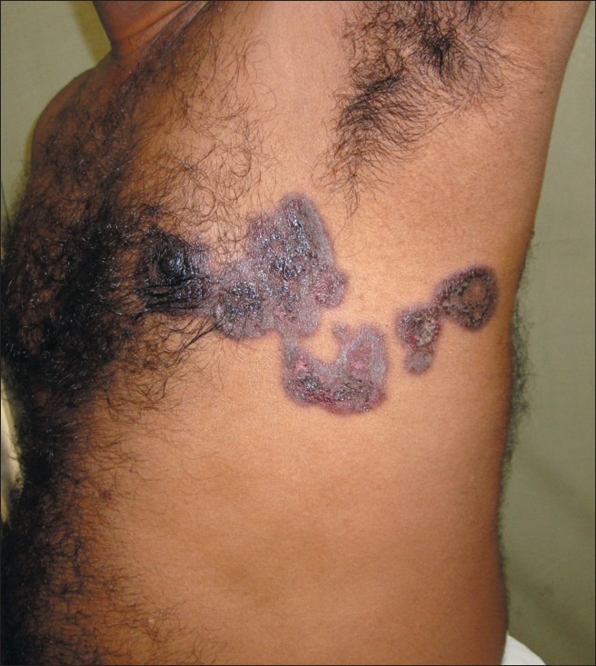
A 38 year old man presented with multiple fluid filled lesions on the left side of the chest and dark colored lesions on the right hand of 3 days duration, associated with itching and burning. He also complained of erosions over the lips and the genitalia. He had been treated with levofloxacin and paracetamol for fever and sorethroat seven days ago. The hyperpigmented lesions on the hand had also occurred 3 months ago following intake of levofloxacin for upper respiratory tract infection. There was no history of chest trauma, herpes zoster or application of topical medications. On examination there were multiple, sharply demarcated hyperpigmented macules, a few of them surmounted by flaccid bullae, extending from the nipple to the midback in a zosteriform configuration, corresponding to the left T4, T5 dermatome [Figure 1]. Two smaller hyperpigmented macules were also seen over the dorsum of the right hand. No other similar lesion was seen over the trunk or limbs. Erosions and crusting were noted over the lips while the glans showed erosions with preputial edema. Histopathology from the hyperpigmented macule showed perivascular mononuclear cell infiltration, pigment incontinence and scattered melanophages consistent with fixed drug eruption (FDE). Hemogram was normal, while culture from throat swab grew streptococci sensitive to erythromycin. He was treated with tablet paracetamol, azithromycin, topical mupirocin with fluticasone and a short course of systemic steroids.
Figure 1.

Sharply demarcated hyperpigmented macules exhibiting a zosteriform pattern
FDE is characterized by a sudden onset of edematous, dusky-red macules/plaques on the skin and mucous membranes, accompanied by burning and/or itching with the characteristic hallmark being reappearance of the lesions precisely over the previously affected sites on re-use of the offending drug.[1] Our patient developed FDE to levofloxacin at multiple new sites in addition to the lesions noticed on the right hand during the first exposure. Although paracetamol is known to produce bullous FDE, our patient gave history of taking the drug over-the-counter and was even continued on paracetamol during this episode. A rechallenge with levofloxacin could not be done as the patient refused it.
Site predilection of the offending drug to induce FDE is well known and has been documented as with tetracycline commonly causing lesions on the genital mucosa, naproxen on the lips, co-trimoxazole on the lips and genitalia, and metamizole on the trunk and extremities.[2,3] Site predilection of FDE occurrence have been reported with lesions preferentially occurring over previous herpes zoster, BCG vaccination, burn scar, venipuncture site, insect bite, herpes simplex or cellulitis and have been attributed to a Koebner-like phenomenon or recall phenomenon or an isotopic response.[2] Preferential localization of FDE at viscerocutaneous reflex zones has been documented and is attributed to reflex alteration in blood flow and vascular permeability.[2,4] Our patient had a history of upper respiratory tract infection with a positive throat swab culture but no specific features of internal organ pathology that could possibly explain the occurrence in a viscerocutaneous reflex zone.
Genetic susceptibility for the occurrence of FDE is known,[3] and the co-occurrence of linear and disseminated drug eruption have been explained on the basis of clonal population of cells being either homozygous or hemizygous for a gene predisposing to the disease.[5] Dermatomal distribution of FDE, as in our patient, has been contemplated with FDE to cephazolin occurring along S1 nerve root and FDE to trimethoprim occurring along C8 dermatome.[6] Though the cause for the distribution of multiple lesions of FDE occurring in a peculiar, unilateral, dermatomal pattern is inexplicable, the zosteriform appearance was nevertheless quite characteristic and unique in our patient.
References
- 1.Sehgal V N. Fixed drug eruption (FDE): Changing scenario of incriminating drugs. Int J Dermatol. 2006;45:897–908. doi: 10.1111/j.1365-4632.2006.02853.x. [DOI] [PubMed] [Google Scholar]
- 2.Ozkaya E. Fixed drug eruption: State of the art. J Dtsch Dermatol Ges. 2008;6:181–8. doi: 10.1111/j.1610-0387.2007.06491.x. [DOI] [PubMed] [Google Scholar]
- 3.Dhar S, Sharma VK. Fixed drug eruption to ciprofloxacin. Br J Dermatol. 1996;134:156–8. [PubMed] [Google Scholar]
- 4.Hauser W. Lokalisationsproblem bei Hautkrankheiten. In: Korting GW, editor. Dermatologie in Praxis und Klinik. Vol. 8. Stuttgart: Thieme; 1980. pp. 8.60–8.93. [Google Scholar]
- 5.Happle R, Effendy I. Coexisting linear and disseminated drug eruption: A clinical clue to the understanding of the genetic basis of drug eruptions. Eur J Dermatol. 2001;11:89. [PubMed] [Google Scholar]
- 6.Ozkaya-Bayazit E, Baykal C. Trimethoprim-induced linear fixed drug eruption. Br J Dermatol. 1997;137:1028–9. doi: 10.1111/j.1365-2133.1997.tb01584.x. [DOI] [PubMed] [Google Scholar]


