Abstract
Background
Ischemia/reperfusion injury (IRI) is common in general surgery and organ transplantation, and in the case of liver it triggers pro-inflammatory innate immune cascade and hepatic necrosis, leading to increased incidence of early and late organ rejection. IL-22, an inducible cytokine of T-cell origin and a member of the IL-10 superfamily, acts on target tissues via IL-22 receptor (IL-22R1).
Methods
Partial hepatic warm ischemia was induced in C57Bl/6 wild-type (WT) and type-1 IFN receptor (IFNAR)-deficient (KO) mice for 90 min followed by 6-24 h of reperfusion. WT mice were treated at 30 min prior to the ischemia insult with recombinant IL-22 (rIL-22) or anti-IL-22 neutralizing antibody (IL-22 Ab); PBS and IgG served as respective controls.
Results
IL-22 was detected at 24 h but not 6 h of liver IRI. The expression of IL-22R1 was increased by 6 h of reperfusion in WT but not IFNAR KO mice that were protected from IRI. Treatment of WT mice with rIL-22 decreased sAST levels, ameliorated cardinal histological features of IR damage (Suzuki’s score) and diminished leukocyte sequestration, along with the expression of IL-22R1 and pro-inflammatory cytokines. IL-22 Ab did not appreciably affect IRI but increased IL-22R1 transcription in the liver. Administration of IL-22 protein exerted hepatoprotection via STAT3 activation.
Conclusions
This is the first report investigating immune modulation by T cell-derived IL-22 in liver injury due to warm ischemia and reperfusion. Treatment with IL-22 protein may represent a novel therapeutic strategy to prevent liver IRI in transplant recipients.
Keywords: IL-22, Liver Transplantation, Ischemia/Reperfusion Injury, Inflammation
Introduction
Ischemia/reperfusion injury (IRI) in the liver is a major complication of hemorrhagic shock, liver resection and transplantation (1, 2). IRI resulting from donor organ retrieval, cold storage and warm ischemia during the surgery often leads to primary organ non-function and/or increased incidence of rejection episodes requiring re-transplantation. Mechanistically, liver IRI represents a continuum of local immune processes that include endothelial activation, increased expression of adhesion molecules, Kupffer cell/neutrophil activation, and cytokine release, followed by ultimate endothelial cell and hepatocyte death (3, 4). We have characterized TLR4-dependent innate immune mechanisms that initiate liver IRI cascade (5, 6). However, activated Kupffer cells release superoxide radicals, TNF-α and IL-1, which promote NF-κB activation, resulting in the recruitment of activated T cells (7). Indeed, we and others have shown that by expressing co-stimulation molecules and releasing pro-inflammatory cytokines, activated Th cells are crucial in the pathophysiology of liver IRI (7-9).
IL-22, an inducible cytokine of the IL-10 superfamily, is produced by select T cells (Th17, Th22, γ/δ, NKT) (10). Its biological activity, unlike other cytokines, does not serve the communication between immune cells, but rather signals directly to the tissue. Its tissue action is through a heterodimer IL-10R2/IL-22R1 complex. In contrast to IL-10R2, which is ubiquitously expressed and largely dispensable, the expression of IL-22R1 is restricted to epithelial cells including hepatocytes, and has not been detected in cells of the hematopoietic lineage.
By increasing tissue immunity in barrier organs such as skin, lungs and the gastrointestinal tract, IL-22 has been associated with a number of human diseases and to contribute to the pathogenesis of psoriasis, rheumatoid arthritis and Crohn’s disease (10-13). However, parallel studies in murine models of mucosal defense against pulmonary bacterial infection, inflammatory bowel disease or acute/chronic liver failure indicate that IL-22 may exert immunoregulatory pathologic vs. protective functions, depending on the context in which it is expressed (14-19). Moreover, HepG2/Hep3B cells transfected with IL-22 grew more rapidly, and were resistant to serum starvation compared with cells devoid of IL-22, suggesting that IL-22 may serve as hepatocyte survival factor (16). Thus, advancing our appreciation of the IL-22-IL-22R1 biology may yield novel therapeutic targets in multiple human diseases.
Although IL-22 is believed to orchestrate innate – adaptive immune cross-regulation and may facilitate protection, its function in liver IRI pathology remains to be elucidated. Here, we report on the role of IL-22 in the mechanism of hepatocellular damage vs. hepatoprotection in a well-defined mouse model of in-situ liver warm ischemia followed by reperfusion.
Results
Distinct kinetics of IR- vs
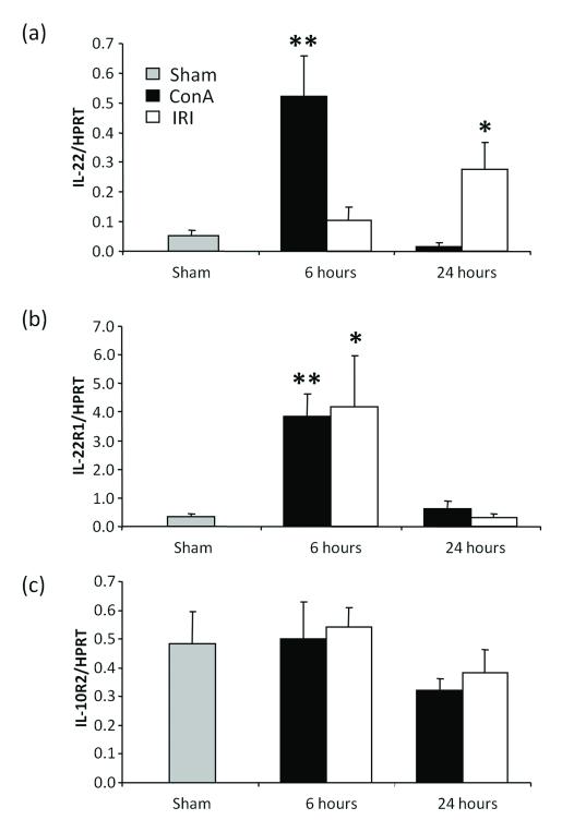
ConA-induced IL-22 expression in the liver. Mouse livers subjected to 90 min of partial warm ischemia were analyzed for IL-22 expression by qRT-PCR at 6h and 24h of reperfusion (Fig. 1a). Unlike at 6h, significantly increased mRNA levels coding for IL-22 were detected at 24 h (p<0.05). Livers from ConA-induced T-cell hepatitis model served as positive controls. In agreement with published data (16, 17), markedly increased IL-22 mRNA levels at 6 h (p<0.005) returned to baseline by 24 h after ConA challenge (Fig. 1a).
Figure 1.
IL-22 signaling in ConA and IRI models in WT mice at 6 h and 24 h of reperfusion. Quantitative RT-PCR-assisted analysis of (a) IL-22; (b) IL-22R1; and (c) IL-10R2 expression in mouse liver tissue subjected to ConA (N=6/group; 15 μg/g i.v.) or IR-triggered damage (90 min ischemia: N=6/group; Sham N=4/group). (Statistical comparisons between experimental and sham samples *: p<0.05, **: p<0.005). (a) Mice subjected to ConA injection expressed high levels of IL-22 at 6 h of infusion, before returning to baseline at 24 h, while IRI mice had low levels of IL-22 at 6 h and a modest increase by 24 h. (b) IL-22R1 expression was elevated in both models at 6 h, and returned to baseline at 24 h. (c) IL-10R2 was not affected by ConA or IRI.
IL-22R1 transcription correlates with the hepatocellular damage
We detected an increase in IL-22R1 expression by qRT-PCR in both ConA (p<0.005) and IRI (p<0.05) models at 6 h, with a return to baseline after 24 h (Fig.1b). In contrast, IL-10R2 expression was comparable between sham, ConA, and IRI models throughout (Fig. 1c).
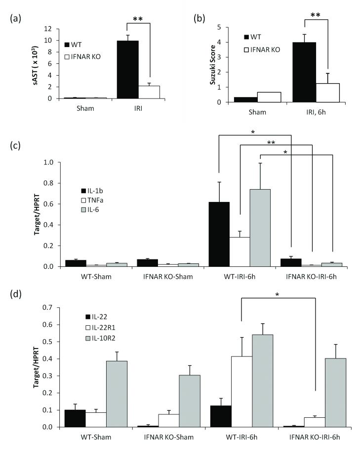
Separate cohorts of WT and IFNAR KO mice were subjected to 90 min of warm ischemia. Consistent with our previous findings (18), disruption of IFNAR signaling significantly reduced sAST levels (Fig 2a), diminished the Suzuki score of IR-liver damage (Fig 2b) and ameliorated local inflammation response, measured by hepatic IL-1β, TNFα and IL-6 levels (Fig. 2c) by 6 h of reperfusion, as compared with WT controls. No changes in IL-22 or IL-10R2 expression were noted by qPCR between the groups (Fig. 2d). In contrast, IL-22R1 levels were selectively and significantly (p<0.05) decreased in IFNAR KO mice.
Figure 2.
Livers in WT and IFNAR KO mice at 6 h of reperfusion following 90 min of warm ischemia (Ischemia: N=8/group; Sham N=4/group). (a) Peripheral serum AST levels; (p<0.005). (b) Liver tissue histology (H&E staining; x4 or x40 magnification; representative liver sections shown). (c) Suzuki’s based grading of the hepatocellular damage. RT-PCR analysis of liver specimens subjected to ischemia of d) pro-inflammatory cytokines and (e) IL-22/IL-22 receptor complex genes. Experiment was performed twice with similar results (Statistical comparisons are between WT and IFNAR KO groups. *: p<0.05, **: p<0.005).
Exogenous rIL-22 lessens IR-triggered hepatocellular damage
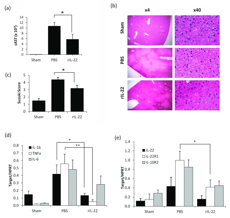
We next investigated the ability of exogenous rIL-22 to affect liver IRI. Indeed, pretreatment with rIL-22 at 30 min prior to ischemic insult significantly decreased sAST levels at 6 h of reperfusion (the peak of liver damage in this model) as compared with PBS controls (Fig 3a, p<0.05). Consistent with systemic liver enzyme levels data, the Suzuki scoring of hepatocyte congestion, vacuolization and early zonal necrosis were selectively decreased in rIL-22 treatment group compared with the PBS group (3.2 ± 0.4 vs. 4.4 ± 0.3; Fig 3b,c, p<0.05).
Figure 3.
The effects of rIL-22 pretreatment (-30 min) in liver IRI model (6 h of reperfusion (PBS and rIL-22: N=8/group, Sham N=4/group). (a) Peripheral serum AST levels (p<0.05). (b) Liver tissue histology (H&E staining; x4 or x40 magnification; representative liver sections shown). (c) Suzuki’s based grading of the hepatocellular damage. RT-PCR analysis of liver specimens subjected to ischemia of (d) pro-inflammatory cytokines and (e) IL-22/IL-22 receptor complex. Experiment was performed twice with similar results (Statistical comparisons are between rIL-22 and PBS groups. *: p<0.05, **: p<0.005)
Exogenous rIL-22 decreases IR-liver inflammatory response and IL-22R1 transcription
By 6 h of reperfusion, we detected decreased expression of IL-1β (p<0.05) and TNFα (p<0.005) in rIL-22-but not PBS-treated livers (Fig. 3d). IL-22 and IL-10R2 levels remained comparable in sham, PBS, and rIL-22 treatment groups (Fig. 3e). However, unlike in controls, infusion of rIL-22 significantly (p<0.05) decreased IL-22R1 mRNA expression levels.
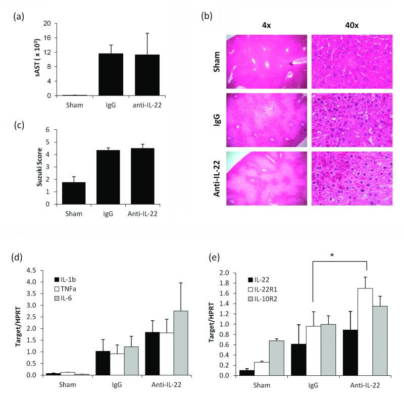
IL-22 neutralization in WT mice does not alter liver IRI but increases IL-22R1 transcription. We then analyzed whether neutralization of native IL-22 levels at 30 min prior to the ischemia insult may affect liver IRI cascade. As shown in Fig. 4a, by 6h of reperfusion sAST levels were similar in IgG and anti-IL-22 Ab groups. In addition, liver IRI Suzuki scores were comparable between both groups, exhibiting moderate to severe damage, with congestion and early necrotic changes (Fig 4b,c). Similarly, anti-IL-22 treatment did not affect the expression of IL-1β, TNFα, and IL-6 (Fig 4d). Interestingly, the expression of mRNA coding for IL-22R1 but not for IL-10R2 increased selectively in animals conditioned with anti-IL-22 Ab as compared with those given control IgG (Fig 4e).
Figure 4.
The effects of IL-22 neutralization (-30 min) in liver IRI (6 h of reperfusion, N=IgG and Anti-IL-22 Ab: 6/group, Sham N=4/group). (a) Peripheral serum AST levels. (b) Liver tissue histology (H&E staining; x4 or x40 magnification; representative liver sections shown). (c) Suzuki’s grading of the hepatocellular damage. RT-PCR analysis of liver specimens subjected to ischemia of (d) pro-inflammatory cytokines and (e) IL-22/IL-22 receptor complex (Statistical comparisons are between anti-IL-22 Ab and IgG groups. *: p<0.05)
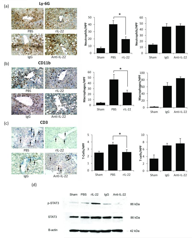
Exogenous rIL-22, but not anti-IL-22 Ab, reduces neutrophil, macrophage, and T cell sequestration in IR-livers. We performed immunohistochemical staining for cells that migrated to IR-livers at 6 h. As shown in Fig. 5a, rIL-22 treatment significantly decreased the number of hepatic neutrophils (Ly-6G), as compared with controls (19.6±3.3 vs. 40.3±5.1; p<0.05). Similarly, liver-accumulating macrophages (CD11b+) were significantly reduced after infusion of rIL-22 as compared with PBS-controls (Fig. 5b; 22.9 ± 4.4 vs. 46.7 ± 9.2; p<0.05). Although relatively few CD3+ T cells could be found in liver samples, their numbers decreased further after treatment with rIL-22, as compared with PBS-controls (Figure 5c: 2.6 ± 0.34 vs. 3.6 ± 0.32; p<0.05). No differences were found in liver accumulation of neutrophils (Fig. 5a), macrophages (Fig. 5b) and T cells (Fig. 5c) between anti-IL-22 Ab and control IgG treatment groups.
Figure 5.
The effects of rIL-22 pretreatment vs. IL-22 neutralization (6 h of reperfusion following 90 min of warm ischemia). Immunohistochemical staining of liver specimens for (a) neutrophils (Ly-6G); (b) macrophages (CD11b) and (c) T-cells (CD3). Left panels: representative liver sections (N=8/group for PBS and rIL-22, N=6/group for IgG and anti-IL-22 Ab) (x40 magnification). Right panels: Cell quantification /HPF±SEM. (*: p<0.05 for PBS vs. rIL-22 and IgG vs. anti-IL-22 Ab). Western blot-assisted detection of liver p-STAT3 and STAT3 in mice treated with PBS, rIL-22, IgG, or neutralizing IL-22 Ab (d). Beta-actin was used as internal control. Representative of two separate experiments is shown.
IL-22 functions in liver IRI via STAT3 phosphorylation
We studied p-STAT-3 expression in our liver IRI experimental system by Western blots. As shown in Fig. 5d, significantly elevated (p<0.05) levels of p-STAT3 were found in rIL-22 (0.29 ± 0.094 AU) as compared with PBS (0.069 ± 0.03 AU) group. Conversely, significantly decreased (p<0.05) STAT3 phosphorylation was detected in anti-IL-22 (0.026 ± 0.009 AU) as compared with control IgG (0.16 ± 0.051 AU) treated recipients.
Discussion
IL-22 is a cytokine with unique properties and therapeutic potential (10, 19-21). Although classified as a T cell-derived interleukin, IL-22 does not communicate between leukocytes, but instead it exerts action on target tissues that express functional IL-22R1. Our present findings complement data from other liver models of T cell hepatitis (15-17), lipogenesis/steatosis (22), chronic alcohol injury (23) and intestinal ulcerative colitis (24) by documenting the beneficial effects of exogenous rIL-22 in a model of partial hepatic warm ischemia followed by reperfusion. We have utilized a mouse model of ischemia and reperfusion, which is a well-established, highly reproducible model of acute local liver injury, and is ideal for furthering the study of IL-22 signaling.
Liver expression of IL-22 increases sharply in T-cell hepatitis at 6 h (16), so by using this as a positive control we first demonstrated that its levels remained very low and within sham-controls by 6 h of reperfusion, a period of the maximal hepatocellular damage in our liver model of 90 min warm ischemia (5, 8). The very low expression of IL-22 at this early time point of reperfusion is not surprising considering warm hepatic IRI is an innate-dominated immune response (5). Indeed, we consistently detect <5×10⁶ of mononuclear cells in mouse livers subjected to warm IRI (vs. <0.5×10⁶ in sham-control livers). Low IL-22 levels in our model were confirmed after its neutralization, which did not yield any significant differences in liver inflammation, histological damage, or leukocyte sequestration. In contrast, IL-22 neutralization (16) or IL-22 gene ablation (17) worsened liver damage in more stringent and fulminant Con A-induced T cell-mediated-hepatitis models.
IL-22 receptor consists of tissue-specific IL-22R1 and broadly expressed IL-10R2. (10). Tissues that lack IL-22R1 will not be a target for IL-22 under currently known molecular pathways. Unlike in ConA and IR liver injury models, we found low IL-22R1 expression in IFNAR KO mice that are resistant against hepatic IRI. Increased IL-22R1 was reported in mouse models of ConA hepatitis (16), Crohn’s disease (24, 25) and in human psoriatic skin lesions (26). These findings imply local IL-22R1 expression increases in stressed inflamed tissues. Future use of liver-specific IL-22 transgenic mice (20) and mice that lack IL-22R1 selectively on their hepatocytes (R. Sabat, personal communication) will be critical to address the true importance of IL-22R1 – IL-22 signaling in the liver.
Consistent with the pathogenic role of IL-22 neutralization to exacerbate hepatocellular damage (16, 17), livers in IL-22 transgenic mice were found to regenerate faster after partial hepatectomy, in association with increased expression of metallothionein 1 and 2, known to play an important role in liver regeneration (27). Hence, IL-22 overexpression is likely crucial in recovery after the liver damage. In our studies, ischemic livers subjected to 24 h of reperfusion demonstrated increased IL-22 expression, accompanied by more defined areas of necrosis and hepatocyte recovery. We believe that IL-22 may be essential in liver regeneration and tissue repair following IR insult, and pre-treatment with rIL-22 likely accelerates this process.
In parallel with increased IL-22R1 levels, IR-livers in WT mice expressed increased pro-inflammatory cytokine levels, and exhibited significant histological damage when compared with IFNAR KO mice. As cytokine assays were performed on serum samples, the results indicate that LPS did not contaminate the experimental preparations. Given the ability of IL-22 to promote hepatocyte survival, and augmented IL-22R1 expression in inflamed stressed tissue (20), we hypothesized that IL-22 may improve hepatic IRI pathology. Indeed, pre-treatment with rIL-22 significantly decreased serum AST levels, reduced hepatic sequestration of leukocytes, and expression of pro-inflammatory cytokines. Histological examination revealed improved tissue architecture after rIL-22, as evidenced by the Suzuki score, although some ischemic damage was still evident.
Little is known about the expression and upstream signaling of IL-22R1, although the downstream phosphorylation of STAT3 has been well described (16, 20). Indeed, deletion of STAT3 in hepatocytes abolished IL-22 mediated protection in alcoholic liver injury (16). Our data confirm an increase in STAT3 phosphorylation in livers after rIL-22 pretreatment, as well as significantly decreased STAT3 activation following IL-22 neutralization. Our data also show that IL-22R1 transcription increased sharply in settings of stress-induced liver inflammation. IL-22R1 expression decreased after treatment with rIL-22 and increased after IL-22 neutralization. However whether this is due to diminished local inflammation or direct negative IL-22 feedback mechanism remains to be determined. Recently described IL-22 transgenic/liver-specific STAT3 KO bigenic mouse in which IL-22 gene is overexpressed while STAT3 gene is deleted in hepatocytes (23) would be invaluable tool to address some of these key questions
Although IL-22 can decrease some injury from IR by modulating the inflammation response, it is not a completely preventative hepatoprotective mechanism in WT recipients, unlike near-complete injury prevention seen in mice deficient if Type-I IFN signaling (18). The ischemic injury, characterized by local metabolic disturbances of glycogen consumption, lack of oxygen supply, and adenosine triphosphate (ATP) depletion is followed by a brisk production of pro-inflammatory cytokine programs upon reperfusion (4). During early ischemic insult by 6 h of reperfusion, hepatocytes undergo a spectrum of damages, ranging from mild to severe, accompanied by increased expression of IL-22R1 and pro-inflammatory cytokine programs. As the reperfusion continues, hepatocytes either recover or progress to irreversible necrosis. After treatment with rIL-22, the damaged hepatocytes express IL-22R1 which is bound by rIL-22 to form the IL-22 receptor complex, thereby stimulating downstream signaling pathways that promote hepatocyte regeneration/survival. Moderately damaged hepatocytes, which previously would have continued to necrosis without IL-22, are now able to recover. Mildly damaged hepatocytes recover with or without therapy, whereas those severely damaged proceed to necrosis regardless of the treatment.
In conclusion, this is the first report that documents benefits of T cell-derived IL-22 immune modulation in stress-induced liver damage due to warm ischemia and reperfusion. Very low endogenous IL-22 levels at 6 h of IRI increase by 24 h of reperfusion when liver recovers from the ischemic insult. Hepatocyte IL-22R1 transcription during IR-liver inflammation correlates with the extent of hepatocellular damage. Exogenous IL-22 protein decreased local inflammation, diminished leukocyte sequestration, and IRI severity, whereas IL-22 neutralization did not appreciably alter IR-pathology. Thus, IL-22 treatment should be considered as a novel therapeutic option to prevent liver IRI in transplant recipients.
Materials and Methods
Animals
Male wild-type (WT; Harlan Laboratories, Indianapolis, IN) and type-I IFN receptor deficient (IFNAR KO; Dr. G. Cheng, UCLA) mice were used (C57BL/6; age 8-12 weeks). Animals, housed in the UCLA animal facility under specific pathogen-free conditions, received humane care according to the "Guide for the Care and Use of Laboratory Animals" prepared by the National Academy of Sciences (NIH publication 86-23 revised 1985).
Warm hepatic IRI model
We employed a warm hepatic IRI model, as described (5, 8). In brief, mice were injected with heparin (100 ug/kg i.v.) and an atraumatic clamp was placed to interrupt arterial and portal venous blood supply to the cephalad liver lobes. After 90 min the clamp was removed and the liver reperfused. Sham controls underwent the same procedure, but without vascular occlusion. Mice were sacrificed at 6 h and 24 h of reperfusion, at which time peripheral serum samples were obtained, and liver lobes subjected to ischemia were collected for RT-PCR, histologic, and immunohistochemical analyses. To study the role of IL-22, animals were treated i.v. at 30 min prior to the ischemia insult with rIL-22 (5 μg in 200 μl PBS; BioLegends, San Diego, CA) or goat anti-mouse IL-22 Ab (AF582; 50 μg in 200 μl PBS; R&D Systems, Minneapolis, MN). Controls received PBS (200 μl) or normal goat IgG (AB-1080C, 50 μg in 200 μl PBS R&D Systems), respectively. All treated mice were sacrificed at 6 h of reperfusion; serum and tissue samples were collected. The aspartate aminotransferase (AST) levels were screened in serum samples by an autoanalyzer (ANTECH Diagnostics, Los Angeles, CA).
ConA-induced hepatitis model
Mice were injected i.v. with ConA (15 μg/g; Sigma, St. Louis, MO) or PBS, as described (16, 17) and sacrificed at 6 h or 24 h for sera/liver sample analyses.
Liver histology/immunohistochemistry
Liver specimens were fixed in 10% buffered formalin and embedded in paraffin. Liver sections (4 μm) were stained with hematoxylin and eosin, and analyzed blindly using Suzuki’s histological criteria of liver damage (28).
For immunohistochemistry, snap-frozen liver cryostat sections (4 μm) were fixed in acetone. Endogenous peroxidase activity was inhibited by peroxidase blocking agent (Dako, Carpinteria, CA), and sections were blocked with 10% normal goat serum. Primary Abs (BD Biosciences) against CD11b (M1/70), Ly-6G (1A8) and CD3 (17A2) were diluted to 1/50, 1/200, and 1/50 in 3% normal goat serum, respectively. Secondary goat anti-rat IgG (Vector laboratories, Burlingame, CA) was diluted at 1/200. Sections were incubated with immunoperoxidase (ABC kit, Vector), washed, and developed with a 3,3′-diaminobenzidine kit (Vector). Negative controls were prepared by omission of the primary Ab. Sections were evaluated by counting positive staining cells (H&E) in portal triads of five high-power fields (HPFs) per slide, and results are expressed as average number of positive cells/HPF.
Quantitative RT-PCR
RNA was extracted from snap-frozen liver tissue samples using the TRIzol technique (Invitrogen, Carlsbad, CA) (6, 7). Five μg RNA was reverse-transcribed into cDNA using oligo-dT primers with Superscript III First-Strand Synthesis System (Invitrogen). Quantitative PCR was performed using the DNA Engine with Chromo 4 Detector (MJ Research, Waltham, MA). In a final volume of 20 μL, the following were added: 1X SuperMix (Platinum SYBR Green qPCR SuperMix-UDG with ROX, Invitrogen), complementary cDNA, and 0.2 μM of each primer. Amplification conditions were: 50°C (2 min), 95°C (5 min) followed by 45 cycles of 95°C (15 sec) and 60°C (30 sec). Primers were used to detect IL-1β, TNFα, IL-6, IL-22, IL-22R1 and IL-10R2. Replication levels were calculated using a 1:5 standard curve dilution and HPRT replication was utilized as a standard housekeeping gene.
Western blots
Protein was extracted from liver tissue with protein lysis buffer (50 mM HEPES, 10 mM MgCl2, 1 mM EDTA, 1 mM EGTA, 0.08 mM Sodium Molybdate, 2 mM Sodium Pyrophosphate, 0.01% Triton X100) with Protease Inhibitor cocktail (Sigma) and PhosSTOP phosphatase inhibitor (Roche Diagnostics, Indianapolis, IN). Proteins were prepared in Loading Buffer (EC-886, National Diagnostics, Atlanta, GA), subjected to 12% sodium dodecyl sulfate polyacrylamide gel electrophoresis in TRIS/Glycing/SDS buffer (Bio-Rad, Hercules, CA) and transferred to PVDF membrane in TRIS/Glycine buffer. Primary Ab blotting was performed for p-STAT3, STAT3 and β-actin (Cell Signaling, Beverly, MA). Secondary Ab included anti-rabbit HRP- and anti-rat HRP-linked IgG. Detection was performed with the Super Signal West Pico chemiluminescent substrate system (Thermo Fisher Scientific, Rockford, IL). The relative protein quantities were determined by densitometer, and expressed in absorbance units (AU).
Statistical analysis
All values are expressed as mean ± SEM. Data were analyzed with an unpaired two-sided Student t-test. P<0.05 was considered statistically significant. Unless stated otherwise, all statistical comparisons were between experimental groups (WT vs. IFNAR KO, PBS vs. rIL-22, IgG vs. anti-IL-22 Ab). Results from sham experiments are displayed for reference.
Acknowledgements
This work was supported by NIH Grants DK 062357-07; DK 062357-06S1; DK 083408-01A1 and The Dumont Research Foundation.
Abbreviations
- AST
aspartate aminotransferase
- IRI
ischemia-reperfusion injury
- IL
interleukin
- IFNAR
type-I Interferon Receptor
- HPRT
hypoxanthine-guanine-phosphoribosyltransferase
Footnotes
Authors’ contribution: PJC - participated in research design, performance of the research, data anaysis and writing the paper; YU - performed microsuirgery procedures and participated in anaysis; WC and MA – performed immunohistological stainings ; CL – provided immunohistological expertise ; RS – discussant and advisor ; RWB - participated in discussion and provided partial funding; JWKW – overall adviser, facilitated the final manuscript vesrion, sponsored the project. The authors declare no conflict of interest.
This is a PDF file of an unedited manuscript that has been accepted for publication. As a service to our customers we are providing this early version of the manuscript. The manuscript will undergo copyediting, typesetting, and review of the resulting proof before it is published in its final citable form. Please note that during the production process errors may be discovered which could affect the content, and all legal disclaimers that apply to the journal pertain.
References
- 1.Vardanian AJ, Busuttil RW, Kupiec-Weglinski JW. Molecular Mediators of Liver Ischemia and Reperfusion Injury: A Brief Review. Mol Med. 2008;14:337. doi: 10.2119/2007-00134.Vardanian. [DOI] [PMC free article] [PubMed] [Google Scholar]
- 2.Land W, Messmer K. The impact of ischemia/reperfusion injury on specific and non-specific early and late chronic events after organ transplantation. Transplant Rev. 1996;10:108. [Google Scholar]
- 3.Fondevila C, Busuttil RW, Kupiec-Weglinski JW. Hepatic ischemia/reperfusion injury- -a fresh look. Exp Mol Pathol. 2003;74:86. doi: 10.1016/s0014-4800(03)00008-x. [DOI] [PubMed] [Google Scholar]
- 4.Zhai Y, Busuttil RW, Kupiec-Weglinski JW. Liver ischemia and reperfusion injury: New insights into mechanisms of innate–adaptive immune-mediated tissue inflammation. Am J Transplant. 2011;11:1563. doi: 10.1111/j.1600-6143.2011.03579.x. [DOI] [PMC free article] [PubMed] [Google Scholar]
- 5.Zhai Y, Shen XD, O’Connell R, et al. Cutting edge: TLR4 activation mediates liver ischemia/reperfusion inflammatory response via IFN regulatory factor 3-dependent MyD88-independent pathway. J Immunol. 2004;173:7115. doi: 10.4049/jimmunol.173.12.7115. [DOI] [PubMed] [Google Scholar]
- 6.Shen XD, Ke B, Zhai Y, et al. Toll-like receptor and heme-oxygenase-1 signaling in hepatic ischemia/reperfusion injury. Am J Transplant. 2005;5:1793. doi: 10.1111/j.1600-6143.2005.00932.x. [DOI] [PubMed] [Google Scholar]
- 7.Caldwell CC, Tschoep J, Lentsch AB. Lymphocyte function during hepatic ischemia/reperfusion injury. J Leukoc Biol. 2007;82:457. doi: 10.1189/jlb.0107062. [DOI] [PubMed] [Google Scholar]
- 8.Shen X, Wang Y, Gao F, et al. CD4 T cells promote tissue inflammation via CD40 signaling without de novo activation in a murine model of liver ischemia/reperfusion injury. Hepatology. 2009;50:1537. doi: 10.1002/hep.23153. [DOI] [PMC free article] [PubMed] [Google Scholar]
- 9.Kuboki S, Sakai N, Tschop J, Edwards MJ, Lentsch AB, Caldwell CC. Distinct contributions of CD4+ T cell subsets in hepatic ischemia/reperfusion injury. Am J Physiol Gastrointest Liver Physiol. 2009;296:G105. doi: 10.1152/ajpgi.90464.2008. [DOI] [PMC free article] [PubMed] [Google Scholar]
- 10.Wolk K, Witte E, Witte K, Warszawska K, Sabat R. Biology of interleukin-22. Semin Immunopathol. 2010;32:17. doi: 10.1007/s00281-009-0188-x. [DOI] [PubMed] [Google Scholar]
- 11.Ma HL, Liang S, Li J, et al. IL-22 is required for Th17 cell-mediated pathology in a mouse model of psoriasis-like skin inflammation. J Clin Invest. 2008;118:597. doi: 10.1172/JCI33263. [DOI] [PMC free article] [PubMed] [Google Scholar]
- 12.Ikeuchi H, Kuroiwa T, Hiramatsu N, et al. Expression of interleukin-22 in rheumatoid arthritis: potential role as a proinflammatory cytokine. Arthritis Rheum. 2005;52:1037. doi: 10.1002/art.20965. [DOI] [PubMed] [Google Scholar]
- 13.Wolk K, Witte E, Hoffmann U, et al. IL-22 induces lipopolysaccharide-binding protein in hepatocytes: A potential systemic role of IL-22 in Crohn’s disease. J Immunol. 2007;178:5973. doi: 10.4049/jimmunol.178.9.5973. [DOI] [PubMed] [Google Scholar]
- 14.Aujla SJ, Chan YR, Zheng M, et al. IL-22 mediates mucosal host defense against Gram-negative bacterial pneumonia. Nat Med. 2008;14:275. doi: 10.1038/nm1710. [DOI] [PMC free article] [PubMed] [Google Scholar]
- 15.Zenewicz LA, Yancopoulos GD, Valenzuela DM, Murphy AJ, Stevens S, Flavell RA. Innate and adaptive Interleukin-22 protects mice from inflammatory bowel disease. Immunity. 2008;29:947. doi: 10.1016/j.immuni.2008.11.003. [DOI] [PMC free article] [PubMed] [Google Scholar]
- 16.Radaeva S, Sun R, Pan HN, Hong F, Gao B. Interleukin-22 (IL-22) plays a protective role in T cell-mediated murine hepatitis: IL-22 is a survival factor for hepatocytes via STAT3 activation. Hepatology. 2004;39:1332. doi: 10.1002/hep.20184. [DOI] [PubMed] [Google Scholar]
- 17.Zenewicz LA, Yancopoulos GD, Valenzuela DM, Murphy AJ, Karow M, Flavell RA. IL-22 but not IL-17 provides protection to hepatocytes during acute liver inflammation. Immunity. 2007;27:647. doi: 10.1016/j.immuni.2007.07.023. [DOI] [PMC free article] [PubMed] [Google Scholar]
- 18.Zhai Y, Qiao B, Gao F, et al. Type I, but not Type II, Interferon is critical in liver injury induced after ischemia and reperfusion. Hepatology. 2008;47:199. doi: 10.1002/hep.21970. [DOI] [PubMed] [Google Scholar]
- 19.Wolk K, Sabat R. Interleukin 22: A novel T- and NK-cell derived cytokine that regulates the biology of tissue cells. Cytokine Growth Factor Rev. 2006;17:367. doi: 10.1016/j.cytogfr.2006.09.001. [DOI] [PubMed] [Google Scholar]
- 20.Park O, Wang H, Weng H, et al. In Vivo consequences of liver-specific Interleukin-22 expression in mice: Implications for human liver disease progression. Hepatology. 2011;54:252. doi: 10.1002/hep.24339. [DOI] [PMC free article] [PubMed] [Google Scholar]
- 21.Wolk K, Kunz S, Witte E, Friedrich M, Asadullah K, Sabat R. IL-22 increases the innate immunity of tissues. Immunity. 2004;21:241. doi: 10.1016/j.immuni.2004.07.007. [DOI] [PubMed] [Google Scholar]
- 22.Yang L, Zhang Y, Wang L, et al. Amelioration of high fat diet induced liver lipogenesis and hepatic steatosis by interleukin-22. J Hepatol. 2010;53:339. doi: 10.1016/j.jhep.2010.03.004. [DOI] [PubMed] [Google Scholar]
- 23.Ki SH, Park O, Zheng M, et al. Interleukin-22 treatment ameliorates alcoholic liver injury in a murine model of chronic-binge ethanol feeding: role of signal transducer and activator of transcription 3. Hepatology. 2010;52:1291. doi: 10.1002/hep.23837. [DOI] [PMC free article] [PubMed] [Google Scholar]
- 24.Sugimoto K, Ogawa A, Mizoguchi E, et al. IL-22 ameliorates intestinal inflammation in a mouse model of ulcerative colitis. J Clin Invest. 2008;118:534. doi: 10.1172/JCI33194. [DOI] [PMC free article] [PubMed] [Google Scholar]
- 25.Brand S, Beigel F, Olszak T, et al. IL-22 is increased in active Crohn’s disease and promotes pro-inflammatory gene expression and intestinal epithelial cell migration. Am J Physiol Gastrointest Liver Physiol. 2006;290:G827. doi: 10.1152/ajpgi.00513.2005. [DOI] [PubMed] [Google Scholar]
- 26.Tohyama M, Hanakawa Y, Shirakata Y, et al. IL-17 and IL-22 mediate IL-20 subfamily cytokine production in cultured keratinocytes via increased IL-22 receptor expression. Eur J Immunol. 2009;39:2779. doi: 10.1002/eji.200939473. [DOI] [PubMed] [Google Scholar]
- 27.Cherian MG, Kang YJ. Metallothionein and liver cell regeneration. Exp Biol Med. 2006;231:138. doi: 10.1177/153537020623100203. [DOI] [PubMed] [Google Scholar]
- 28.Suzuki S, Toledo-Pereyra LH, Rodriguez FJ, Cejalvo D. Neutrophil accumulation as an important factor in liver ischemia and reperfusion injury. Modulating effects of FK506 and cyclosporine. Transplantation. 1993;55:1265. doi: 10.1097/00007890-199306000-00011. [DOI] [PubMed] [Google Scholar]


