
FIGURE 3.
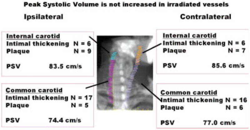
The number, type, and location of lesions identified by ultrasound. For peak systolic volume (PSV) (ipsilateral vs contralateral), the P value was not found to be significant for each vessel using the Student t test and the signed rank test.