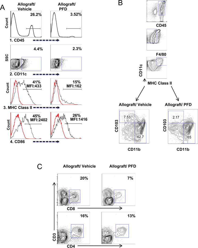
Figure 2.
A. PFD treatment reduces activation of DC in vivo
CD45+ and CD11c+ cells were isolated based on flow cytometry from both untreated and PFD treated mouse lung allografts seven days after transplantation. 1.) Anti-CD45 marker was used to determine total leukocytes in allografts. There is a decrease in percentage of CD45+ cells in transplanted lung with PFD treatment. 2.) CD11c+ expression on gated CD45+ cells showed a decrease of DCs in PFD treated allografts. 3. and 4.) To determine activation of DCs, the percentage/MFI of MHC class II and CD86 expression on the gated CD11c+ cells were analyzed and the results reveal a decrease in activation of DCs in PFD treated allografts. Illustrated are representative analyses of the MHC class II and CD86 with isotype controls for 4 PFD and untreated transplant samples.
B. PFD treatment reduce CD11b− CD103+ DC
Highly purified CD11c+ cells were sorted by magnetic cells sorting from both untreated and PFD treated mouse lung allografts seven days after transplantation. CD45+F4/80− CD11c+ MHC Class II+ cells were gated and surface expression of CD11b and CD103 analyzed. The result showed a decrease in CD11b−CD103+ cells but not CD11b high cells. Illustrated are representative analyses of each for 4 individual PFD treated and untreated transplant samples. Illustrated are representative analyses for 4 PFD and untreated transplant samples.
C. PFD treatment reduce lung CD8 T cells
Seven days after transplantation, single cell suspensions from lung allografts of both untreated and PFD treated mouse recipients were stained with anti-CD45, anti-CD3, anti-CD4 and anti-CD8. There was a decrease in the percentage of CD3+CD8+ T cells but not CD3+CD4+ T cells. Illustrated are representative analyses for 4 PFD and untreated transplant samples.

