Abstract
Cardiac myxoma is a benign (non-malignant) neoplasm that represents the most common primary tumour of the heart. We present the case of a 36 year old woman with background hypertension who presented with features of left ventricular failure and seizures, and was found during transthoracic echocardiography to have left atrial myxoma protruding through the mitral valve orifice. She subsequently had excision of the atrial myxoma. The usefulness of early transthoracic echocardiography in any patient presenting with features of heart failure even when the aetiology seems obvious cannot be over-emphasised.
Keywords: atrial, myxoma, mitral
Introduction
Cardiac myxoma is a benign (non-malignant) neoplasm that represents the most common primary tumour of the heart. Although the left atrium is the most commonly involved site of origin in 75% of cases, it can arise from any of the cardiac chambers.1–3 Although most cases of cardiac myxomas occur sporadically, familial lesions and lesions associated with a clinical complex have also been reported.4,5 Most affected patients with cardiac myxoma present with at least one feature of a classically described triad that includes constitutional symptoms (such as fever, weight loss or symptoms resembling connective tissue disease), obstructive symptoms and embolic events.6
Case Report
A 36 year-old woman was hospitalized after presenting with mild exertional dyspnoea, orthopnoea, easy fatiguability and pre-syncope one month before admission. She also had palpitations one week before admission and two episodes of seizures on the day of admission.
The only significant past medical history was hypertension which was diagnosed one year prior to admission and for which she was commenced on low dose thiazide diuretic.
Systemic examination revealed a pulse rate of 106 beats per minute which was of small volume and there were few ectopics. The blood pressure was 110/80 mmHg. The respiratory rate was 32 breaths/ minute and there were fine bibasal crepitations.
Full blood count showed an haemoglobin concentration of 11.2 gm/dL and white blood cell count of 12,800/mm3 with a neutrophil count of 73% and lymophocyte count of 19/mm3.
Chest radiograph showed features of left atrial enlargement and upper lobe diversion while electrocardiography showed sinus tachycardia with a heart rate of 110 beats/min, occasional premature ventricular complexes and left atrial enlargement.
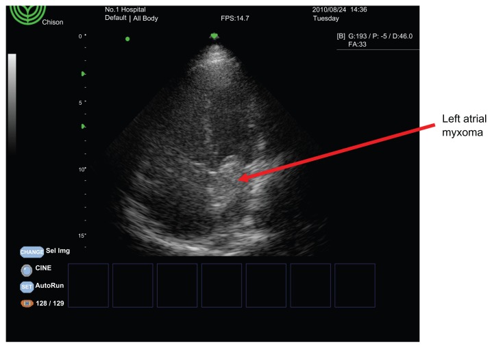
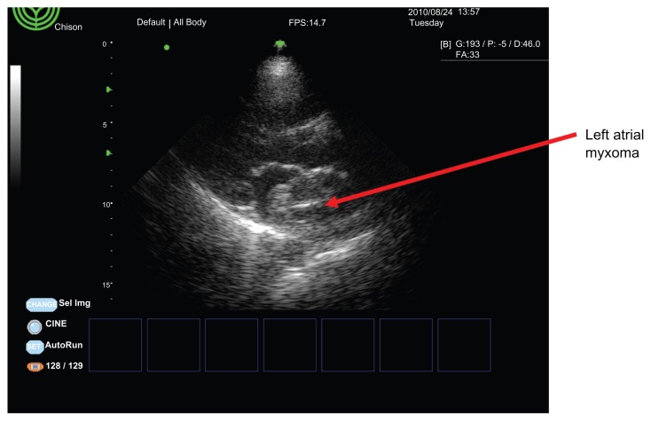
Transthoracic echocardiography showed a large mobile left atrial mass measuring 40.8 mm by 25.0 mm and attached to the inter-atrial septum (Figs. 1 and 2). The tumour was prolapsing across the mitral valve orifice in diastole causing a functional stenosis. There was a mild mitral regurgitation and moderate tricuspid regurgitation with a mild pulmonary hypertension. The systolic function was preserved with an estimated left ventricular ejection fraction of 58%.
Figure 1.
Apical for-chamber view showing the left atrial myxoma.
Figure 2.
Parasternal long-axis view showing the left atrial myxoma.
Patient had the tumour excised and the histology showed myxomatous bluish pink background with stellate cells and compressed blood vessels, with no atypical malignant cells in keeping with myxoma.
Discussion
Primary cardiac tumours occur infrequently with an incidence of 0.0017%–0.19% in autopsy series performed in non-selected populations.1–2 Cardiac myxomas are more common among women with a female-to-male ratio of 2:1 and occur more frequently between ages 30–60 years.1–2 Incidentally our patient is female and falls within this age bracket at 36 years.
Left atrial myxomas become symptomatic when they obstruct the mitral valve, embolize or cause systemic effects.7 The symptoms of exertional dyspnoea, orthopnoea, easy fatigue and pre-syncope in our patient are most likely due to the obstruction of the mitral valve orifice by the tumour mass. On the other hand, seizures experienced by our patients can be partly attributable to obstruction of the mitral valve orifice with reduced blood and oxygen flow to the brain. It can also be explained by arrhythmias (patient had palpitations with premature ventricular complexes on 12-lead ECG). Remotely, it can be due to embolization to the brain.
Although more than half of left atrial myxoma shows obstructive symptoms, it is only in 10% of patients that it will cause severe stenosis just like in our patient. The severity of obstruction is determined by the size, location and mobility of the myxoma.8 Although the myxoma size in our case is average covering only about 50%–60% of the left atrium, its mobility and proximity to the mitral valve orifice must have made the obstructive effect to be severe.
The early echocardiographic examination played a very important role in the diagnosis and management of this patient emphasizing the need for early echocardiographic examination of any patient presenting with features of heart failure even when you think the aetiology of heart failure is obvious.
Acknowledgements
Our appreciation goes to every member of staff of Cardiology Unit, Department of Medicine, University of Abuja Teaching Hospital, Gwagwalada, Abuja.
Footnotes
Author Contributions
DBO drafted the manuscript while MHM,OO,ZH and HO helped in the case summary and KS proof read the manuscript. All authors reviewed and approved of the final manuscript.
Competing Interests
Author(s) disclose no potential conflicts of interest.
Disclosures and Ethics
As a requirement of publication author(s) have provided to the publisher signed confirmation of compliance with legal and ethical obligations including but not limited to the following: authorship and contributorship, conflicts of interest, privacy and confidentiality and (where applicable) protection of human and animal research subjects. The authors have read and confirmed their agreement with the ICMJE authorship and conflict of interest criteria. The authors have also confirmed that this article is unique and not under consideration or published in any other publication, and that they have permission from rights holders to reproduce any copyrighted material. Any disclosures are made in this section. The external blind peer reviewers report no conflicts of interest.
Funding
Author(s) disclose no funding sources.
References
- 1.Reynen K. Cardiac myxomas. N Engl J Med. 1995;333:1610–7. doi: 10.1056/NEJM199512143332407. [DOI] [PubMed] [Google Scholar]
- 2.Goswami KC, Shrivastava S, Bahl VK, Saxena A, Manchanda SC, Wasir HS. Cardiac myxomas: clinical and echocardiographic profile. Int J Cardiol. 1998;63:251–9. doi: 10.1016/s0167-5273(97)00316-1. [DOI] [PubMed] [Google Scholar]
- 3.Ojji Dike B, Ajiduku Stella S, Omonua Omonuyi O, Abdulkareem Lukman L, Parsonage Will. A probable right atrial myxoma prolapsing through the tricuspid value into the right ventricle: a case report. Cases Journal. 2008;1:386. doi: 10.1186/1757-1626-1-386. [DOI] [PMC free article] [PubMed] [Google Scholar]
- 4.McCarthy PM, Piehler JM, Schaff HV, et al. The significance of multiple, recurrent and ‘complex’ cardiac myxomas. J Thorac Cardiovasc Surg. 1986;91:389–96. [PubMed] [Google Scholar]
- 5.Pinede L, Duhaunt P, Loire R. Clinical presentation of left atrial cardiac myxoma. A series of 112 consecutive cases. Medicine (Baltimore) 2001;80:159–72. doi: 10.1097/00005792-200105000-00002. [DOI] [PubMed] [Google Scholar]; Parissis JT, Zezas S, Sfiras N, Kastellanos S. An atypical left atrial myxoma causing intracavitary pressure gradient and typical diastolic transmitral flow of severe mitral stenosis. Int J Cardiol. 2005;102:165–7. doi: 10.1016/j.ijcard.2004.05.071. [DOI] [PubMed] [Google Scholar]
- 6.Bajraktari G, Emini M, Berisha V, et al. Giant left atrial myxoma in an elderly patient: natural history over a 7-year period. J Clin Ultrasound. 2006;34:461–3. doi: 10.1002/jcu.20265. [DOI] [PubMed] [Google Scholar]
- 7.Panidis IP, Mintz GS, Mc Allister M. Haemodynamic Consequences of left atrial myxomas as assessed by Doppler ultrasound. Am Heart J. 1986;111:927–31. doi: 10.1016/0002-8703(86)90644-7. [DOI] [PubMed] [Google Scholar]
- 8.Carney JA. The Carney complex (myxomas, spotty pigmentation, endocrine overactivity and schannomas) Dermatol Clin. 1995;13:19–26. [PubMed] [Google Scholar]


