Summary
Background:
Blow-out fracture of the orbit is a common injury. However, not many cases are associated with massive subcutaneous emphysema. Even fewer cases are caused by minor trauma or are associated with barotrauma to the orbit due to sneezing, coughing, or vomiting. The authors present a case of blow-out fracture complicated by extensive subcutaneous and mediastinal emphysema that occurred without any obvious traumatic event.
Case Report:
A 43-year-old man presented to the Emergency Department with a painful right-sided exophthalmos that he had noticed in the morning immediately after waking up. The patient also complained of diplopia. Physical examination revealed exophthalmos and crepitations suggestive of subcutaneous emphysema. The eye movements, especially upward gaze, were impaired. CT showed blow-out fracture of the inferior orbital wall with a herniation of the orbital soft tissues into the maxillary sinus. There was an extensive subcutaneous emphysema in the head and neck going down to the mediastinum. The patient did not remember any significant trauma to the head that could explain the above mentioned findings. At surgery, an inferior orbital wall fracture with a bony defect of 3×2 centimeter was found and repaired.
Conclusions:
Blow-out fractures of the orbit are usually a result of a direct trauma caused by an object with a diameter exceeding the bony margins of the orbit. In 50% of cases, they are complicated by orbital emphysema and in 4% of cases by herniation of orbital soft tissues into paranasal sinuses. The occurrence of orbital emphysema without trauma is unusual. In some cases it seems to be related to barotrauma due to a rapid increase in pressure in the upper airways during sneezing, coughing, or vomiting, which very rarely leads to orbital wall fracture. Computed tomography is the most accurate method in detecting and assessing the extent of orbital wall fractures.
Keywords: blow-out fracture, complications, computed tomography
Background
Blow-out fracture of the orbit is a common injury. However, not many cases are associated with massive subcutaneous emphysema. Even fewer cases are caused by minor trauma or are associated with barotrauma to the orbit due to sneezing, coughing, or vomiting. The authors present a case of blow-out fracture complicated by extensive subcutaneous and mediastinal emphysema that occurred without any obvious traumatic event.
Case Report
A 43-year-old patient was admitted to the Department of Cranio-Maxillofacial Surgery of Medical University of Warsaw due to a right-sided exophthalmos, diplopia and pain located in the right orbit. The problem was noticed on awakening, after the night’s rest. The patient, however could not recall any incident that might have caused the above mentioned symptoms.
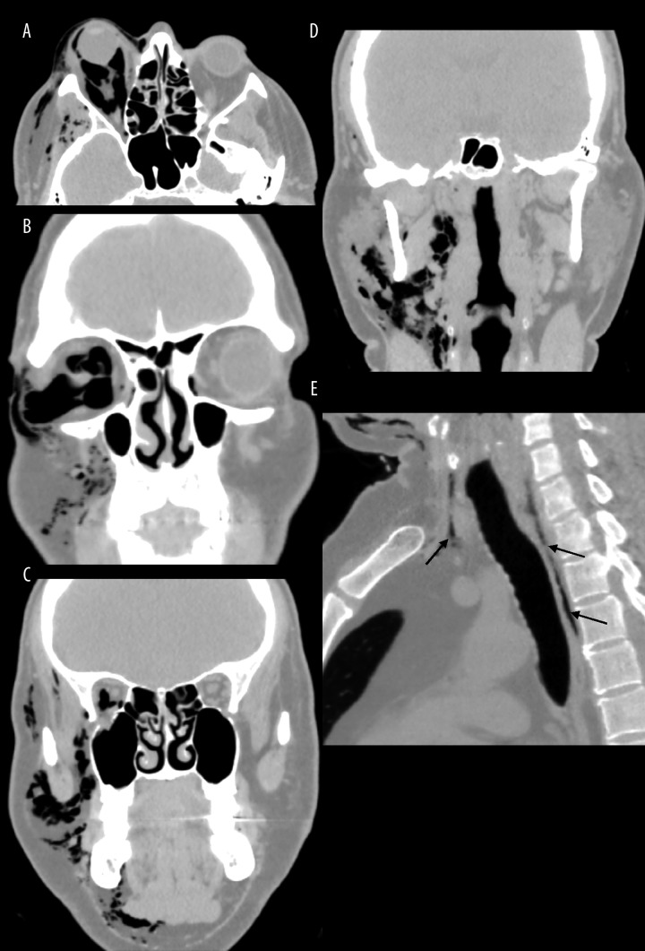
Physical examination revealed severe right-sided exophthalmos with upgaze restriction of the right eye, accompanied by diplopia. Additionally, extensive subcutaneous emphysema was palpable in the right palpebrae, cheek and submandibular area on that side. A spiral CT of the facial skeleton was performed using 1-mm slices with soft tissue and bone algorithm. A large intraorbital emphysema with exophthalmos was found (the distance from corneal apex to the line connecting orbital rims was 29 mm (normal range up to 21 mm)) (Figure 1). Air was entering soft tissues of temporal and subtemporal fossa, buccal space, masticator space, parotid gland, para- and retropharyngeal space, carotid space and along the visceral space of the neck to the anterior and posterior mediastinum (Figure 2). Multiplanar reconstructions in the “bone” window showed a right-sided blow-out fracture of the lower orbital wall with slight herniation of intraorbital adipose tissue into the maxillary sinus (Figure 3).
Figure 1.

Axial image in soft tissue window. There is a large right-sided exophthalmos.
Figure 2.
Axial image and multiplanar reformations in lung window show a large intraorbital emphysema (A,B) extending into the spaces of the neck (C,D) and the superior mediastinum (arrows in E).
Figure 3A–C.
Coronal and sagittal reformatted images in bony window show a fracture of the inferior orbital wall on the course of the infraorbital canal with a formation of a bony trapdoor (arrows). There is a small protrusion of the retrobulbar fat into the maxillary sinus.
Basing on a physical examination and additional tests, an isolated right orbital floor fracture was diagnosed (IOFF according to H. Wanyura classification) [1]. Ophthalmology consultation confirmed the imbalance of oculomotor muscles in the right eye, being an indication for surgical treatment.
Under general anesthesia with endotracheal intubation, a transconjunctival incision was performed in the lower recess of the conjunctival sac of the right eye. Right orbital emphysema was decompressed. The lower margin and the floor of the right orbit was exposed subsequently. No fractures fissures of the lower orbital margin were not visualized, while in the orbital floor, a bone defect (25×20 mm in size) was found with a fracture fissure descending from the medial wall of the right orbit, posteriorly to the lacrimal sac and running down to the inferior orbital fissure in posterior 2/3 of the orbital floor and toward the back, to the orbital process of palatine bone. Orbital tissue hernia trapped within the bone defect was released. Orbital floor was reconstructed using a synthetic resorbable implant, 20 x 30 mm in size. At the end of operation, conjunctival sac was sutured.
Postoperative healing proceeded without complications. As a result of surgery, diplopia resolved completely and the patient regained full mobility of the right eye.
Discussion
Blow-out fracture is characterized by a damage to the orbital walls with intact orbital margins and bone fragments displaced outside the orbit [2,3]. It is usually due to a trauma caused by an object with a diameter exceeding the bony margins of the orbit [4]. There is a number of theories explaining the mechanism of this type of fracture. The most probable is the hydraulic theory, assuming that the intraorbital pressure rises during trauma and the acting forces are transmitted by compression and subsequent decompression of the eye ball [2,4].
Blow-out fractures usually occur as a consequence of transport-related injuries or battery. These fractures most often involve the inferior (36.7%) and medial (31%) orbital wall. Orbital roof is rarely affected [3]. They can be coexistent with orbital soft tissue (muscles and fat) entrapment [2,3]. It is estimated that orbital soft tissue hernia occurs in approximately 4% of cases, whereas the oculomotor muscle incarceration is found in 2.6% of cases [3]. In orbital floor fracture the inferior rectus muscle is affected, while the medial wall fracture involves the medial rectus muscle. Around 50% of blow-out fracture cases are complicated with orbital emphysema [5,6] due to pathological accumulation of air within orbit caused by the fracture of paranasal sinus walls (usually ethmoid) accompanied by a rapture of mucous membrane [3,7–9].
Orbital emphysema in about 63% of cases occurs as a result of blunt orbital or facial trauma [3]. Other causes include postoperative complications, infection, esophageal rupture and barotrauma [3,10]. Cases of spontaneous orbital emphysema caused by sneezing, cough or nose blowing are very rare [7,11–13]. The pressure evoked by forceful nose blowing rises to 66 mmHg, as estimated. Sneezing with the oral and nasal passages blocked may evoke a pressure up to 176 mmHg [9].
Orbital emphysema without impaired vision is not a life-threatening condition and usually resolves spontaneously within 2 weeks due to formation of fibrous tissue between the paranasal sinus and the orbit. In the meantime, patients are told to avoid sneezing, nose blowing, vomiting, coughing or any other activities that may lead to increased pressure in the nasal cavity, including diving and flying by plane [7,9,11,14–16]. Nevertheless, orbital emphysema may also lead to serious complications, particularly in case of compressive orbital emphysema when air enters the orbit but cannot leave it freely [5]. The increase of intraorbital pressure followed by intrabulbar hypertension may cause an occlusion of central retinal artery and optic nerve ischemia (particularly, if intrabulbar pressure exceeds 65–70 mmHg), which may result in permanent or transient loss of vision [6,8,11,17]. From that reason, every orbital fracture needs eye examination assessing visual acuity, pupillary reactions and ocular motility as well as fundoscopic examination [6,8,11]. Other complications of orbital emphysema include diplopia, ocular motility disorders and infection spreading by direct continuity from paranasal sinuses [7,10,14,18]. Diplopia with restricted ocular movements and incarceration of intraorbital soft tissues is an indication for surgical treatment within 2 weeks [19].
Orbital emphysema is usually related to recent craniofacial trauma, particularly involving paranasal sinuses [3,6,14]. The occurrence of orbital emphysema, however, may be delayed. It is usually directly associated with sneezing, nose blowing and cough, when air enters the orbit via bone defect and mucous membrane rupture, as a result of the increased pressure inside nasal cavities [7,10,12,14,16]. There were reports of orbital emphysema occurring immediately after nose blowing in a patient who had suffered from orbital trauma 5 months earlier without ocular symptoms at that time [17], as well as a case of orbital emphysema while plane landing in a patient with a history of trauma 2 days earlier [20].
There were also cases of spontaneous orbital emphysema after forceful nose blowing, sneezing or cough, without a history of previous trauma, often as a result of lamina papyracea (orbital lamina) rupture [2,7,11–13,19,21]. The orbital lamina, as the thinnest orbital wall with a thickness of 0.25 mm, is a part the most vulnerable to barotraumas [2,4,6,11,15,17]. Additionally, possible developmental defects of this structure can promote orbital emphysema in case of increased pressure inside the nasal cavity. Chronic paranasal sinusitis can additionally reduce its resistance [6,11,17].
Orbital emphysema coexistent with massive subcutaneous emphysema and emphysema within the neck region and mediastinum is a very rare condition [22,23]. Mediastinal emphysema usually develops due to tracheal, bronchial, pulmonal or esophageal rupture [22], and rarely as a result of facial trauma [24]. The natural spreading of air is along fascial and soft tissue from mediastinum towards the neck and head. Opposite direction of spreading from orbit and paranasal sinuses towards mediastinum requires increased pressure in the upper respiratory tract as it happens during forceful nose blowing, sneezing and cough in patients with past fractures of orbit or paranasal sinus [22–24].
The case reported hereby is unusual considering the coexistence of extensive subcutaneous emphysema and also the lack of evident trauma history. The patient asserted that he had noticed exophthalmos immediately after awakening without any direct cause. Scrupulous history taking revealed a mild head injury of the temporal area on the previous night. It seems rather unlikely that orbital floor fracture was a result of this trauma, not involving orbital area itself. Other possible etiology includes previous trauma, remaining asymptomatic until increased pressure in the upper respiratory tract occurred [7,19]. However, a spontaneous orbital floor fracture due to forceful nose blowing or cough cannot be excluded. So far only 3 cases of orbital emphysema due to orbital floor fracture spontaneously occurring after forceful nose blowing have been described [7,19,21]. One of them was reported in a 70-year-old woman. It was suggested that fracture of the inferior orbital wall in this case was a result of impaired resistance due to aging processes [21]. Other cases were reported in patients around 40-year-old, with coexistent inflammatory changes in maxillary sinuses. It has to be emphasized that no inflammatory changes of paranasal sinuses potentially leading to resistance reduction of orbital walls were found in our patient.
References:
- 1.Samolczyk-Wanyura D, Wanyura H. Kliniczno-anatomopatologiczna klasyfikacja złamań górnego masywu twarzy. Czas Stomatol. 1991;64(12):848–55. [Google Scholar]
- 2.Suzuki H, Furukawa M, Takahashi E, et al. Barotraumatic blowout fracture of the orbit. Auris Nasus Larynx. 2001;28(3):257–59. doi: 10.1016/s0385-8146(00)00122-x. [DOI] [PubMed] [Google Scholar]
- 3.Lee HJ, Jilani M, Frohman L, et al. CT of orbital trauma. Emerg Radiol. 2004;10(4):168–72. doi: 10.1007/s10140-003-0282-7. [DOI] [PubMed] [Google Scholar]
- 4.Al-Shammari L, Majithia A, Adams A, et al. Tension pneumo-orbit treated by endoscopic, endonasal decompression: case report and literature review. J Laryngol Otol. 2008;122(3):8. doi: 10.1017/S002221510700165X. [DOI] [PubMed] [Google Scholar]
- 5.Key SJ, Ryba F, Holmes S, et al. Orbital emphysema – the need for surgical intervention. J Craniomaxillofac Surg. 2008;36(8):473–76. doi: 10.1016/j.jcms.2008.04.004. [DOI] [PubMed] [Google Scholar]
- 6.Muhammad JK, Simpson MT. Orbital emphysema and the medial orbital wall: a review of the literature with particular reference to that associated with indirect trauma and possible blindness. J Craniomaxillofac Surg. 1996;24(4):245–50. doi: 10.1016/s1010-5182(96)80008-4. [DOI] [PubMed] [Google Scholar]
- 7.García de Marcos JA, del Castillo-Pardo de Vera JL, Calderón-Polanco J. Orbital floor fracture and emphysema after nose blowing. Oral Maxillofac Surg. 2008;12(3):163–65. doi: 10.1007/s10006-008-0119-3. [DOI] [PubMed] [Google Scholar]
- 8.Harmer SG, Ethunandan M, Zaki GA, et al. Sudden transient complete loss of vision caused by nose blowing after a fracture of the orbital floor. Br J Oral Maxillofac Surg. 2007;45(2):154–55. doi: 10.1016/j.bjoms.2005.06.008. [DOI] [PubMed] [Google Scholar]
- 9.Vairaktaris E, Moschos MM, Vassiliou S, et al. Delayed appearance of diplopia due to orbital emphysema after repair of orbital fractures. Oral Surg Oral Med Oral Pathol Oral Radiol Endod. 2008;106(1):e8–10. doi: 10.1016/j.tripleo.2008.03.014. [DOI] [PubMed] [Google Scholar]
- 10.Lu TC, Ko PC, Ma MH, et al. Delayed orbital emphysema as the manifestation of isolated medial orbital wall fracture. J Emerg Med. 2006;31(2):223–24. doi: 10.1016/j.jemermed.2005.09.017. [DOI] [PubMed] [Google Scholar]
- 11.Rosh AJ, Sharma R. Orbital emphysema after nose blowing. J Emerg Med. 2008;34(3):327–29. doi: 10.1016/j.jemermed.2007.05.030. [DOI] [PubMed] [Google Scholar]
- 12.Castelnuovo P, Mauri S, Bignami M. Spontaneous compressive orbital emphysema of rhinogenic origin. Eur Arch Otorhinolaryngol. 2000;257(10):533–36. doi: 10.1007/s004050000289. [DOI] [PubMed] [Google Scholar]
- 13.Dunn C. Surgical emphysema following nose blowing. J Laryngol Otol. 2003;117(2):141–42. doi: 10.1258/002221503762624620. [DOI] [PubMed] [Google Scholar]
- 14.Taguchi Y, Sakakibara Y, Uchida K, et al. Orbital emphysema following nose blowing as a sequel of a snowboard related head injury. Br J Sports Med. 2004;38(5):e28. doi: 10.1136/bjsm.2003.007112. [DOI] [PMC free article] [PubMed] [Google Scholar]
- 15.Chiu WC, Lih M, Huang TY, et al. Spontaneous orbital subcutaneous emphysema after sneezing. Am J Emerg Med. 2008;26(3):381.e1–2. doi: 10.1016/j.ajem.2007.05.021. [DOI] [PubMed] [Google Scholar]
- 16.Papadimitriou P, Ntomouchtsis A, Antoniades K. Delayed traumatic occular emphysema: a case report. Oral Surg Oral Med Oral Pathol Oral Radiol Endod. 2006;102(6):e18–20. doi: 10.1016/j.tripleo.2006.05.003. [DOI] [PubMed] [Google Scholar]
- 17.Lee SL, Mills DM, Meyer DR, et al. Orbital Emphysema. Ophthalmology. 2006;113(11):2113.e1–2. doi: 10.1016/j.ophtha.2006.06.013. [DOI] [PubMed] [Google Scholar]
- 18.Ben Simon GJ, Bush S, Selva D, et al. Orbital Cellulitis: A Rare Complication after Orbital Blowout Fracture. Ophthalmology. 2005;112(11):2030–34. doi: 10.1016/j.ophtha.2005.06.012. [DOI] [PubMed] [Google Scholar]
- 19.Rahmel BB, Scott CR, Lynham AJ. Comminuted orbital blowout fracture after vigorous nose blowing that required repair. Br J Oral Maxillofac Surg. 2010;48(4):e21–22. doi: 10.1016/j.bjoms.2010.02.004. [DOI] [PubMed] [Google Scholar]
- 20.Monaghan AM, Millar BG. Orbital emphysema during air travel: a case report. J Cranio Max Fac Surg. 2002;30(6):367–68. doi: 10.1054/jcms.2002.0321. [DOI] [PubMed] [Google Scholar]
- 21.Oluwole M, White P. Orbital floor fracture following nose blowing. Ear Nose Throat J. 1996;75(3):169–70. [PubMed] [Google Scholar]
- 22.Ashley M, Jones C. Pneumomediastinum: an unusual radiographic finding following mid-facial trauma injury. Injury. 1997;28(3):229–30. doi: 10.1016/s0020-1383(96)00179-9. [DOI] [PubMed] [Google Scholar]
- 23.Barıs T, Atılay Y, Tülay E, et al. Extensive Subcutaneous Emphysema and Pneumomediastinum Associated With Blowout Fracture of the Medial Orbital Wall. J Trauma. 2008;64(5):1366–69. doi: 10.1097/01.ta.0000235507.60878.3d. [DOI] [PubMed] [Google Scholar]
- 24.Griffey RT, Ledbetter S. Pneumomediastinum and Subcutaneous Emphysema After Isolated Blunt Facial Trauma. J Trauma. 2008;65(5):1201. doi: 10.1097/01.ta.0000244374.14523.a6. [DOI] [PubMed] [Google Scholar]


