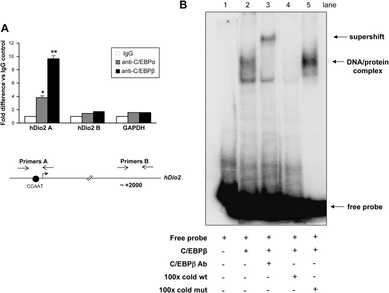
Fig. 4.
A, Quantitative chromatin immunoprecipitation. JEG3 cells were cross-linked with 1% formaldehyde and lysed. The lysates were sonicated and immunoprecipitated with control IgG, C/EBPα, and C/EBPβ antisera. At the end of the incubation, the immunocomplexes were washed extensively and eluted, the cross-linking was reversed, and the DNA was purified, precipitated, and resuspended in Tris-EDTA buffer. Quantitative PCR was performed using primers amplifying Dio2 CCAAT sequence (primers A), an intronic region of Dio2 gene (primers B), and the unrelated GAPDH gene. ANOVA test: *, C/EBPα vs. control, P < 0.05; **, C/EBPβ vs. control, P < 0.001. Results represent the average value ± sd of three different experiments, each performed in triplicate. hDio2, Human Dio2. B, EMSA. 32P-Labeled Dio2 CCAAT oligonucleotide probe, recombinant flag-tagged C/EBPβ, anti-C/EBPβ antibody, 100-fold molar excess of cold wild-type (wt) and cold mutated (mut) oligonucleotides were added to the reaction, as indicated. The free probe is indicated. The indicated retarded bands represent the complex between recombinant C/EBPβ and the Dio2 CCAAT probe (DNA-protein complex) or the supershift upon addition of antisera (supershift).

