Abstract
The relationship between trunk and lower limb kinematics in healthy females versus males is unclear since trunk kinematics in the frontal and transverse planes have not been systematically examined with lower limb kinematics. The aim of this study was to investigate the existence of different multi-joints movement strategies between genders during a single leg squat. We expected that compared to males, females would have greater trunk and pelvis displacement due to less trunk control and display hip and knee movement consistent with medial-collapse (i.e. greater hip adduction, hip medial rotation, knee abduction, knee lateral rotation) on the weight-bearing limb.
9 females and 10 males participated in the study. Kinematic data were collected using an 8-camera, 3D-motion-capture-system. Trunk relative to pelvis, pelvis relative to the laboratory, hip and knee angles in three planes (sagittal, frontal and transverse) were examined at two time events relevant to knee joint mechanics: 45° of knee flexion and peak knee flexion. Females flexed their trunk less than males and rotated their trunk and pelvis toward the weight-bearing limb more than males. Females also displayed greater hip adduction and knee abduction than males.
Taken together these results suggest that females and males used different movement strategies during a single leg squat. Females displayed a trunk and pelvic movement pattern that may put them at risk of knee injury and pain.
Introduction
Previous authors have documented differences in multi-planar lower limb kinematics between genders in weight-bearing activities and athletic maneuvers. Specifically, females show more knee abduction and hip adduction [1, 2] and less hip and knee flexion than males [3] in landing and single leg squat. Females also demonstrate greater hip internal rotation and knee external rotation than males during running and single leg landing [4, 5]. Some authors have reported that tibial abduction and external rotation can strain the ACL [6] and, in particular, tibial external rotation is believed to increase lateral retropatella contact surface, which is a factor linked to patellofemoral pain (PFP) syndrome [7]. These movement pattern differences between females and males may account for the higher rate of anterior cruciate ligament (ACL) injuries [8] and PFP syndrome in females compared to males [9].
Beyond differences in lower limb mechanics, little is known about trunk movements during weight-bearing activities and athletic maneuvers, or about how the trunk and lower limbs are coordinated during these tasks. Recent studies documented that poor neuromuscular control of the trunk predicts knee injuries in females but not in males [10]. Furthermore, video analysis of movements during basketball games showed that females who sustained an ACL injury laterally bent their trunk more toward the side of the injured knee compared to males [11]. Previous authors have also found that females land with more erect posture than males [12] and that less trunk flexion is associated with less hip and knee flexion during drop landing [13], which then could expose females to greater risk for ACL rupture or tear [13].
Previous literature lacks studies in which trunk and pelvis motion are simultaneously examined with lower extremity movements. The identification of different movement strategies occurring at several points along the kinematic chain in healthy females versus males should provide new insights about the contribution of trunk and pelvis kinematics to knee injury and pain development in female clinical populations. Hence, the aim of the current study was to examine gender differences in multi-joint movement strategies in the three planes of motion during a single leg squat.
We hypothesized that, compared to males, females would have less trunk flexion in the sagittal plane and greater trunk and pelvis displacement in the frontal and transverse planes, due to less trunk control. We also expected that compared to males, females would display hip and knee movements consistent with medial collapse (i.e. greater hip adduction, hip medial rotation, knee abduction and lateral rotation [14]) on the weight-bearing limb.
Methods
Subjects
Nineteen healthy volunteers (9 females and 10 males) took part in this study (Table 1). Subjects reported no unresolved or recent musculoskeletal injuries, surgeries or pain. All subjects reported some type of occasional moderate physical activity. None of them were athletes or physically active on a regular basis. The study was approved by the Institutional Review Board (IRB) of Saint Louis University and all subjects read and signed an informed consent form before participating. The dominant leg was assessed by asking subjects which leg they would kick a ball with [1]. All subjects were right leg dominant.
Table 1.
Means, (SD) and Effect Size (Cohen’s d) of subjects demographic and anthropometric data.
| Females | Males | T-test p-value | Effect size | |
|---|---|---|---|---|
| Age (yr) | 26.89 (5.77) | 28.70 (6. 05) | P= 0.51 | 0.31 |
| Height (m) | 1.68 (0.03) | 1.77 (0.06) | P= 0.001* | 1.77 |
| Weight (kg) | 67.68 (9.84) | 75.96 (6.16) | P= 0.051 | 0.95 |
| BMI (kg/m2) | 24.11 (3.68) | 24.32 (1.78) | P= 0.88 | 0.07 |
Asterisks represent significant differences from T-test performed between genders (p< 0.05).
Experimental setting
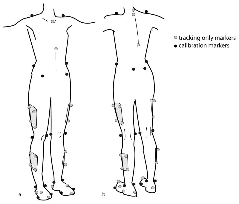
Kinematic data (120Hz) were collected using an 8-camera, 3D motion capture system (Vicon Nexus, Los Angeles, CA) and a 6-degrees-of-freedom model/marker set (Visual3D, C-motion, Inc.) (Figure 1). Before data collection a calibration trial was collected for each subject. The experimenter demonstrated the task to each subject by performing a squat with the non-weight-bearing knee flexed (lower leg behind the body). Subjects performed 3 repetitions of a single right leg squat while keeping their arms out to their sides. Data collection occurred in one session.
Figure 1.
Schematic of the marker placements: (a) front, (b) back. Markers were placed on the upper body (acromia, C7, T2, T10, jugular notch, xiphoid process), pelvis (iliac crests, anterior superior iliac spines, posterior superior iliac spines), lower limbs (medial and lateral femoral epicondyles, 4 marker thigh clusters, medial and lateral malleoli, 4 marker shank cluster) and feet (heel, 2nd toe tip, 1st and 5th metatarsal head, foot dorsum, lateral aspect of the each foot). The markers on the femoral epicondyles were placed to approximate the knee flexion/extension axis.
The 6-degrees-of-freedom model
The 6-degrees-of-freedom model incorporated the trunk, pelvis, thigh, shank and foot. For the pelvis, the CODA model (Charnwood Dynamics Ldt., UK) was used. For each of the segments, the frontal plane was defined first. The frontal plane of the trunk was represented by the two iliac crest markers (proximal end in relation to the pelvis) and the two acromion markers (distal end in relation to the pelvis). The frontal plane for the thigh was defined by the hip joint center (proximal endpoint) and the two femoral epicondyle markers (distal end). The hip joint center was calculated as previously described [15]. The frontal plane for the shank was defined by the thigh distal endpoint (proximal end) and the two malleolus markers (distal end). The frontal plane of the foot was defined by the two malleolus markers (proximal end) and the projection on the floor of the two malleolus markers (distal end). The local coordinate system of each segment was located at the proximal endpoint of each segment. The frontal plane defined the orientation of the x axis. The z axis was aligned so that it passed through the proximal endpoint and the distal endpoint of the segments. The y-axis was oriented orthogonal to both x and z axes.
Data analysis
Data were processed in Vicon for marker labelling and in Visual3D (C-Motion, Inc.) to apply the 6-degrees-of-freedom model. Marker trajectories were lowpass filtered (6Hz, 4th order Butterworth filter) and then imported into Matlab R2010b (The MathWorks, Inc). Two time events relevant in weight-bearing tasks were selected between the start of movement (SOM) and the end of movement (EOM): 45° of knee flexion (45KF), a common point analyzed in the descent phase, and peak knee flexion (PKF), the point when the knee was at maximum loading. The SOM was defined as the first time point at the start of the descent phase at which the angular velocity of the knee joint in the sagittal plane was greater than zero, and EOM was defined as the last time point at the end of the ascent phase at which the angular velocity of the knee joint in the sagittal plane was less than zero. Visual inspection of each repetition ensured the algorithm accuracy.
In the sagittal, frontal and transverse planes, the following joint angles were calculated at 45KF and PKF: trunk (trunk relative to pelvis), hip (femur relative to pelvis), and knee (tibia relative to femur). The pelvis angle relative to the lab (global coordinate system) was calculated to shed light on the contribution of the pelvis segment to the net trunk and net hip angles. For the hip and knee, angles were expressed in the reference frame of the proximal segment and positive values represent flexion, adduction and medial rotation. For the trunk and pelvis, positive values represent flexion, lateral flexion toward the non-weight-bearing limb and transverse rotation toward the non-weight-bearing limb. The time spent to perform the squat was also calculated as the difference between the knee joint EOM and the SOM time points. Dependent measures were averaged across repetitions for each subject. A 2-tailed, independent samples T-test was peformed on the trunk, pelvis, hip and knee angles in the three planes of motion (x, y, z) at 45KF and PKF. The alpha level was set at p ≤0.05.
Reliability testing
Between-day intra-rater reliability of the dependent measures was calculated using the intraclass correlation coefficient (ICC(3,3)). Data on 19 subjects were collected on two occasions, approximately 4.52 ±1.89 days apart. The ICCs was calculated so that the standard error of measurement (SEM) could be also estimated.
Results
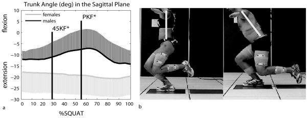
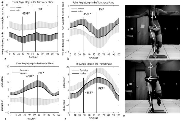
Males were taller and heavier than females but body mass index was not different between genders (Table 1). Dependent measures showed good to excellent reliability [16] in each of the three planes of motion (Supplementary Table). Females flexed their trunk less than males at both the 45KF (p= 0.013) and PKF (p= 0.006) position (Figure 2). At 45KF females laterally flexed their trunk toward the weight-bearing limb while males laterally flexed their trunk toward their non-weight-bearing limb, although this difference was not statistically significant (p= 0.095). Females rotated their trunk in the transverse plane toward the weight-bearing limb less than males (p= 0.039, Figure 3a). In the transverse plane at 45KF females rotated the pelvis toward the weight-bearing limb (in the same direction of the trunk) while males toward the non-weight-bearing limb (opposite direction of the trunk) (p= 0.004, Figure 3b).
Figure 2.
(a) Time series curves of the trunk angle in the sagittal plane normalized as % squat cycle and averaged across subjects for each gender. 45KF and PKF represent the time points where 45 knee flexion and peak knee flexion occurred. Thick lines represent the means. Error bars represent the SD at each time point. Asterisks refer to significant differences (p<0.05). (b) Descent squat phase in one female and one male subject.
Figure 3.
Time series curves of the trunk angle (a) and pelvis angle (b) in the transverse plane and of the hip angle (c) and knee angle (d) in the frontal plane normalized as % squat cycle and averaged across subjects for each gender. 45KF and PKF represent the time points where 45 knee flexion and peak knee flexion occurred. Thick lines represent the means. Error bars represent the SD at each time point. Asterisks refer to significant differences (p<0.05). (e) Descent squat phase in one female and one male subject.
Compared to males, females presented greater hip adduction at both 45KF (p= 0.035) and PKF (p= 0.013) and presented greater knee abduction at both 45KF (p= 0.009) and PKF (p= 0.0008) (Figure 3c and 3d). Females also performed the squat in less time (2.36s ±0.79s) than males 3.18s ±0.83; p= 0.041). All the statistically significant differences were greater than the SEM (Supplementary Table 1). Means, standard deviations (SD) and effect sizes are provided in Table 2. In Supplementary Table 2 the means and SD of two females and males with similar height and weight are reported. The data reported in Supplementary Table 2 shows a similar trend in movement pattern to the data from the entire female and male sample (except for trunk flexion at 45KF). Thus, the kinematic differences across groups are unlikely to be due simply to the differences in height and weight between females and males.
Table 2.
Mean (SD), P-value and Effect Size for each kinematic variable.
| 45KF | PKF | ||||||||
|---|---|---|---|---|---|---|---|---|---|
| Females | Males | P-value | Effect size | Females | Males | P-value | Effect size | ||
| Trunk | sagittal | −19.12 (8.87) | −11.49 (6.58) | 0.013* | 0.98 | −19.28 (9.24) | −7.04 (7.91) | 0.006* | 1.42 |
| frontal | −0.74 (3.24) | 1.64 (2.61) | 0.095 | 0.81 | −4.12 (5.22) | −4.75 (3.68) | 0.76 | 0.14 | |
| transverse | −0.96 (2.27) | −3.56 (2.74) | 0.039* | 1.03 | −0.34 (3.10) | −2.21 (3.39) | 0.23 | 0.57 | |
| Pelvis | sagittal | 22.71 (9.98) | 20.70 (6.13) | 0.59 | 0.24 | 26.77 (11.71) | 30.19 (11.31) | 0.53 | 0.29 |
| frontal | 0.49 (2.40) | −0.58 (2.58) | 0.39 | 0.40 | 3.02 (2.33) | 3.05 (3.52) | 0.98 | 0.01 | |
| transverse | −1.49 (1.46) | 1.17 (1.96) | 0.004* | 1.54 | −4.23(3.79) | −4.05 (3.09) | 0.91 | 0.05 | |
| Hip | sagittal | 41.15 (11.98) | 40.74 (9.85) | 0.94 | 0.04 | 59.09 (15.47) | 72.39(21.88) | 0.15 | 0.70 |
| frontal | 9.69 (3.50) | 6.15 (3.24) | 0.035* | 1.05 | 17.28 (2.62) | 13.53 (3.22) | 0.013* | 1.28 | |
| transverse | 0.35 (4.23) | 1.21 (4.39) | 0.68 | 0.19 | −1.04 (4.40) | −0.70 (3.87) | 0.86 | 0.08 | |
| Knee | sagittal | 45.39 (0.17) | 45.31 (0.09) | 0.24 | 0.55 | 69.77 (7.27) | 76.43 (10.15) | 0.12 | 0.75 |
| frontal | −0.89 (3.95) | 3.34 (2.14) | 0.009* | 1.33 | −1.25(4.77) | 7.004 (4.11) | 0.0008* | 1.85 | |
| transverse | 6.72 (3.12) | 6.44 (5.34) | 0.89 | 0.06 | 4.10 (4.89) | 7.76 (6.06) | 0.17 | 0.66 | |
Asterisks indicate significant differences (P<0.05). For the hip and knee, angles were expressed in the reference frame of the proximal segment and positive values represent flexion, adduction and medial rotation. For the trunk and pelvis, positive values represent flexion, lateral flexion toward the non-weight bearing limb and transverse rotation toward the non-weight bearing limb.
Discussion
The primary new finding of the present study was that females and males used different movement strategies at all the levels of the kinematic chain (i.e. trunk, pelvis, hip and knee) to complete a squat on a single leg. During the descent phase of the squat, females showed a more erect posture (less trunk flexion) than males. It has been argued that this posture may expose females to the risk of ACL injuries by increasing the demand on the quadriceps to maintain the control of the center of mass [12]. In drop landing, for example, the vertical ground reaction force vector falls between the hip and the knee in the sagittal plane resulting in flexion moments at the hip and knee joints [17]. Bending the trunk forward moves the vertical ground reaction force vector farther from the hip joint center, thereby increasing the demand on the hip extensors and decreasing the demand on the knee extensors [17]. One reason females in our study maintained a more erect posture than men may have been because they lacked the hip extensor strength to control the forward displacement of the center of mass in the descent phase. As a result females had to rely on the quadriceps, a strategy that could place the ACL at risk for injury [17]. Speculatively, our finding that females perform the task in less time than males could be explained by the fact that females might have had less hip muscles strength than males. Previous authors found that by asking the subjects to flex their trunk forward, hip and knee flexion also increased in drop landing [13]. These findings suggest that trunk flexion is a primary strategy that, if employed, contributes to safer hip and knee kinematics in the sagittal plane for energy absorption in drop landing. In our study we did not find significant differences in hip and knee flexion between genders, perhaps due to the use of a different task (single leg squat). A single leg squat is not a high acceleration task and likely does not require the same degree of hip and knee flexion as a drop landing. On the other hand, a unique finding in the current study is that females maintained a more erect posture and displayed greater hip adduction and knee abduction than males [13]. Previous authors failed to find an association between hip and knee frontal plane angles and trunk flexion, [13] likely because they did not compare kinematics between genders. Our finding is important because higher knee abduction occurring together with decreased trunk flexion has been proposed to be a risk factor for ACL injury [12]. The association between hip and knee frontal plane angles and trunk flexion found in our study is new. The causal relationship between trunk sagittal plane motion and hip and knee frontal plane motion, however, needs further investigation.
In the transverse plane, females rotated their trunk toward the weight-bearing limb to a lesser degree than males. Trunk rotation in the females also occurred in the direction of pelvis rotation while males rotated their pelvis toward the non-weight-bearing limb. During gait, the trunk and pelvis move in phase in opposite directions [18] in order to maintain the head still in space. In this way, the central nervous system can gain reliable visual and vestibular input to maintain postural stability [19]. Our data may suggest that females adopt a trunk-pelvis movement strategy that does not comply with the effort to keep the head still in space for postural stability. However, by keeping an upright posture and not displacing the non-weight-bearing-limb posteriorly, trunk rotation and pelvis rotation may not have been needed to counteract each other for maintenance of postural stability in females.
The gender difference in pelvis motion identified in our study is another unique finding. Previously it had been postulated that trunk stability is strictly dependent on pelvis stability and that trunk muscles cannot completely compensate for poor pelvis control [17]. However, previous studies examining gender differences in lower limb kinematics have not focused on pelvis movements. We found that in the transverse plane females rotated their pelvis more toward the weight-bearing limb while males rotated more toward the non-weight-bearing limb. The rotation toward the weight-bearing limb in females could be a consequence of decreased hip lateral rotator muscle strength, which was found during isometric strength testing by previous investigators [20]. The rotation could also be due to altered muscle activation of the hip lateral rotators, which was measured with surface EMG during single leg landing by previous authors [21].
The presence of greater knee abduction and greater hip adduction suggest that females move toward the direction of medial collapse during the squat. These findings are in agreement with findings from previous studies of healthy subjects performing a single leg squat [2, 11, 22] and in females affected by PFP syndrome [23]. Our findings suggest that females employ a different movement strategy at multiple levels of the kinematic chain while performing a weight-bearing task. Such a strategy can be more hazardous for the knee joint, which could expose females to a greater risk of knee injury and knee pain. Future studies examining clinical female populations are needed in order to understand if, and how, the trunk and pelvis contribute to lower extremity injuries and pain, and whether rehabilitation strategies should incorporate trunk and pelvis movement retraining.
Some results of our study differ from previous work. In the transverse plane, there were no significant differences at the hip and knee between genders as we would have expected based on previous literature on PFP syndrome [7, 24]. In the frontal plane there were no significant differences in pelvis motion between genders as would be expected in the presence of less hip abductor strength in females, which has been reported by previous authors [25, 26]. However, previous work has focused on people with a history of knee injury or knee pain. Another difference from previous studies is that in our study trunk lateral flexion was not significantly different between genders (P= 0.095), however, this is likely because of our small sample size. Females and males laterally flex the trunk in the opposite direction. Females flex more to the side of the weight-bearing limb while males flex more to the side of the non-weight-bearing limb. The higher lateral flexion on the side of the weight-bearing limb was previously found during ongoing ACL injuries [11]. The lateral trunk flexion on the side of the supporting limb may decrease the hip adduction load but increase the knee abduction load [11]. Such loading could potentially increase the strain on the ACL and increase the lateral forces acting at the patella [17]. Larger samples and other types of tasks that can better challenge lateral trunk movements, however, are needed to better understand trunk movement in the frontal plane between genders.
A limitation of this study is the lack of measures of strength and of muscle activation patterns. Hip abduction and external rotator muscle weakness has been found to contribute to higher hip adduction and internal rotation in PFP syndrome [24, 27]. However, it is still not clear if, and how, muscle weakness or different muscle activation strategies during the ongoing movement contribute to PFP [28]. Another limitation of this study is that we do not know if our results are specific overall to the squat task and to young, lean, healthy subjects.
Overall these results demonstrate that healthy females use a different movement strategy during a single leg squat than healthy males and that females showed a multi-segmental movement pattern involving the trunk and pelvis that could expose them to knee injury and pain development.
Supplementary Material
Acknowledgments
This project was funded in part by the National Institute of Child Health and Human Development (NICHD, R15HD059080, and R15HD059080-01A1S1)
Footnotes
Publisher's Disclaimer: This is a PDF file of an unedited manuscript that has been accepted for publication. As a service to our customers we are providing this early version of the manuscript. The manuscript will undergo copyediting, typesetting, and review of the resulting proof before it is published in its final citable form. Please note that during the production process errors may be discovered which could affect the content, and all legal disclaimers that apply to the journal pertain.
References
- 1.Ford KR, Myer GD, Hewett TE. Valgus knee motion during landing in high school female and male basketball players. Med Sci Sports Exerc. 2003;35(10):1745–1750. doi: 10.1249/01.MSS.0000089346.85744.D9. [DOI] [PubMed] [Google Scholar]
- 2.Willson JD, Ireland ML, Davis I. Core strength and lower extremity alignment during single leg squats. Med Sci Sports Exerc. 2006;38(5):945–52. doi: 10.1249/01.mss.0000218140.05074.fa. [DOI] [PubMed] [Google Scholar]
- 3.Pollard CD, Sigward SM, Powers CM. Limited hip and knee flexion during landing is associated with increased frontal plane knee motion and moments. Clin Biomech. 2010;25(2):142–146. doi: 10.1016/j.clinbiomech.2009.10.005. [DOI] [PMC free article] [PubMed] [Google Scholar]
- 4.Gomez E, DeLee JC, Farney WC. Incidence of injury in Texas girls’ high school basketball. Am J Sports Med. 1996;24(5):684–687. doi: 10.1177/036354659602400521. [DOI] [PubMed] [Google Scholar]
- 5.Bjordal JM, Arnøy F, Hannestad B, Strand T. Epidemiology of anterior cruciate ligament injuries in soccer. Am J Sports Med. 1997;25(3):341–345. doi: 10.1177/036354659702500312. [DOI] [PubMed] [Google Scholar]
- 6.Fung DT, Zhang L-Q. Modeling of ACL impingement against the intercondylar notch. Clin Biomech. 2003;18(10):933–941. doi: 10.1016/s0268-0033(03)00174-8. [DOI] [PubMed] [Google Scholar]
- 7.Salsich G, Perman W. Patellofemoral joint contact area is influenced by tibiofemoral rotation alignment in individuals who have patellofemoral pain. J Orthop Sports Phys Ther. 2007;37(9):521–8. doi: 10.2519/jospt.2007.37.9.521. [DOI] [PubMed] [Google Scholar]
- 8.Arendt E, Dick R. Knee injury patterns among men and women in collegiate basketball and soccer. Am J Sports Med. 1995;23(6):694–701. doi: 10.1177/036354659502300611. [DOI] [PubMed] [Google Scholar]
- 9.Fulkerson JP, Arendt EA. Anterior knee pain in females. Clin Orthop Relat Res. 2000;372:69–73. doi: 10.1097/00003086-200003000-00009. [DOI] [PubMed] [Google Scholar]
- 10.Zazulak BT, Hewett TE, Reeves NP, Goldberg B, Cholewicki J. Deficits in neuromuscular control of the trunk predict knee injury risk. Am J Sports Med. 2007;35(7):1123–1130. doi: 10.1177/0363546507301585. [DOI] [PubMed] [Google Scholar]
- 11.Hewett TE, Torg JS, Boden BP. Video analysis of trunk and knee motion during non-contact anterior cruciate ligament injury in female athletes: lateral trunk and knee abduction motion are combined components of the injury mechanism. Br J Sports Med. 2009;43(6):417–422. doi: 10.1136/bjsm.2009.059162. [DOI] [PMC free article] [PubMed] [Google Scholar]
- 12.Griffin LY, Agel J, Albohm MJ, Arendt EA, Dick RW, Garrett WE, Garrick JG, Hewett TE, Huston L, Ireland ML, Johnson RJ, Kibler WB, Lephart S, Lewis JL, Lindenfeld TN, Mandelbaum BR, Marchak P, Teitz CC, Wojtys EM. Noncontact anterior cruciate ligament injuries: risk factors and prevention strategies. J Am Acad Orthop Surg. 2000;8(3):141–150. doi: 10.5435/00124635-200005000-00001. [DOI] [PubMed] [Google Scholar]
- 13.Blackburn JT, Padua DA. Influence of trunk flexion on hip and knee joint kinematics during a controlled drop landing. Clin Biomech. 2008;23(3):313–319. doi: 10.1016/j.clinbiomech.2007.10.003. [DOI] [PubMed] [Google Scholar]
- 14.Powers C. The influence of altered lower-extremity kinematics on patellofemoral joint dysfunction: a theoretical perspective. J Orthop Sports Phys Ther. 2003;33(11):639–46. doi: 10.2519/jospt.2003.33.11.639. [DOI] [PubMed] [Google Scholar]
- 15.Bell AL, Pederson DR, Brand RA. A comparison of the accuracy of several hip center location prediction methods. J Biomech. 1990;23:617–621. doi: 10.1016/0021-9290(90)90054-7. [DOI] [PubMed] [Google Scholar]
- 16.Shrout PE, Fleiss JL. Intraclass correlations: uses in assessing rater reliability. Psychol Bull. 1979;86(2):420–428. doi: 10.1037//0033-2909.86.2.420. [DOI] [PubMed] [Google Scholar]
- 17.Powers C. The influence of abnormal hip mechanics on knee injury: a biomechanical perspective. J Orthop Sports Phys Ther. 2010;40(2):42–51. doi: 10.2519/jospt.2010.3337. [DOI] [PubMed] [Google Scholar]
- 18.Perry J, Burnfield JM. Normal Pathological Function. 2. Thorofare: Slack Incoporated; 2010. Gait Analysis. [Google Scholar]
- 19.Pozzo T, Berthoz A, Lefort I. Head stabilization during various locomotor tasks in humans. I. Normal subjects. Exp Brain Res. 1990;82:97–106. doi: 10.1007/BF00230842. [DOI] [PubMed] [Google Scholar]
- 20.Leetun DT, Ireland ML, Willson JD, Ballantyne BT, Davis IM. Core stability measures as risk factors for lower extremity injury in athletes. Med Sci Sports Exerc. 2004;36(6):926–34. doi: 10.1249/01.mss.0000128145.75199.c3. [DOI] [PubMed] [Google Scholar]
- 21.Zazulak B, Ponce P, Straub S, Medvecky M, Avedisian L, Hewett TE. Gender comparison of hip muscle activity during single leg-squat landing. J Orthop Sports Phys Ther. 2005;35:292–299. doi: 10.2519/jospt.2005.35.5.292. [DOI] [PubMed] [Google Scholar]
- 22.Jacobs CA, Uhl TL, Mattacola CG, Shapiro R, Rayens WS. Hip abductor function and lower extremity landing kinematics: sex differences. J Athl Train. 2007;42(1):76–83. [PMC free article] [PubMed] [Google Scholar]
- 23.Salsich GB, Long-Rossi F. Do females with patellofemoral pain have abnormal hip and knee kinematics during gait? Physiother. Theory Pract. 2010;26(3):150–159. doi: 10.3109/09593980903423111. [DOI] [PMC free article] [PubMed] [Google Scholar]
- 24.Powers CM, Ward SR, Fredericson M, Guillet M, Shellock FG. Patellofemoral kinematics during weight-bearing and non-weight-bearing knee extension in persons with lateral subluxation of the patella: a preliminary study. J Orthop Sports Phys Ther. 2003;33(11):677–85. doi: 10.2519/jospt.2003.33.11.677. [DOI] [PubMed] [Google Scholar]
- 25.Hewett TE, Myer GD, Ford KR, Heidt RS, Colosimo AJ, McLean SG, van den Bogert AJ, Paterno MV, Succop P. Biomechanical measures of neuromuscular control and valgus loading of the knee predict anterior cruciate ligament injury risk in female athletes. Am J Sports Med. 2005;33(4):492–501. doi: 10.1177/0363546504269591. [DOI] [PubMed] [Google Scholar]
- 26.Willson JD, Davis IS. Lower extremity mechanics of females with and without patellofemoral pain across activities with progressively greater task demands. Clin Biomech. 2008;23(2):203–211. doi: 10.1016/j.clinbiomech.2007.08.025. [DOI] [PubMed] [Google Scholar]
- 27.Dierks TK, Manal TJ, Hamill I, Davis S. Proximal and distal influences on hip and knee kinematics in runners with patellofemoral pain during a prolonged run. J Orthop Sports Phys Ther. 2008;38(8):448–456. doi: 10.2519/jospt.2008.2490. [DOI] [PubMed] [Google Scholar]
- 28.Heiderscheit B. Lower extremity injuries: is it just about hip strength? J Orthop Sports Phys Ther. 2010;40(2):39–41. doi: 10.2519/jospt.2010.0102. [DOI] [PMC free article] [PubMed] [Google Scholar]
Associated Data
This section collects any data citations, data availability statements, or supplementary materials included in this article.


