Summary
Background and objectives
Circulating troponin T levels are frequently elevated in patients undergoing long-term dialysis. The pathophysiology underlying these elevations is controversial.
Design, setting, participants, & measurements
In 70 prevalent hemodialysis (HD) patients, HD-induced myocardial stunning was assessed echocardiographically at baseline and after 12 months. Nineteen patients were not available for the follow-up analysis. The extent to which predialysis troponin T was associated with the occurrence of HD-induced myocardial stunning was assessed as the primary endpoint.
Results
The median troponin T level in this hemodialysis cohort was 0.06 ng/ml (interquartile range, 0.02–0.10). At baseline, 64% of patients experienced myocardial stunning. These patients showed significantly higher troponin T levels than patients without stunning (0.08 ng/ml [0.05–0.12] versus 0.02 ng/ml [0.01–0.05]). Troponin T levels were significantly correlated to measures of myocardial stunning severity (number of affected segments: r=0.42; change in ejection fraction from beginning of dialysis to end of dialysis: r=−0.45). In receiver-operating characteristic analyses, predialytic troponin T achieved an area under the curve of 0.82 for the detection of myocardial stunning. In multivariable analysis, only ultrafiltration volume (odds ratio, 4.38 for every additional liter) and troponin T (odds ratio, 9.33 for every additional 0.1 ng/ml) were independently associated with myocardial stunning. After 12 months, nine patients had newly developed myocardial stunning and showed a significant increase in troponin T over baseline (0.03 ng/ml at baseline versus 0.05 ng/ml at year 1).
Conclusions
Troponin T levels in HD patients are associated with the presence and severity of HD-induced myocardial stunning.
Introduction
Patients with ESRD undergoing long-term hemodialysis (HD) are at a substantially increased risk for death, with 1-, 3-, and 5-year mortality rates around 15%, 35%, and 55%, respectively (1,2). The latest U.S. Renal Data System Annual Report also showed an annual mortality rate of 221 deaths per 1000 patient-years in the United States (3). Cardiovascular events are a major cause of these dire mortality figures; more than 40% of all patients with ESRD die of cardiac causes (3). Recently, recurrent episodes of myocardial ischemia and transient segmental left ventricular wall-motion abnormalities have been established to occur commonly during standard thrice-weekly HD (4–7). These repeated episodes of myocardial stunning can eventually lead to myocardial hibernation, myocardial remodeling, scarring, and irreversible loss of contractile function (8) and are becoming increasingly appreciated as a principal pathophysiologic foundation of excess cardiovascular mortality in HD patients.
Cardiac troponins, structural proteins unique to the heart, are sensitive and specific biochemical markers of myocardial damage (9,10). In addition, cardiac troponin levels, as measured by fully automated standard assays, are superior to all other clinically available biomarkers for the diagnosis of acute myocardial ischemia (11–13). However, circulating levels of cardiac troponins are frequently elevated in long-term dialysis patients even in the absence of acute coronary syndromes (14–16).
We therefore aimed to assess the association between the presence and extent of HD-induced myocardial ischemia and troponin T levels in unselected patients undergoing maintenance HD.
Materials and Methods
Patient Population
Overall, 138 long-term HD patients were screened for enrollment in a single hospital-based HD unit. In short, 19 patients were excluded for ethical considerations (legal guardianship, inability to speak or comprehend written English, age < 18 years), and 17 patients were excluded because they experienced an acute heart failure episode or an acute myocardial infarction within the preceding 4 weeks. One patient was excluded as a result of inadequate image quality. In total, 70 patients consented and were recruited for a 12-month observational cohort study. Because patients were undergoing dialysis as part of a conventional thrice-weekly treatment regimen (4-hour dialysis sessions), all study sessions were conducted after the first 2-day interdialytic period. The study was carried out according to the principles of the Declaration of Helsinki and was approved by the local ethics committee. Written informed consent was obtained from all participating patients.
Echocardiographic Assessment
Patients were assessed at baseline and after 12 months to evaluate the presence and extent of HD-induced myocardial stunning. Two-dimensional echocardiography was performed by a single experienced technician while patients were in the left lateral decubitus position (1.5–3.6 MHz 3S probe; Vivid 3, GE Medical Systems, Sonigen, Germany). Images were recorded before HD began (rest), during the final 15 minutes of the HD session (peak stress), and 30 minutes after HD (recovery). Standard apical two-chamber and four-chamber views were digitally recorded for subsequent analysis using regional wall-motion analysis software (CMS-View and Echo-CMS; MEDIS, Leiden, the Netherlands) (17–19). In short, the left ventricle was divided into 10 segments, and HD-induced myocardial stunning was defined as a >20% reduction in segmental wall motion at peak stress during HD versus wall motion at rest, with evidence of functional recovery in the postdialysis period. More than two segments needed to be affected. Ejection fraction was calculated using the biplane Simpson method. Changes in ejection fraction were calculated by subtracting ejection fraction before the start of the study session from the ejection fraction at peak dialytic stress.
HD Details
Dialysis was performed using Hospal Integra monitors (Hospal, Mirandola, Italy) with low-flux polysulfone dialyzers, either 1.8 m2 or 2.0 m2, per individual patients’ usual prescriptions (LOPS 18/20; Braun Medical Ltd, Sheffield, United Kingdom). Dialysate composition was sodium, 138 mmol/L; potassium, 1 mmol/L; calcium, 1.25 mmol/L; magnesium, 0.5 mmol/L; bicarbonate, 32 mmol/L; glucose, 5.6 mmol/L; and acetate, 3 mmol/L. All treatments were of 4 hours’ duration, and anticoagulation was provided with unfractionated heparin. The dialysate flow rate was 500 mL/min, and dialysate temperature was set at 37°C. For each session, net fluid removal was set on an individual basis according to ideal dry weight. Blood pump speed varied between 250 and 450 mL/min, depending on the patient’s vascular access. BP was measured before dialysis and then serially every 15 minutes during HD using an automated digital oscillometric device (Model UA-767, A&D Instruments, Japan).
Measurement of Hematologic and Biochemical Variables
Predialysis blood samples were drawn immediately after insertion of access needles and were collected into lithium heparin and EDTA tubes. Biochemical analysis was performed on a multichannel autoanalyzer. Cardiac troponin T was measured using a fourth-generation electrochemiluminescence assay (Elecsys, Roche Diagnostics, Lewes, United Kingdom). According to Roche, the lower limit of detection of the assay is 0.01 µg/L, the 99th-percentile cutoff is <0.01 µg/L, and the 10% coefficient of variation is 0.035 µg/L. All treating physicians were blinded to troponin T levels measured for study purposes. However, clinically indicated troponin T measurements (e.g., episodes of acute chest pain) were allowed during the study period.
Outcomes
The extent to which predialysis troponin T was associated with the occurrence of HD-induced myocardial stunning was assessed as the primary endpoint.
Statistical Analyses
The statistical analyses were performed using the SPSS/PC (version 19.0, SPSS Inc., Chicago, IL) software package. A statistical significance level of 0.05 was used. Discrete variables are expressed as counts (percentage) and continuous variables as means ± SDs or medians and interquartile ranges (IQRs). The comparison between patients with and without myocardial stunning was done with a chi-squared test and a Fisher exact test for categorical variables and a t test for continuous variables (if normally distributed) or Mann-Whitney test (if not normally distributed). Comparisons between troponin T values at various time points for individual patients were done using Wilcoxon test for paired samples. The Spearman rank correlation was used to perform correlation analyses. All hypothesis testing was two-tailed. The diagnostic accuracy of troponin T was evaluated using receiver-operating characteristic (ROC) curve analysis. Univariate and multivariable logistic regression analyses were applied to identify detectors of HD-induced myocardial stunning. To assess whether troponin T levels improve the detection of HD-induced myocardial stunning over the information provided by clinical measures, ROC curves for the multivariable model with and without the inclusion of troponin T were constructed. Areas under ROC curves (AUCs) were compared using MedCalc software (version 12.2, MedCalc Software, Mariakerke, Belgium). The variable inclusion method was used for multivariable regression analysis. Multivariable analysis included all significant candidate variables (P<0.05) established in univariate analysis.
Results
Baseline Characteristics
The demographic characteristics and biologic variables of the overall study cohort are displayed in Table 1. At baseline, 64% of patients (45 of 70) experienced myocardial stunning during their routine dialysis session. These patients were older, experienced greater interdialytic weight gain, and were more likely to have diabetes mellitus and intradialytic hypotension (Table 1). In addition, HD-induced myocardial stunning was more frequent in patients with coronary artery disease than in patients without coronary artery disease. However, this trend was not statistically significant.
Table 1.
Baseline characteristics
| Characteristic | All Patients (n=70) | Patients Not Developing HD-Induced Stunning | Patients Developing HD-Induced Stunning | P Value |
|---|---|---|---|---|
| Age (yr) | 63.1±13.7 | 59.4±3 | 65.6±1.9 | 0.03 |
| Men:women (n) | 47:23 | 19:6 | 28:17 | 0.29 |
| Dialysis measures | ||||
| Kt/V urea | 1.3±0.2 | 1.3±0.3 | 1.3±0.2 | 0.88 |
| ultrafiltration volume (L) | 2.03±0.6 | 1.59±0.75 | 2.13±0.91 | 0.01 |
| dialysis vintage (mo) | 45.2±32.3 | 48.1±34.8 | 43.5±31.0 | 0.57 |
| Dialysis access, n (%) | ||||
| AV fistula | 57 (81) | 23 (90) | 34 (75) | 0.12 |
| tunneled catheter | 10 (14) | 1 (4) | 9 (20) | 0.07 |
| Causes, n (%) | ||||
| diabetes mellitus | 22 (31) | 4 (16) | 18 (40) | 0.06 |
| glomerular disease | 12 (17) | 4 (16) | 8 (18) | 1.00 |
| Adult polycystic kidney disease | 7 (10) | 2 (8) | 5 (11) | 1.00 |
| urologic | 6 (9) | 3 (12) | 3 (7) | 0.66 |
| other | 14 (29) | 8 (32) | 6 (13) | 0.12 |
| unknown | 9 (13) | 4 (16) | 5 (11) | 0.71 |
| Comorbid conditions, n (%) | ||||
| coronary artery disease | 24 (34) | 5 (20) | 19 (42) | 0.07 |
| diabetes mellitus | 28 (40) | 4 (16) | 24 (53) | <0.01 |
| arterial hypertension | 42 (60) | 15 (60) | 27 (60) | 1.00 |
| active smoker | 11 (16) | 4 (15) | 7 (16) | 1.00 |
| Echocardiography measures | ||||
| left ventricular mass index (g/m2) | 85±31 | 90±32 | 82±30 | 0.36 |
| left ventricular ejection fraction (%) | 55±10 | 56±9 | 54±11 | 0.51 |
| change in ejection fraction at peak dialytic stress (%) | −1±10 | 7±7 | 5±8 | <0.01 |
| Laboratory measures | ||||
| predialysis systolic BP (mmHg) | 145±24 | 140±16 | 148±27 | 0.19 |
| intradialytic hypotension, n (%) | 25 (36) | 5 (20) | 20 (44) | 0.04 |
| hemoglobin (g/dl) | 11.2±1.2 | 11.5±1 | 11.0±1.3 | 0.15 |
| sodium (mEq/L) | 138±3 | 139±4 | 138±3 | 0.18 |
| creatinine (µmol/L) | 690±205 | 742±227 | 662±189 | 0.13 |
| urea | 20.6±5.3 | 19.9±5.1 | 20.9±5.4 | 0.33 |
| troponin T (ng/ml)a | 0.06 (0.02–0.10) | 0.02 (0.01–0.05) | 0.08 (0.05–0.12) | <0.01 |
Data expressed with a plus/minus sign are the mean ± SD. HD, hemodialysis; AV, arteriovenous.
Values in parentheses are interquartile range.
Troponin T Levels in Patients Experiencing HD-Induced Myocardial Stunning
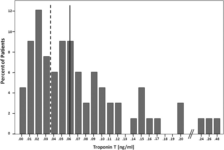
The median troponin T level in this HD cohort was 0.06 ng/ml (IQR, 0.02–0.10 ng/ml). In 81% of patients, troponin T levels were elevated above the 99th percentile of a healthy reference population (0.01 ng/ml). Sixty-three percent of patients showed troponin T levels >0.03 ng/ml, the cutoff commonly used to detect acute myocardial infarction in the setting of acute chest pain (9,10). Figure 1 shows the distribution of troponin T values in the study population.
Figure 1.
Histogram showing the frequency distribution of troponin T levels in 70 prevalent hemodialysis patients. The dashed line separates values above and below 0.03 ng/ml (commonly used to detect acute myocardial infarction in the setting of acute chest pain), and the solid line separates values above and below 0.06 ng/ml (median value of this hemodialysis population).
Patients experiencing HD-induced myocardial stunning had significantly elevated troponin T values compared with patients who did not have stunning. Additionally, troponin T levels were significantly correlated with measures of severity of HD-induced myocardial stunning (number of affected segments: r=0.42, P=0.01; change in ejection fraction from beginning of dialysis to end to dialysis: r=−0.45, P<0.01; i.e., higher troponin T levels were associated with a decrease in ejection fraction during dialysis), but not with age (P=0.13), Kt/V urea (P=0.47), the number of intradialytic hypotensive episodes (P=0.37), or ultrafiltration volume (P=0.10).
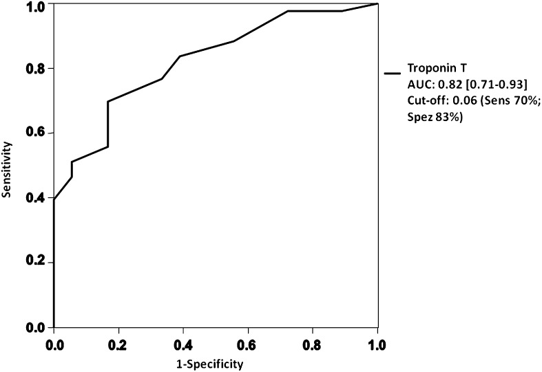
To evaluate the potential of troponin T measurements to detect HD-induced myocardial stunning, ROC analyses were performed. Troponin T levels drawn at the beginning of the dialysis session achieved an AUC of 0.82 (95% confidence interval [CI], 0.71–0.93) (Figure 2). The best calculated troponin T cut-point of 0.06 ng/ml that maximized the product of sensitivity × specificity achieved a sensitivity of 0.70 and a specificity of 0.83. The diagnostic accuracy of troponin T persisted in patients without known coronary artery disease (AUC, 0.84 [95% CI, 0.71–0.96]).
Figure 2.
Receiver-operating characteristic curve displaying the potential predialytic troponin T values to detect hemodialysis-induced myocardial stunning. AUC, area under the receiver-operating characteristic curve. Sens, sensitivity; Spec, specificity.
In univariate logistic regression analysis, age (odds ratio [OR], 1.05 [95% CI, 1.01–1.09]; P=0.02), diabetes mellitus (OR, 4.38 [95% CI, 1.40–13.76]; P=0.01), ultrafiltration volume (OR, 2.54 for every additional liter [95% CI, 1.22–5.31]; P=0.01), intradialytic hypotension (OR, 1.97 [95% CI, 1.05–3.69]; P=0.04), and troponin T values (OR, 16.38 for every additional 0.1 ng/ml [95% CI, 2.84–94.43]; P<0.01) were significantly associated with HD-induced myocardial stunning (Table 2).
Table 2.
Detection of hemodialysis-induced myocardial stunning in univariate and multivariable regression analyses
| Predictor | Odds Ratio (95% CI) | P Value |
|---|---|---|
| Univariate analysis | ||
| age | 1.05 (1.01–1.09) | 0.02 |
| male sex | 1.93 (0.64–5.76) | 0.24 |
| history of coronary artery disease | 2.11 (0.71–6.31) | 0.18 |
| history of diabetes mellitus | 4.38 (1.40–13.76) | 0.01 |
| history of arterial hypertension | 1.38 (0.51–3.74) | 0.53 |
| left ventricular ejection fraction | 0.98 (0.93–1.03) | 0.42 |
| dialysis vintage | 0.99 (0.98–1.01) | 0.57 |
| interdialytic hypotension (per every additional episode) | 1.97 (1.05–3.69) | 0.04 |
| ultrafiltration volume (for every additional liter) | 2.54 (1.22–5.31) | 0.01 |
| troponin T (for every 0.1-ng/ml increase) | 16.38 (2.84–94.43) | <0.01 |
| troponin T level > 0.06 ng/ml | 10.96 (3.11–38.62) | <0.01 |
| Multivariable analysis | ||
| age | 1.05 (0.99–1.11) | 0.08 |
| history of diabetes mellitus | 2.95 (0.59–14.53) | 0.18 |
| interdialytic hypotension (per every additional episode) | 1.95 (0.80–4.80) | 0.14 |
| ultrafiltration volume (for every additional liter) | 4.38 (1.01–18.24) | 0.04 |
| troponin T (for every 0.1-ng/ml increase) | 9.33 (1.63–53.43) | 0.01 |
CI, confidence interval.
In multivariable analysis, only ultrafiltration volume (OR, 4.38 for every additional liter [95% CI, 1.01–18.24]; P=0.04) and troponin T (OR, 9.33 for every additional 0.1 ng/ml [95% CI, 1.63–53.43]; P=0.01) remained as independent diagnostic factors. When we entered categorical (>0.06 ng/ml) instead of continuous troponin T values in the multivariable analysis, troponin T values remained the only independent diagnostic factor (OR, 6.90 [95% CI, 1.65–28.79]; P=0.01). ROC curve analyses describing the diagnostic accuracy of the multivariable model before and after the inclusion of troponin T (i.e., addition of troponin T to age, history of diabetes mellitus, presence of intradialytic hypotension, ultrafiltration volume) revealed a significantly larger AUC for the model containing troponin T (AUCTroponin, 0.88 [95% CI, 0.77–0.95], versus AUCNo Troponin, 0.81 [95% CI, 0.70–0.90]; P=0.01).
Troponin T Values in Patients Developing Stunning during 12-Month Observation Period
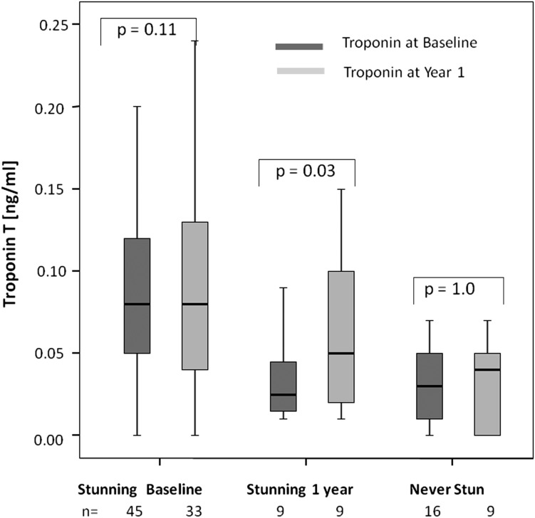
After the 12-month observation period, 51 patients were available for the follow-up assessment. Nineteen patients were not available for the follow-up analysis (patients who had died [n=9], undergone kidney transplantation [n=6], changed dialysis modality [n=3], or withdrawn consent [n=1]). During the 12-month observation period, 2 patients with stunning at baseline showed a reduction in the number of affected segments (decrease of one and three affected segments, respectively) but continued to show regional wall-motion abnormalities, whereas 9 patients newly developed HD-induced myocardial stunning. Age, sex, comorbid conditions, dialysis vintage or adequacy, or ultrafiltration volume at baseline did not significantly differ between patients who developed stunning and those who never developed stunning. Patients developing myocardial stunning showed a significant increase in troponin T values over their baseline value (0.03 ng/ml [IQR, 0.01–0.05 ng/ml] at baseline versus 0.05 ng/ml [IQR, 0.03–0.11 ng/ml] at 1 year; P=0.02). The median troponin T increase in the patients developing stunning was 0.03 ng/ml. Troponin T values did not significantly change in patients stunning at neither or both timepoints (Figure 3). Again, troponin T levels were significantly higher in patients who developed stunning after 12 months compared with patients not stunning at 12 months (stunning, 0.06 ng/ml [IQR, 0.03–0.11 ng/ml] versus 0.05 ng/ml [IQR, 0.01–0.07 ng/ml]; P=0.04) regardless of baseline stunning.
Figure 3.
Boxplots displaying troponin T levels at baseline (dark) and at year 1 (light). All 33 patients with stunning at baseline and being followed up after 1 year also had stunning at year 1. Five patients without stunning at baseline were not followed up for 1 year and are counted as “never stunning.” P values are derived from Wilcoxon test for paired samples and include only patients sampled at both time points. The top of the box represents the 75th percentile, the bottom of the box represents the 25th percentile, and the line in the middle represents the 50th percentile (median). The whiskers represent the highest and lowest values (excluding outliers).
Discussion
Cardiac troponins are commonly elevated in patients undergoing long-term HD, even in the absence of any signs and symptoms that could suggest acute myocardial infarction (14–16). However, the pathophysiology underlying these elevations is still controversial. Previous hypotheses have suggested that left ventricular hypertrophy and coronary artery disease may induce elevated troponin values in dialysis patients (20,21). However, subsequent studies were not able to substantiate these assumptions (21,22). In a prospective multicenter study enrolling 224 patients undergoing long-term HD, deFilippi et al. found no correlation between increasing troponin T quartiles and the frequency of left ventricular hypertrophy (21). Similarly, Abbas and coworkers did not find a significant independent relationship between left ventricular mass index and troponin T concentrations (22). It also seems unlikely that coronary artery disease per se triggers elevated troponin T levels in HD patients. In a study assessing 194 clinically indicated coronary angiographies, Obialo and colleagues found elevated troponin T levels in up to 50% of all dialysis patients without overt coronary artery disease (23). Similarly, in a study comparing the diagnostic abilities of dobutamine stress echocardiography and troponin T with that of coronary angiography, troponin T did not significantly detect coronary artery disease in patients with ESRD (24). Our results extend and corroborate these results by specifically investigating the association of HD-induced myocardial stunning with troponin T levels.
Short intermittent HD treatments exert significant hemodynamic effects, and 20%–30% of HD sessions are complicated by episodes of significant intradialytic hypotension (25). These hemodynamic changes, in combination with a high prevalence of left ventricular hypertrophy (26), increased arterial stiffness (27), diastolic dysfunction with high vulnerability to rapid volume changes (28), myocyte/capillary mismatch (29), and the subsequently reduced coronary flow reserve even in the absence of coronary vessel stenoses (30,31), render HD patients at a markedly increased risk for myocardial ischemia. A series of recent studies was able to show that standard thrice-weekly HD is able to induce transient episodes of myocardial ischemia (4–7). In a study using H215O positron emission tomography to measure myocardial blood flow during dialysis, we demonstrated that HD precipitates reductions in myocardial blood flow, at peak dialytic stress, to the levels consistent with the development of overt myocardial ischemia. Of note, this study was conducted after all patients had undergone coronary angiography to exclude the presence of coronary artery disease (4). The same study also confirmed that the observed reduction in myocardial blood flow correlates directly with the occurrence of segmental left ventricular dysfunction measured by simultaneous echocardiography. The occurrence of HD-induced myocardial stunning was subsequently confirmed in a study of unselected, prevalent HD patients (5).
Most important, we found the widespread elevation of troponin T in patients with ESRD to correlate closely with the severity of myocardial stunning. It is noteworthy that there was no correlation between troponin T levels and age, intradialytic hypotension, and ultrafiltration volume, factors currently believed to underlie the pathophysiology of HD-induced stunning (5). It is currently unclear whether intradialytic hypotension is purely a trigger for HD-induced myocardial stunning or could also be its clinical correlate. Also, while there was a trend to more patients with coronary artery disease in the stunning group, coronary artery disease was not independently associated with the occurrence of HD- induced stunning. In fact, in a substudy of 12 patients undergoing prestudy coronary angiography, 3 patients without coronary artery disease (i.e., no coronary stenosis ≥70% diameter during coronary angiography) experienced HD-induced myocardial stunning, whereas one patient with proven two-vessel coronary artery disease did not display myocardial stunning. Consequently, only troponin T levels and ultrafiltration volume remained independently associated with myocardial stunning in multivariable regression analysis. These observations suggest that troponin T levels truly reflect the presence of intradialytic ischemic episodes, rather than common comorbid conditions or communal risk factors. This theory is further backed by the significant increase in troponin T levels observed for patients newly developing myocardial stunning during the observation period. In addition, a study by Hung et al. found predialytic troponin values to be markedly increased if the previous dialysis sessions had been marked by intradialytic hypotension compared with troponin levels after stable sessions (32). Our group has subsequently described similar findings (33). These studies further strengthen the theory that predialytic troponin samples correctly quantify the dialytic ischemic burden.
Importantly, modified dialysis regimens such as biofeedback dialysis, cooling of the dialysate, and increased dialysis frequency have recently been shown to reduce HD-induced stunning over standard thrice-weekly dialysis (34–36). Unfortunately, the effects of these therapy modulations on circulating troponin T values were not the primary focus of these short-term studies.
Several limitations merit consideration. First, this was an observational, single-center study. However, because baseline characteristics and mortality rates are similar to those in other published dialysis cohorts, we consider our patient population representative. Similarly, the sample size and the number of cardiovascular endpoints observed during the 1-year follow-up were too limited to allow a reliable multivariable regression analysis on the prognostic potential of troponin T. Second, troponin T values were exclusively measured by the Roche fourth-generation Elecsys troponin T assay. However, more sensitive troponin assays have subsequently become clinically available and have been shown to improve the diagnostic and prognostic performance over less sensitive assays (37,38). Further studies will be needed to establish cutoff values that adequately separate stunning from nonstunning patients for the novel, more sensitive troponin assays. Finally, we exclusively assessed predialytic troponin T values and did not evaluate the association between postdialytic troponin T levels and intradialytic ischemic episodes. Although these levels (especially when drawn 4–6 hours after dialysis) might increase the accuracy of detecting HD-induced stunning, postdialytic blood draws are severely hampered by the effect of hemoconcentration and their lack of practicality. The novel, more sensitive troponin assays might help to clarify the value of postdialytic troponin values.
In conclusion, troponin T levels in patients undergoing long-term HD are associated with the presence and severity of HD-induced myocardial stunning. This mechanism provides a robust pathophysiologic basis for the prognostic importance of elevated troponin levels in the HD population.
Disclosures
None.
Acknowledgments
T.B. was supported by a research grant from the Swiss National Science Foundation, and the study was supported by a grant from Kidney Research UK.
Footnotes
Published online ahead of print. Publication date available at www.cjasn.org.
References
- 1.Saudan P, Kossovsky M, Halabi G, Martin PY, Perneger TV, Western Switzerland Dialysis Study Group : Quality of care and survival of haemodialysed patients in western Switzerland. Nephrol Dial Transplant 23: 1975–1981, 2008 [DOI] [PubMed] [Google Scholar]
- 2.Breidthardt T, Moser-Bucher CN, Praehauser C, Garzoni D, Bächler K, Steiger J, Dickenmann M, Mayr M: Morbidity and mortality on chronic haemodialysis: A 10-year Swiss single centre analysis. Swiss Med Wkly 141: w13150, 2011 [DOI] [PubMed] [Google Scholar]
- 3.National Institutes of Health: USRDS 2008 Annual Data Report. Bethesda, MD, US Renal Data System, 2008 [Google Scholar]
- 4.McIntyre CW, Burton JO, Selby NM, Leccisotti L, Korsheed S, Baker CS, Camici PG: Hemodialysis-induced cardiac dysfunction is associated with an acute reduction in global and segmental myocardial blood flow. Clin J Am Soc Nephrol 3: 19–26, 2008 [DOI] [PMC free article] [PubMed] [Google Scholar]
- 5.Burton JO, Jefferies HJ, Selby NM, McIntyre CW: Hemodialysis-induced cardiac injury: Determinants and associated outcomes. Clin J Am Soc Nephrol 4: 914–920, 2009 [DOI] [PMC free article] [PubMed] [Google Scholar]
- 6.Dasselaar JJ, Slart RH, Knip M, Pruim J, Tio RA, McIntyre CW, de Jong PE, Franssen CF: Haemodialysis is associated with a pronounced fall in myocardial perfusion. Nephrol Dial Transplant 24: 604–610, 2009 [DOI] [PubMed] [Google Scholar]
- 7.Breidthardt T, McIntyre CW: Dialysis-induced myocardial stunning: The other side of the cardiorenal syndrome. Rev Cardiovasc Med 12: 13–20, 2011 [DOI] [PubMed] [Google Scholar]
- 8.Braunwald E, Kloner RA: The stunned myocardium: Prolonged, postischemic ventricular dysfunction. Circulation 66: 1146–1149, 1982 [DOI] [PubMed] [Google Scholar]
- 9.Thygesen K, Alpert JS, White HD, Jaffe AS, Apple FS, Galvani M, Katus HA, Newby LK, Ravkilde J, Chaitman B, Clemmensen PM, Dellborg M, Hod H, Porela P, Underwood R, Bax JJ, Beller GA, Bonow R, Van der Wall EE, Bassand JP, Wijns W, Ferguson TB, Steg PG, Uretsky BF, Williams DO, Armstrong PW, Antman EM, Fox KA, Hamm CW, Ohman EM, Simoons ML, Poole-Wilson PA, Gurfinkel EP, Lopez-Sendon JL, Pais P, Mendis S, Zhu JR, Wallentin LC, Fernández-Avilés F, Fox KM, Parkhomenko AN, Priori SG, Tendera M, Voipio-Pulkki LM, Vahanian A, Camm AJ, De Caterina R, Dean V, Dickstein K, Filippatos G, Funck-Brentano C, Hellemans I, Kristensen SD, McGregor K, Sechtem U, Silber S, Tendera M, Widimsky P, Zamorano JL, Morais J, Brener S, Harrington R, Morrow D, Lim M, Martinez-Rios MA, Steinhubl S, Levine GN, Gibler WB, Goff D, Tubaro M, Dudek D, Al-Attar N, Joint ESC/ACCF/AHA/WHF Task Force for the Redefinition of Myocardial Infarction : Universal definition of myocardial infarction. Circulation 116: 2634–2653, 2007 [DOI] [PubMed] [Google Scholar]
- 10.Apple FS, Wu AH, Jaffe AS: European Society of Cardiology and American College of Cardiology guidelines for redefinition of myocardial infarction: How to use existing assays clinically and for clinical trials. Am Heart J 144: 981–986, 2002 [DOI] [PubMed] [Google Scholar]
- 11.Eggers KM, Oldgren J, Nordenskjöld A, Lindahl B: Diagnostic value of serial measurement of cardiac markers in patients with chest pain: Limited value of adding myoglobin to troponin I for exclusion of myocardial infarction. Am Heart J 148: 574–581, 2004 [DOI] [PubMed] [Google Scholar]
- 12.Hochholzer W, Morrow DA,, Giugliano RP: Novel biomarkers in cardiovascular disease: update 2010. Am Heart J 160: 583–594 [DOI] [PubMed] [Google Scholar]
- 13.Morrow DA, Antman EM, Tanasijevic M, Rifai N, de Lemos JA, McCabe CH, Cannon CP, Braunwald E: Cardiac troponin I for stratification of early outcomes and the efficacy of enoxaparin in unstable angina: a TIMI-11B substudy. J Am Coll Cardiol 36: 1812–1817, 2000 [DOI] [PubMed] [Google Scholar]
- 14.Ooi DS, House AA: Cardiac troponin T in hemodialyzed patients. Clin Chem 44: 1410–1416, 1998 [PubMed] [Google Scholar]
- 15.Ishii J, Nomura M, Okuma T, Minagawa T, Naruse H, Mori Y, Ishikawa T, Kurokawa H, Hirano T, Kondo T, Nagamura Y, Ezaki K, Hishida H: Risk stratification using serum concentrations of cardiac troponin T in patients with end-stage renal disease on chronic maintenance dialysis. Clin Chim Acta 312: 69–79, 2001 [DOI] [PubMed] [Google Scholar]
- 16.Conway B, McLaughlin M, Sharpe P, Harty J: Use of cardiac troponin T in diagnosis and prognosis of cardiac events in patients on chronic haemodialysis. Nephrol Dial Transplant 20: 2759–2764, 2005 [DOI] [PubMed] [Google Scholar]
- 17.Bosch JG, Savalle LH, van Burken G, Reiber JH: Evaluation of a semiautomatic contour detection approach in sequences of short-axis two-dimensional echocardiographic images. J Am Soc Echocardiogr 8: 810–821, 1995 [DOI] [PubMed] [Google Scholar]
- 18.Bosch JG, Mitchell SC, Lelieveldt BP, Nijland F, Kamp O, Sonka M, Reiber JH: Automatic segmentation of echocardiographic sequences by active appearance motion models. IEEE Trans Med Imaging 21: 1374–1383, 2002 [DOI] [PubMed] [Google Scholar]
- 19.Sugioka K, Hozumi T, Yagi T, Yamamuro A, Akasaka T, Takeuchi K, Homma S, Yoshida K, Yoshikawa J: Automated quantification of left ventricular function by the automated contour tracking method. Echocardiography 20: 313–318, 2003 [DOI] [PubMed] [Google Scholar]
- 20.Mallamaci F, Zoccali C, Parlongo S, Tripepi G, Benedetto FA, Cutrupi S, Bonanno G, Fatuzzo P, Rapisarda F, Seminara G, Stancanelli B, Bellanuova I, Cataliotti A, Malatino LS: Troponin is related to left ventricular mass and predicts all-cause and cardiovascular mortality in hemodialysis patients. Am J Kidney Dis 40: 68–75, 2002 [DOI] [PubMed] [Google Scholar]
- 21.deFilippi C, Wasserman S, Rosanio S, Tiblier E, Sperger H, Tocchi M, Christenson R, Uretsky B, Smiley M, Gold J, Muniz H, Badalamenti J, Herzog C, Henrich W: Cardiac troponin T and C-reactive protein for predicting prognosis, coronary atherosclerosis, and cardiomyopathy in patients undergoing long-term hemodialysis. JAMA 290: 353–359, 2003 [DOI] [PubMed] [Google Scholar]
- 22.Abbas NA, John RI, Webb MC, Kempson ME, Potter AN, Price CP, Vickery S, Lamb EJ: Cardiac troponins and renal function in nondialysis patients with chronic kidney disease. Clin Chem 51: 2059–2066, 2005 [DOI] [PubMed] [Google Scholar]
- 23.Obialo CI, Sharda S, Goyal S, Ofili EO, Oduwole A, Gray N: Ability of troponin T to predict angiographic coronary artery disease in patients with chronic kidney disease. Am J Cardiol 94: 834–836, 2004 [DOI] [PubMed] [Google Scholar]
- 24.Sharma R, Pellerin D, Gaze DC, Shah JS, Streather CP, Collinson PO, Brecker SJ: Dobutamine stress echocardiography and cardiac troponin T for the detection of significant coronary artery disease and predicting outcome in renal transplant candidates. Eur J Echocardiogr 6: 327–335, 2005 [DOI] [PubMed] [Google Scholar]
- 25.Bos WJ, Bruin S, van Olden RW, Keur I, Wesseling KH, Westerhof N, Krediet RT, Arisz LA: Cardiac and hemodynamic effects of hemodialysis and ultrafiltration. Am J Kidney Dis 35: 819–826, 2000 [DOI] [PubMed] [Google Scholar]
- 26.Foley RN, Parfrey PS, Harnett JD, Kent GM, Martin CJ, Murray DC, Barre PE: Clinical and echocardiographic disease in patients starting end-stage renal disease therapy. Kidney Int 47: 186–192, 1995 [DOI] [PubMed] [Google Scholar]
- 27.Blacher J, Guerin AP, Pannier B, Marchais SJ, London GM: Arterial calcifications, arterial stiffness, and cardiovascular risk in end-stage renal disease. Hypertension 38: 938–942, 2001 [DOI] [PubMed] [Google Scholar]
- 28.Chesterton LJ, Selby NM, Burton JO, Fialova J, Chan C, McIntyre CW: Categorization of the hemodynamic response to hemodialysis: The importance of baroreflex sensitivity. Hemodial Int 14: 18–28, 2010 [DOI] [PubMed] [Google Scholar]
- 29.Amann K, Breitbach M, Ritz E, Mall G: Myocyte/capillary mismatch in the heart of uremic patients. J Am Soc Nephrol 9: 1018–1022, 1998 [DOI] [PubMed] [Google Scholar]
- 30.Tok D, Gullu H, Erdogan D, Topcu S, Ciftci O, Yildirim I, Muderrisoglu H: Impaired coronary flow reserve in hemodialysis patients: A transthoracic Doppler echocardiographic study. Nephron Clin Pract 101: c200–c206, 2005 [DOI] [PubMed] [Google Scholar]
- 31.Ragosta M, Samady H, Isaacs RB, Gimple LW, Sarembock IJ, Powers ER: Coronary flow reserve abnormalities in patients with diabetes mellitus who have end-stage renal disease and normal epicardial coronary arteries. Am Heart J 147: 1017–1023, 2004 [DOI] [PubMed] [Google Scholar]
- 32.Hung SY, Hung YM, Fang HC, Yeh JH, Hung GC, Wu CJ, Chou KJ, Chung HM: Cardiac troponin I and creatine kinase isoenzyme MB in patients with intradialytic hypotension. Blood Purif 22: 338–343, 2004 [DOI] [PubMed] [Google Scholar]
- 33.Selby NM, Fluck RJ, Taal MW, McIntyre CW: Effects of acetate-free double-chamber hemodiafiltration and standard dialysis on systemic hemodynamics and troponin T levels. ASAIO J 52: 62–69, 2006 [DOI] [PubMed] [Google Scholar]
- 34.Selby NM, Lambie SH, Camici PG, Baker CS, McIntyre CW: Occurrence of regional left ventricular dysfunction in patients undergoing standard and biofeedback dialysis. Am J Kidney Dis 47: 830–841, 2006 [DOI] [PubMed] [Google Scholar]
- 35.Selby NM, Burton JO, Chesterton LJ, McIntyre CW: Dialysis-induced regional left ventricular dysfunction is ameliorated by cooling the dialysate. Clin J Am Soc Nephrol 1: 1216–1225, 2006 [DOI] [PubMed] [Google Scholar]
- 36.Jefferies HJ, Virk B, Schiller B, Moran J, McIntyre CW: Frequent hemodialysis schedules are associated with reduced levels of dialysis-induced cardiac injury (myocardial stunning). Clin J Am Soc Nephrol 6: 1326–1332, 2011 [DOI] [PMC free article] [PubMed] [Google Scholar]
- 37.Reichlin T, Hochholzer W, Bassetti S, Steuer S, Stelzig C, Hartwiger S, Biedert S, Schaub N, Buerge C, Potocki M, Noveanu M, Breidthardt T, Twerenbold R, Winkler K, Bingisser R, Mueller C: Early diagnosis of myocardial infarction with sensitive cardiac troponin assays. N Engl J Med 361: 858–867, 2009 [DOI] [PubMed] [Google Scholar]
- 38.Omland T, de Lemos JA, Sabatine MS, Christophi CA, Rice MM, Jablonski KA, Tjora S, Domanski MJ, Gersh BJ, Rouleau JL, Pfeffer MA, Braunwald E, Prevention of Events with Angiotensin Converting Enzyme Inhibition (PEACE) Trial Investigators : A sensitive cardiac troponin T assay in stable coronary artery disease. N Engl J Med 361: 2538–2547, 2009 [DOI] [PMC free article] [PubMed] [Google Scholar]


