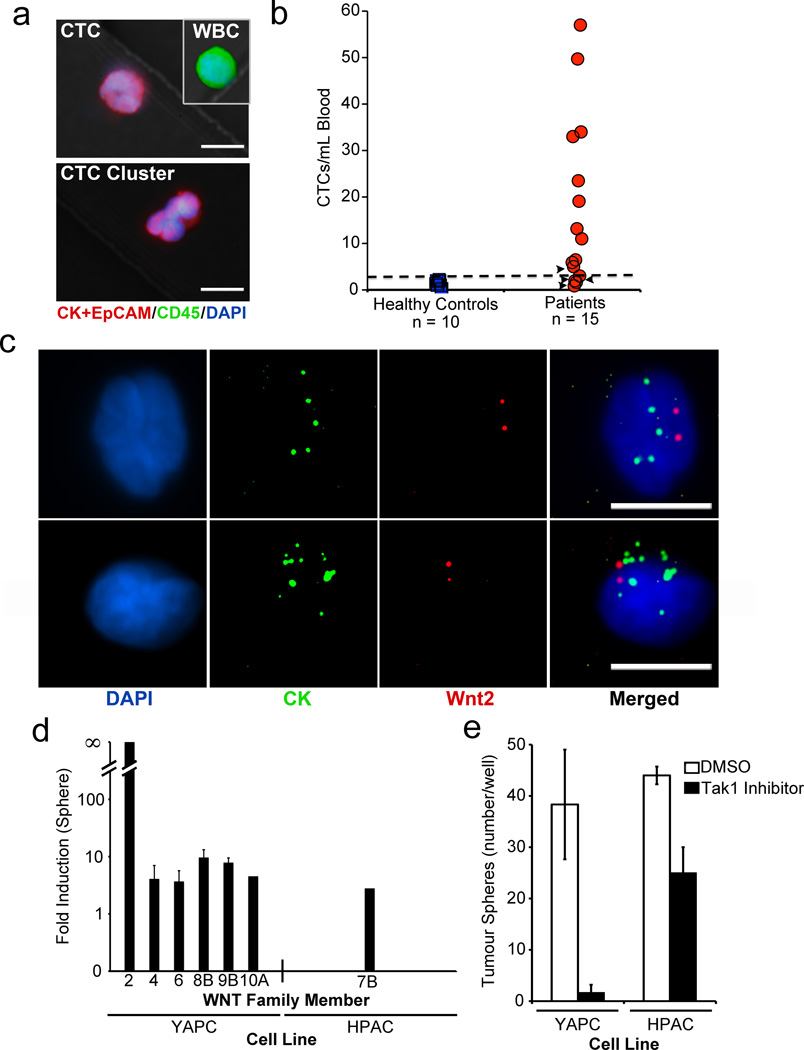
Figure 4. Detection of Wnt2 mRNA expression and non-canonical Wnt signature in human pancreatic CTCs.
a, Immunofluorescence staining of human pancreatic CTC, leukocyte (WBC), and CTC cluster captured on anti-human EpCAM HbCTC-Chip (DAPI nuclear stain, blue; CK and EpCAM cocktail, red; CD45, green). b, Enumeration of CK+EpCAM positive human CTCs (CTCs/mL) captured from patients with metastatic pancreatic cancer. Arrowheads indicate lower CTC numbers in patients responding to therapy. Blood samples from healthy donors were used to establish the threshold of ≥ 3 CK+EpCAM+/CK45− cells/mL (dashed line). c, RNA-ISH analysis of human pancreatic CTCs, showing co-expression of mRNAs for cytokeratins (CK) 8, 18, 19 and 23 (pooled probes in green) and Wnt2 (red). (DAPI, blue). (Scale bars = 10 µm). d, Induction of Wnt transcripts in pancreatic cancer cells grown as non-adherent tumour spheres compared to standard conditions (fold increase). e, Quantification of tumour spheres with or without Tak1 inhibitor (3µM 5-Z-7-Oxozeanol). (n=3; mean ± s.d.)

