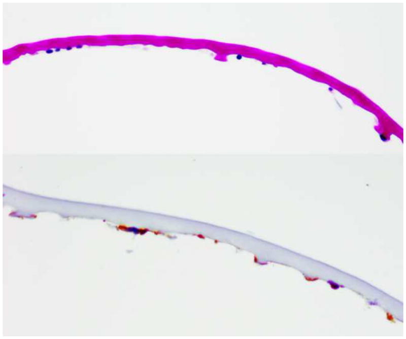
Figure 3.

Descemet’s membrane is thickened and contains posterior nodular excrescences with thickened Descemet’s membrane (Top image) with multilayered, scattered endothelial cells positive for cytokeratins AE1,3 and MAK 6 immunohistochemical satins (Bottom image). (Periodic acid schiff, 100X)
