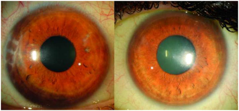
Figure 6.

Slit lamp photo of right and left cornea at final evaluation. The right cornea cornea (left image) shows complete resolution of corneal edema with corneal clarity, while the left cornea (right image) shows resolution of central corneal edema with deep paracentral stromal haze in the region corresponding to final endothelial cell repopulation.
