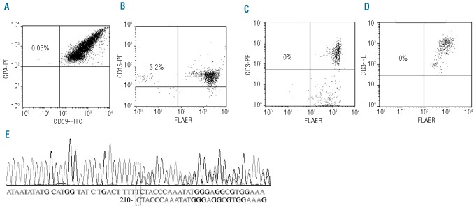
Figure 3.
PNH cell analysis in a patient (M1) with MDS. (A) Erythrocytes stained with PE conjugated anti-glycophorin A and FITC conjugated anti-CD59. (B) Granulocytes stained with PE conjugated anti-CD15 and FLAER. (C) T lymphocytes stained with PE conjugated anti-CD3 and FLAER. (D) T lymphocytes stained with PE conjugated anti-CD3 and FLAER after 14 days culturing in a T-lymphocyte enrichment medium with proaerolysin selection. (E) PIG-A DNA sequencing analysis of a single myeloid CFU-G grown under proaerolysin selection indentified a deletion mutation of “T” at 210 bp of exon 2, which caused a frameshift mutation, and a premature translation termination in the middle of exon 2.

