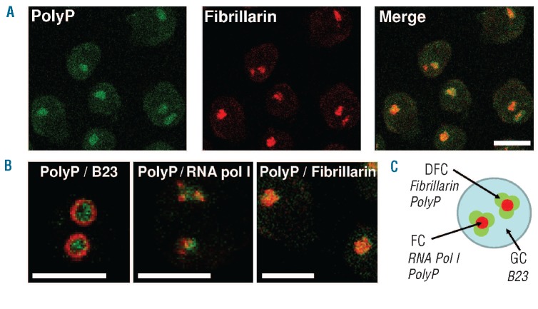
Figure 4.
Confocal immunofluorescence analysis in U266 MC. (A) The figure shows the colocalization of polyP and fibrillarin (nucleolar marker). (B) Analysis of subnucleolar markers and polyP localization: the figure shows merged images of the localization of polyP (green) and the following nucleolar proteins (in red): B23 (granular component), RNA pol I (fibrillar center), and fibrillarin (dense fibrillar component). Bars: 10 μm. Representative experiments are shown (n=3). (C) Descriptive scheme of polyP distribution within the nucleolus: in accordance with the results shown in 4B, a model describing polyP distribution in the subnucleolar compartments was devised. GC: granular component, FC: fibrillar center, DFC: dense fibrillar component. (Adapted from Lam et al.30).

