Summary
The current trend of antimony (Sb) unresponsiveness in the Indian subcontinent is a major impediment to effective chemotherapy of visceral leishmaniasis (VL). Although contributory mechanisms studied in laboratory raised Sb-R parasites include an up regulation of drug efflux pumps and increased thiols, their role in clinical isolates is not yet substantiated. Accordingly, our objectives were to study the contributory role of thiols in generation of Sb unresponsiveness in clinical isolates. Promastigotes were isolated from VL patients who were either Sb responsive (n = 2) or unresponsive (n = 3). Levels of thiols as measured by HPLC and flow cytometry showed higher basal levels of thiols and a faster rate of thiol regeneration in Sb unresponsive strains as compared with sensitive strains. The effects of antimony on generation of reactive oxygen species (ROS) in normal and thiol depleted conditions as also their H2O2 scavenging activity indicated that in unresponsive parasites, Sb mediated ROS generation was curtailed which could be reversed by depletion of thiols and was accompanied by a higher H2O2 scavenging activity. Higher levels of thiols in Sb unresponsive field isolates from patients with VL protects parasites from Sb mediated oxidative stress, thereby contributing to the antimony resistance phenotype.
Keywords: visceral leishmaniasis, antimonial resistance, thiols, oxidative stress, reactive oxygen species
Introduction
The trypanosomatids, members of the order Kinetopastida include parasitic protozoa such as Leishmania spp that are responsible for Leishmaniasis (Guerin et al. 2002; Herwaldt 1999). The disease endemicity extends to over 88 countries, 72 being in the developing world while 13 belong to the category of least developed countries (Murray et al. 2005). Globally, the population at risk is 350 million, overall prevalence being 12 million, 2 million new cases occur annually and the Disability adjusted Life years (DALY) burden is considered to be 860,000 men and 1.2 million women (Leishmaniasis. Seventeenth Programme Report. Progress 2003-2004). Estimated annual incidence of the visceral form is around 500,000 in 61 countries with 90% of these cases being confined to 5 countries namely India (especially the state of Bihar and its adjoining states), Bangladesh, Nepal (Terai region), Sudan and North Eastern Brazil (Guerin et al. 2002).
An increasing incidence of unresponsiveness to sodium antimony gluconate (SAG) is a critical issue in the current, prolonged epidemic in India where over 50-65% of the estimated 250,000 annual cases are non-responsive to this first-line agent (Croft et al. 2006). Mechanism(s) by which Leishmania spp acquire resistance to antimony is a subject of intense research and to serve as models for resistance, drug resistant strains of Leishmania tarentolae have been generated in vitro by step-wise exposure to increasing concentrations of antimonials or arsenicals (Borst and Ouellette, 1995). A diminished biological reduction of SbV to SbIII has been demonstrated in L. donovani amastigotes resistant to antimony (Shaked-Mishan et al. 2001). Aquaglyceroporin1 (AQP1) have recently been demonstrated to mediate uptake of SbIII in Leishmania spp. and their overexpression generates hypersensitivity to SbIII (Marquis et al. 2004). Resistance to antimonials also required an increased synthesis of trypanothione (a bis glutathionyl-spermidine conjugate), the major intracellular thiol of these parasites (Haimeur et al. 2000; Fairlamb and Cerami, 1995). Further corroborative evidence was provided by amplification of GSH1 gene coding for γ-glutamylcysteine synthetase ( γ-GCS, Grondin et al. 1997) along with overexpression of ornithine decarboxylase (ODC, Haimeur et al. 1999), rate limiting steps in glutathione and spermidine synthesis respectively, and necessary for trypanothione overproduction (Fairlamb and Cerami, 1995). It was proposed that following formation of SbIII-thiol complexes (spontaneously or enzymatically), an enhanced extrusion of trivalent heavy metal-thiol conjugates occurs at a rate sufficient to outmatch the influx (Dey et al. 1994), attributed to an increased expression of P-glycoprotein A or PgpA (Ouellette and Borst, 1991), a member of the family of ATP-binding cassette (ABC) proteins, several of which are implicated in drug resistance (Lee, 2004; Gottesman, 2002).
In view of the alarming increase in antimonial resistance in the Indian subcontinent, the next important step would be to elucidate whether these mechanisms are operative in field strains. Singh et al. (2007) using DNA microarray have reported amplification of multidrug resistant associated protein A (MRPA) and γ-GCS at a transcriptional level in antimony resistant clinical isolates. Mukherjee et al. (2007) have reported amplification of MRPA, γ-GCS, and ODC both at a genetic and transcriptional level in one or more antimony resistant clinical isolates and established that in field isolates, antimony resistance is a multifactorial phenomenon. Accordingly, this study was undertaken in field isolates from the same geographical region, wherein we have focused on the functional aspects of overexpression of γ-GCS and ODC, rate limiting enzymes of GSH and polyamine biosynthesis respectively. We have been able to establish that in antimony-resistant strains, a higher content of cellular thiols was accompanied with both a decreased generation of reactive oxygen species (ROS) and a higher scavenging activity of ROS that collectively contribute to decreased antimonial responsiveness.
Materials and Methods
Hydrogen peroxide (H2O2, 5%) was obtained from Merck (India). Pentavalent (SbV) and trivalent (SbIII) antimony were kindly provided by Dr SL Croft, London School of Tropical Medicine & Hygiene, UK. All other chemicals unless otherwise mentioned, were obtained from Sigma Chemicals Co. (St. Louis, MO, USA).
Parasite culture
Promastigotes of Indian Leishmania donovani strains MHOM/IN/83/AG83, MHOM/IN/90/GE1F8R along with three untyped strains 2001, NS2, and 41 were isolated from patients with visceral leishmaniasis and routinely cultured at 24°C in M-199 medium supplemented with 10% heat inactivated fetal calf serum (HIFCS) and Penicillin/Streptomycin referred to as Medium A. For experimental purposes, log phase promastigotes were obtained by subculturing every 72-96 hrs, inoculum being 1 × 10⁶/ml.
Previous studies in the amastigote-macrophage model have established that MHOM/IN/83/AG83 and 2001 are antimony sensitive or Sb-S referred to in this study as S1 and S2 respectively whereas NS2, 41 and MHOM/IN/90/GE1F8R, are antimony resistant or Sb-R (Mukherjee et al. 2007; Singh et al. 2007) referred to as R1, R2 and R3 respectively. SAG contains pentavalent antimony (SbV) with m-chlorocresol as preservative which itself is a potent anti-promastigote agent (Roberts and Rainey, 1993). Accordingly, in this study, we have used m-chlorocresol free SbV and SbIII.
Analysis of intracellular thiols using HPLC
Mid log phase promastigotes (5 × 10⁷) were collected by centrifugation (1,600 × g, 10 min, 4 °C) and derivatized with monobromobimane as described previously (Shim and Fairlamb, 1998). Acid soluble thiols were separated by ion paired, reverse phase HPLC on a Beckman Ultrasphere C18 column using a Beckman System Gold instrument fitted with a Gilson-121 fluorometer.
Flow cytometric determination of intracellular non protein thiols using mercury orange (MO)
Non protein thiols were measured using the method described by O’Connor et al. (1988) with slight modifications. The assay was initially standardized for log phase promastigotes (1 × 10⁷/ml) of S1 and R3, representative of Sb-S and Sb-R strains respectively. After washing cells with ice cold phosphate buffered saline (0.02M pH 7.2, PBS), cell pellets were resuspended in mercury orange (100, 250 and 500 μM in acetone) and incubated for 5 min on ice. The cells were then washed thoroughly with ice cold PBS and analyzed for fluorescence in FL3 channel. Based on the above experiment, we choose 500 μM and log phase promastigotes (1 × 10⁷/ml) from 5 strains were labelled with mercury orange (500 μM) and MFC analyzed as described above.
To measure the regeneration rate of intracellular thiols in promastigotes, parasites (1 × 10⁶/ml) were initially exposed to buthionine sulphoximine (3 mM, BSO), an inhibitor of γ-GCS (Griffith and Meister, 1979) for 48 h at 24°C in Medium A. Cells were then washed with PBS, resuspended in Medium A and fluorescence of mercury orange was measured to confirm depletion of thiols. Subsequently, cells were harvested at 0, 30, 60 and 150 minutes and thiol content was assessed. Results were expressed as the % increment of MFC following their incubation in BSO free medium. Similarly, the effect of SbIII (300 μg/ml, 3 h at 37°C) on intracellular thiols was measured in log phase promastigotes (1 × 10⁶/ml) using mercury orange and results expressed as % change of MFC from 0 min.
Effects of pentavalent (SbV) or trivalent (SbIII) antimony on generation of ROS by promastigotes
To study the capacity of SbV and SbIII to generate ROS, log phase promastigotes (1 × 10⁶/ml) were exposed to SbV or SbIII (300 μg/ml) for 3 h at 37°C in medium M199 (serum free). The cells were then washed with PBS, incubated with dichlorodihydrofluorescein diacetate (H2DCFDA, 50 μM) for 45 min at 37°C and analyzed on a FACS Calibur (Becton Dickenson, USA), dead cells being excluded using propidium iodide (PI, 1 μg/ml).
To study whether thiol depletion influenced ROS generated by SbIII, log phase promastigotes from 5 strains were initially treated with BSO (3 mM) to deplete thiols and was confirmed by reduction of fluorescence using mercury orange. These thiol depleted cells (1 × 10⁶/ml) were then exposed to SbIII (300 μg/ml) and the extent of ROS generation was measured using H2DCFDA (50 μM), dead cells being excluded using PI.
ROS scavenging activity of promastigotes
To assess the ROS scavenging activity of promastigotes, log phase parasites (1 × 10⁶/ml) were exposed to H2O2 (1 – 1000 μM) at 37°C in PBS for 1 h. Cells were then washed and incubated at 37°C for 45 min with H2DCFDA (50 μM). Labelled cells were analyzed on a FACS Calibur, dead cells being excluded using PI.
Flow cytometry
Cells (106) from different experimental groups were monitored for their intracellular fluorescence on a flow cytometer (FACS Calibur, Becton Dickenson, San Jose, CA, USA) equipped with an argon-ion laser (15 mW) tuned to 488 nm. Fluorescence of DCF was collected in FL1 channel, equipped with a 530/30 nm band pass filter, PI in FL2 channel having a 585/42-nm band pass filter and mercury orange in FL3 channel equipped with 670 nm long pass filter. Fluorescence was measured in the log mode and expressed as mean fluorescence channel (MFC). Analyses were performed on 10,000 gated events, while data acquisition and analysis was carried out with CellQuest Pro software.
Statistical Analysis
Each experiment was performed at least thrice in duplicates and results expressed as mean ± standard error of the mean (SEM). For significance, Student’s t test was performed, P values < 0.05 were considered significant.
RESULTS
Sb-R strains have higher amounts of cellular thiols than Sb-S strains
Non-protein thiols are established molecules that combat against xenobiotic toxicity and oxidative damage (Meister and Anderson, 1983). In Leishmaniasis, Mukhopadhyay et al. (1996) have shown that in laboratory raised antimonal resistant L. tarentolae, the degree of drug resistance correlated with concomitantly raised thiol levels. Accordingly, we studied whether differences existed between levels of thiols in Sb-S vs. Sb-R field isolates, as measured by HPLC and flow cytometry.
HPLC measurement of thiols, glutathione (GSH) and trypanothione (TSH) indicated that Sb-S strains (S1 and S2) have significantly lower amounts of GSH and TSH than Sb-R strains, R1, R2 and R3 (Table 1) wherein the differences in GSH levels was more pronounced.
Table 1.
Thiol levels in Sb-S and Sb-R strains.
| Strain | Thiol content, nmol (108 cells)-1 | |
|---|---|---|
| GSH | T[SH]2 | |
| S1 | 0.38 ± 0.06 | 2.64 ± 0.21 |
| S2 | 0.328 ± 0.06 | 3.28 ± 0.11 |
| R1 | 0.86 ± 0.007* | 4.48 ± 0.18* |
| R2 | 0.77 ± 0.10* | 4.41 ± 0.13* |
| R3 | 0.97 ± 0.09* | 6.64 ± 0.21* |
Mid log phase promastigotes (5 × 10⁷) from Sb-sensitive strains (S1 and S2) and Sb-resistant strains (R1, R2 and R3) were derivatized with monobromobimane19 and analysed as described by Materials and Methods. Each data point is the mean of at least three determinations ± SD.
P = <0.001 when compared to sensitive group
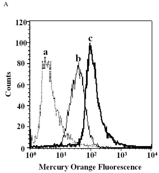
Mercury orange reacts with all sulfhydryl (-SH) groups generating a fluorescent product which is retained within cells. However, as the reaction rate of mercury orange with non protein thiols is much faster than protein thiols, pulse labelling for 5 min on ice allowed MO to react only with non protein –SH groups. Therefore, the level of fluorescence represented the level of cellular non protein thiols (O’Connor et al. 1988). The assay was initially optimized for Leishmania promastigotes using increasing concentrations of MO (100 - 500 μM). At 100 μM of MO, fluorescence from both the Sb-S (S1) and Sb-R strain (R3) was comparable, MFC being 39.66 and 50.84 respectively. Increasing MO to 250 μM, the MFC of R3 became 2 fold higher than S1 being 92.59 vs. 43.95 respectively. A further increase of MO to 500 μM resulted in the MFC of R3 becoming 3 fold higher than S1 being 139.91 vs. 45.15 respectively (Fig. 1A) However, further increase in MO caused no change in both strains (data not shown) and accordingly, 500 μM of MO was subsequently selected.
Figure 1A. Flow cytometric measurement of thiols using Mercury orange (MO).
Log phase promastigotes (unstained, a) of S1 (b) and R3 (c) were exposed to MO (500 μM) in acetone, incubated on ice for 5 minutes, washed and analysed for fluorescence in FL3 channel as described in Materials and Methods.
Measurement of non protein thiols in the five strains, as shown in Fig. 1B clearly indicated that Sb-S strains had lower amounts of thiols, the MFC ± SEM of S1 and S2 being 45.24 ± 4.19 and 48.81 ± 3.8 respectively as compared to the Sb-R strains namely R1 (62.09 ± 2.60), R2 (92.33 ± 3.7) and R3 (110.84 ± 7.63). Collectively, intracellular thiols in Sb-S strains were 1.9 fold lower than Sb-R strains, MFC ± SEM being 47.02 ± 2.67 vs. 88.41 ± 5.64, p< 0.001. The addition of BSO (3 mM, 48 h) caused a dramatic decrease in fluorescence confirming depletion of thiols and the extent of decrease was similar in all strains (Fig. 1B).
Figure 1B. Flow cytometric detection of basal intracellular thiols in Sb-S and Sb-R strains.
Log phase promastigotes both from Sb-S (S1 and S2) and Sb-R (R1, R2 and R3) strains were labelled with Mercury Orange (MO) before (open bars,□) and after treatment with buthionine sulphoximine (3 mM, 48 h, 24°C) (filled bars,■) and fluorescence analyzed as described in Materials and Methods. Data are expressed as MFC ± SEM of at least three independent experiments in duplicates.
The generation of ROS by SbIII is reduced in Sb-R strains
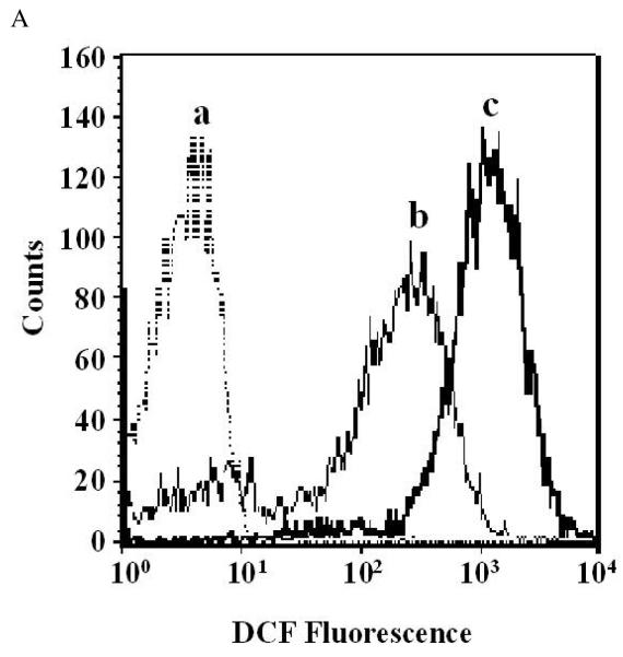
Mehta and Shaha (2006) have shown that antimony exerts its antileishmanial activity by generating ROS which is triggered by loss of mitochondrial membrane potential and uncoupling of oxidative phosphorylation. To examine whether differences exist in the amount of ROS generated by Sb-S vs. Sb-R strains in the presence of SbIII, their oxidative status was measured using H2DCFDA. H2DCFDA, a lipid soluble, membrane permeable compound is cleaved by non-specific esterases to remove the diacetate portion and release H2DCF which in turn is oxidised by intracellular reactive oxygen species (ROS) to produce a fluorescent compound DCF (Wan et al. 1993). Therefore, the fluorescence is directly proportional to the amount of ROS present within cells and H2DCFDA (50 μM) incubated at 37°C for 45 minutes gave the best results (Fig. 2A).
Figure 2A. H2O2 mediated increase of H2DCFDA mediated fluorescence.
Log phase promastigotes of S1 (unstained, a) were exposed to increasing concentrations of H2O2, 100 μM (b) and 1000 μM (c), probed with dichlorodihydrofluorescein diacetate (H2DCFDA) and analysed for dichlorofluorescein (DCF) fluorescence in FL1 channel as described in Materials and Methods.
Initially, the concentration of SbIII and duration of incubation was defined by exposing a Sb-S (S1) and a Sb-R strain (R3) to both, variable concentrations of SbIII (0-600 μg/ml) and time (1-6 h) at 37°C. It was established that maximal ROS generation was achieved with SbIII (300 μg/ml) for 3 h, parasite viability remaining >80% as measured by PI uptake (data not shown).
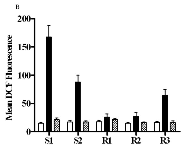
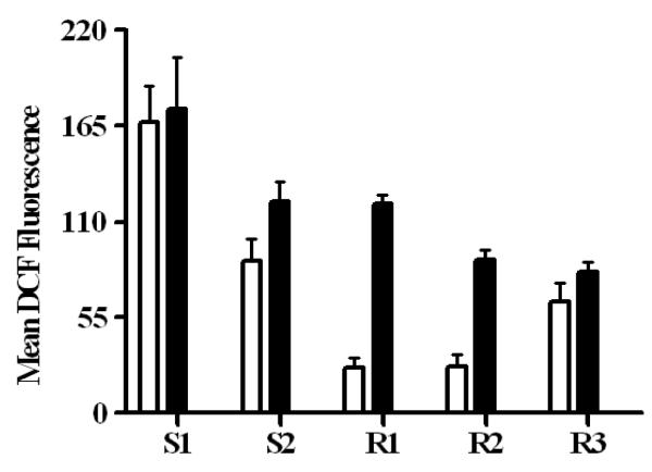
In the Sb-S strains, generation of ROS by SbIII (167.45 ± 20.65) was higher, maximum being in S1 followed by S2 (87.89 ± 12.48) whereas in all three Sb-R strains, the amount of ROS generated was consistently lower, MFC ± SEM being 25.85 ± 5.75, 26.79 ± 6.99 and 64.37 ± 10.54 in R1, R2 and R3 respectively (Fig. 2B). Taken together, ROS production triggered by SbIII was 3.6 fold higher in Sb-S strains than Sb-R strains, MFC ± SEM being 133.95 ± 19.55 vs. 37.17 ± 5.14, p< 0.001. Importantly, promastigotes exposed to SbV (300 μg/ml) for 3 h at 37°C failed to generate any ROS corroborating the earlier reports that SbV is ineffective against promastigotes. Based on this finding we decided to use only SbIII for further experiments.
Figure 2B. Generation of ROS by SbIII and SbV in Sb-S and Sb-R strains.
Log phase promastigotes (open bars,□) both from Sb-S (S1 and S2) and Sb-R (R1, R2 and R3) strains were exposed to either SbIII (filled bars,■) or SbV (hatched bars, ) for 3 h at 37°C, labelled with dichlorodihydrofluorescein diacetate (H2DCFDA) and dichlorofluorescein (DCF) fluorescence was analysed as described in Materials and Methods. Data are expressed as MFC ± SEM of at least three independent experiments in duplicates.
The rate of thiol synthesis in Sb-S strains is slower than in Sb-R strains
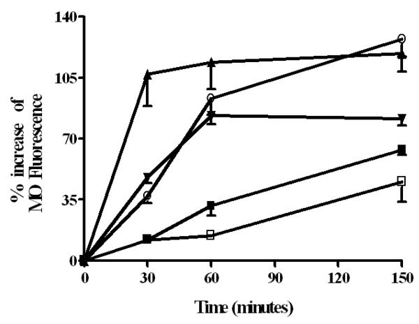
As non protein thiols were higher in Sb-R strains (Fig. 1B) and was associated with a lower generation of ROS by SbIII in these strains (Fig. 2B), it prompted us to study whether Sb-R strains had an up regulation in the biosynthetic machinery of thiols which caused a sustained increase in intracellular thiols. Accordingly, the regeneration rate of thiols in Sb-S and Sb-R strains was studied following its depletion using BSO, an established inhibitor of γ-GCS. It was clearly evident that R3 possessed the most efficient thiol generating machinery as normal thiol levels were again achieved within 30 min, post BSO treatment (Fig. 3). The other two Sb-R strains, R1 and R2 took 60 minutes to reach their normal levels and in fact with R2, levels overshot beyond basal levels. In sharp contrast, in the antimony-S strains S1 and S2, their thiol regenerating machinery was much slower than their Sb-R counterparts, as normal thiol levels were achieved much later at 150 minutes, post- BSO treatment (Fig. 3).
Figure 3. Kinetics of thiol regeneration in Sb-S and Sb-R strains.
Log phase promastigotes from Sb-S strains, S1 (closed squares, -■-) and S2 (open squares, -□-), as also Sb-R strains R1 (inverted closed triangles, -▼-), R2 (open circles, -○-) and R3 (upright closed triangles, -▲-) were depleted of thiols using buthionine sulphoximine (3 mM, 48 h, 24°C); promastigotes were then washed and resuspended in Medium A, harvested at different time points, labelled with Mercury Orange (MO) and fluorescence analysed as described in Materials and methods. Data are expressed as mean ± SEM of % increment of MFC from 0 minute of at least three independent experiments in duplicates.
Depletion of intracellular thiols enhances SbIII mediated generation of ROS in Sb-R strains
To confirm that the higher amount of thiols present in Sb-R strains contributes to the attenuated generation of ROS by SbIII, the amount of ROS generated by SbIII following removal of thiols by BSO was measured. In the absence of BSO, SbIII triggered a higher generation of ROS in S1 and S2 than all three Sb-R strains corroborating with previous data. Following thiol depletion by BSO as confirmed by MO (Fig. 1B), ROS production by SbIII was marginally increased in S1, S2 and R3 (Fig. 4). However, in R1 and R2, depletion of thiols resulted in a dramatic 3 fold increase in ROS generation as compared to their respective control values, p<0.001 (Fig. 4). Taken together, removal of thiols failed to augment SbIII mediated ROS generation in Sb-S strains (133.95 ± 19.55 vs. 148.57 ± 16.85, p = 0.53) whereas in Sb-R strains, depletion of thiols triggered a 2.6 fold increase in fluorescence being 37.17 ± 5.14 vs. 96.64 ± 5.21, p < 0.001.
Figure 4. Effect of thiol depletion on SbIII mediated ROS generation in Sb-S and Sb-R strains.
Log phase promastigotes (open bars,□) from Sb-S (S1 and S2) and Sb-R (R1, R2 and R3) strains following removal of thiols (filled bars,■) were exposed to SbIII (300 μg/ml) for 3 h at 37°C. Cells were labelled with dichlorodihydrofluorescein diacetate (H2DCFDA) and dichlorofluorescein (DCF) fluorescence analysed as described in Materials and Methods. Data were expressed as MFC ± SEM of at least three independent experiments in duplicates.
SbIII depletes thiols primarily in Sb-S strains
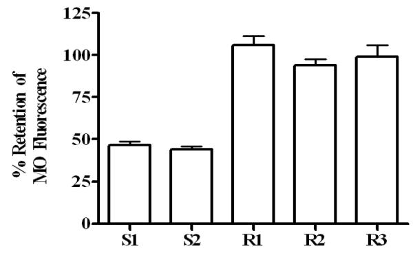
Wyllie et al., (2004) have demonstrated that antimony exerts its leishmanicidal activity by depleting thiols causing an altered redox potential. To study whether the amount of thiols depleted by SbIII varied in Sb-S vs. Sb-R strains, intracellular thiols were measured before and after treatment with SbIII. In Sb-S strains, a 3 h incubation with SbIII resulted in an almost 55% depletion of thiols; conversely in all Sb-R strains, SbIII caused no change in their thiol levels (Fig. 5), the % retention of thiols in Sb-S vs. Sb-R strains being 45.31 ± 5.10 vs. 99.62 ± 13.70 respectively.
Figure 5. Effect of SbIII on thiol levels in Sb-S and Sb-R strains.
Log phase promastigotes from Sb-S (S1 and S2) and Sb-R (R1, R2 and R3) were exposed to SbIII (300 μg/ml) at 37°C for 3 h; they were then harvested, labelled with Mercury Orange (MO) and fluorescence analysed as described in Materials and Methods. Data were expressed as mean ± SEM of % retention of MFC from baseline of at least three independent experiments in duplicates.
Sb-R strains scavenge ROS more efficiently than Sb-S strains
ROS, generated within phagolysosomes contributes significantly to the microbicidal activity of macrophages, H2O2 being one of the major components. With reference to Leishmaniasis, Mookerjee Basu et al. (2006) have established that the leishmanicidal activity of antimony is augmented via enhanced generation of ROS within phagolysosomes. Therefore, it is conceivable that in the antimony resistance phenotype, the Leishmania parasite enhances scavenging of ROS within parasites and/or macrophages.
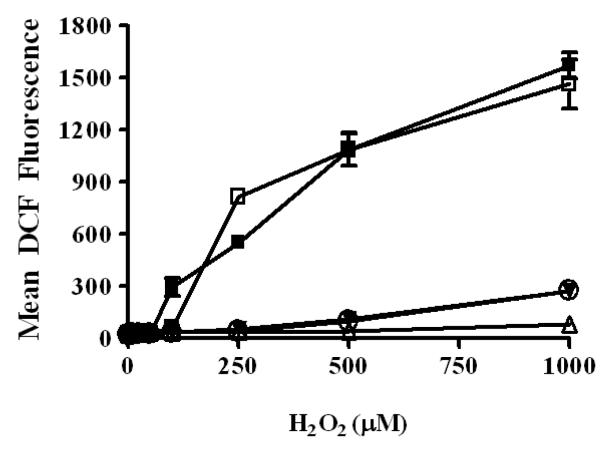
Measurement of ROS scavenged in Sb-S and Sb-R strains revealed that lower concentrations of H2O2 (1-100 μM) showed no detectable fluorescence suggesting that Leishmania parasite irrespective of their chemosensitivity profiles, can effectively scavenge this amount of H2O2. However, with increasing concentrations of H2O2, a dose dependent increase in fluorescence was demonstrated exclusively in Sb-S strains, the average MFC ± SEM of S1 and S2 in the presence of 1 mM H2O2 being 1571.63 ± 77.18 and 1462.68 ± 144.36 respectively (Fig. 6). However, in Sb-R strains, addition of 1 mM H2O2 generated far lower fluorescence, the MFC ± SEM of R1 and R3 being 272.48 ± 34.78 and 269.21 ± 25.46 respectively, indicating that Sb-R strains possess a greater ability to scavenge ROS. With regard to R2, the scavenging of ROS was even more remarkable as with 1 mM of H2O2, the MFC ± SEM was only 79.08 ± 4.24 (Fig. 6). Taken together, Sb-R strains scavenge ROS more efficiently, the fluorescence generated by H2O2 (1 mM) in Sb-S strains was 7 fold higher, the MFC ± SEM being 1517.15 ± 77.4 vs. 206.92 ± 25.9, p< 0.001.
Figure 6. Flow cytometric detection of ROS scavenging activity in Sb-S and Sb-R strains.
Log phase promastigotes from Sb-S strains S1 (closed squares, -■-), S2 (open squares, -□-) and Sb-R strains R1 (inverted closed triangles, -▼-), R2 (upright open triangles, -Δ-) and R3 (open circles, -○-) were exposed to increasing concentration of H2O2 (0 - 1000 μM) at 37°C for 1 h, probed with dichlorodihydrofluorescein diacetate (H2DCFDA) at 37°C for 45 for minutes and dichlorofluorescein (DCF) fluorescence was analysed as described in Materials and Methods. Data were expressed as MFC ± SEM of at least three independent experiments in duplicates.
Discussion
The growing resistance against conventional antimonial drugs is a major problem in the current scenario of Indian visceral leishmaniasis. The primary aim of this study was to investigate the role of anti-oxidative mechanisms contributing to development of antimonial resistance in field isolates. The key findings are that promastigotes from Sb-R field isolates (i) have a higher content of non- protein thiols (ii) curtail SbIII mediated ROS production and (iii) are more efficient scavengers of ROS than their Sb-S counterparts.
Although antimonial compounds have been used as antileishmanial agents for more than 60 years, their mechanism(s) of action is still to be precisely defined. It is an established fact that pentavalent antimony (SbV) is the prodrug that is reduced to trivalent (SbIII) form to be an effective antileishmanial agent (Sereno et al. 1998). This is possibly because promastigotes lack antimony reductase (Shaked-Mishan et al. 2001) leading to ineffectiveness of SbV. Accordingly, to simulate in vivo conditions, we have used SbIII in this study. It has been proposed that antimony acts upon several targets that include influencing the bioenergetics of Leishmania parasites by inhibiting parasite glycolysis, fatty acid beta-oxidation and inhibition of ADP phosphorylation (Sundar and Chatterjee, 2006). It causes non specific blocking of SH groups of amastigote proteins and inhibition of DNA Topoisomerase I (Chakraborty and Majumder, 1988). More recently, it has been demonstrated that antimony can compromise the thiol-redox potential in both forms of the parasite by actively promoting efflux of thiols, Glutathione and Trypanothione and additionally by increasing the proportion of thiols present in their respective disulfide forms. Collectively, antimony acts as a double edged sword by reducing both the intracellular thiol buffering capacity and the thiol redox potential rendering the parasite more susceptible to oxidative stress (Wyllie et al. 2004).
Non protein thiols, such as glutathione, are well established molecules for combating oxidative damage, toxic effects of xenobiotics and also for maintaining cellular redox homeostasis (Meister and Anderson, 1983). Accordingly, augmentation in cellular thiols can attenuate oxidative damage and minimize toxicity of xenobiotics. Trypanosomatids that include Leishmania are unique in that they possess trypanothione [N1,N8- bis (glutathionyl) spermidine], as the major substitute of glutathione present in other systems (Fairlamb et al. 1985). It has been shown that trypanothione is the key intermediate in the regulation of parasite redox homeostasis as well as in defence against xenobiotics and oxidative stress (Wyllie et al. 2004; Fairlamb and Cerami, 1995). Sb-R strains had a significantly higher level of cellular thiols than Sb-S strains as measured by HPLC and flow cytometry (Table 1, Fig. 1A and B). Additionally, these Sb-R strains have a more efficient biosynthetic machinery for regeneration of thiols than their Sb-S counterparts, as following depletion of thiols, they reverted to their normal level of thiols rapidly within 30-60 min. as compared to 150 min. required by Sb-S strains (Fig. 3). Our previous study also demonstrated that although there is no amplification of γ-GCS at genetic level in any strains (S1, S2, R1, R2 and R3), R2 showed upregulation at transcriptional level. With regard to ODC all resistant strains (R1, R2 and R3) shown upregulation both at genomic and protein level.(Mukherjee et al. 2007). Taken together, Sb-R strains have a greater potential to curtail oxidative damage and xenobiotic toxicity by sustaining their intracellular thiols.
Mehta and Shaha, (2006) have demonstrated that trivalent antimony in promastigotes induces mitochondrial dysfunction leads to uncoupling of oxidative phosphorylation ultimately resulting in generation of reactive oxygen species (ROS), the critical effector molecule responsible for parasiticidal activity. In Sb-S strains, it was observed that SbIII triggered a significantly higher amount of ROS production concomitant with a greater proportion of thiol loss than Sb-R strains (Fig. 2B, Fig. 5). This generated a higher level of oxidative stress, accounting for the more potent leishmanicidal activity observed in Sb-S strains. It is known that trivalent antimony is the active form of the drug and absence of DCF fluorescence upon addition of pentavalent antimony to promastigotes corroborated this existing knowledge that pentavalent antimony is ineffective against promastigotes. Additionally, it highlighted that generation of ROS is critical for antimony to mediate its antileishmanial activity. It is very likely that this attenuation of SbIII mediated ROS production in Sb-R strains was achieved by their higher proportion of intracellular thiols (Fig. 1A and B, Table 1). The vital contribution of thiols in generation of antimony resistance was further substantiated by the selective augmentation of ROS production in Sb-resistant strains, R1 and R2 following removal of thiols (Fig. 4). This augmentation in SbIII mediated ROS production following thiol depletion in R1 and R2 negated the possibility of reduced SbIII uptake by AQP1 down regulation contributing to antimonial resistance. However, in R3, the removal of thiols marginally enhanced generation of ROS suggesting that down regulation of AQP1 could well be a contributory factor (Marquis et al. 2004). Alternatively, in R3, it is also possible that the rate of regeneration of thiols is rapid enough to efficiently replenish its thiols (Fig. 3). The enzyme-glutamyl cysteine synthetase (γ-GCS) catalyzes conjugation of L-glutamate with L-cysteine and is the rate limiting step for glutathione biosynthesis (Lu, 2000). The activity of γ-GCS is controlled by a nonallosteric feedback by glutathione, cysteine availability, and by factors that control the transcription and post translational modification of the enzyme (Lu, 2000). Considering that in R3, the rate of thiol regeneration was the fastest and it possessed the highest amount of GSH and T[SH]2 and as Mukherjee et al. (2007) have demonstrated that in these strains, there is only ODC upregulation with no accompanying γ-GCS upregulation, one is tempted to suggest that in Sb-R strains such as R3, modifications in their γ-GCS activity could well be the key factor for these parasites to become resistant; such studies are ongoing.
Intracellular parasitic protozoans of the genus Leishmania evade toxic, free-radical damage inflicted by the phagocytic macrophage via elaboration of enzymatic pathways such as superoxide dismutase (Ghosh et al. 2003) and non-enzymatic pathways like non-protein thiols. Antimonial compounds generated ROS (mainly H2O2) within phagolysosomes of macrophages via phosphorylation of phosphoinositide 3-kinase (PI3K), protein kinase C (PKC) Ras and extracellular-signal regulated kinase (ERK) (Mookerjee Basu et al. 2006). As inhibition of these proteins or addition of a free radical scavenger like N-acetylcysteine inhibited antimony mediated killing of intracellular amastigotes, it emphasised the contribution of ROS in antimony mediated parasiticidal activity. Therefore, it is evident that the efficacy of antimony as an anti-leishmanial agent hinges upon its ability to generate ROS both within the parasite and/or phagolysosomes of infected macrophages. It would be rational to suggest that Leishmania strains having a higher amount of thiols would also possess a higher capacity to scavenge free radicals are more likely to be unresponsive to antimony. Here we demonstrate for the first time, that indeed in promastigotes from Sb-S field strains, the addition of H2O2 (1 mM) triggered an almost 7 fold increase in DCF fluorescence as compared to Sb-R strains (Fig. 6) clearly indicating that the Sb-R strains have a more potent ROS scavenging activity. This is the first demonstration that in field isolates from patients with VL, Leishmania parasites up regulate their antioxidant pathways through raised non-protein thiols leading to a higher ROS scavenging activity thus generating antimonial unresponsiveness.
Acknowledgements
This work received financial assistance from Council of Scientific and Industrial Research, Govt. of India.
Footnotes
Transparency declarations: None to declare.
References
- Borst P, Ouellette M. New mechanisms of drug resistance in parasitic protozoa. Annual Review of Microbiology. 1995;49:427–460. doi: 10.1146/annurev.mi.49.100195.002235. [DOI] [PubMed] [Google Scholar]
- Chakraborty AK, Majumder HK. Mode of action of pentavalent antimonials: specific inhibition of type I DNA topoisomerase of Leishmania donovani. Biochemical and Biophysical Research Communications. 1988;152:605–611. doi: 10.1016/s0006-291x(88)80081-0. [DOI] [PubMed] [Google Scholar]
- Croft SL, Sundar S, Fairlamb AH. Drug resistance in leishmaniasis. Clinical Microbiology Reviews; 2006;19:111–126. doi: 10.1128/CMR.19.1.111-126.2006. [DOI] [PMC free article] [PubMed] [Google Scholar]
- Dey S, Papadopoulou B, Haimeur A, Roy G, Grondin K, Dou D, Rosen BP, Ouellette M. High level of arsenite resistance in Leishmania tarentolae is mediated by an active extrusion system. Molecular and Biochemical Parasitology. 1994;67:49–57. doi: 10.1016/0166-6851(94)90095-7. [DOI] [PubMed] [Google Scholar]
- Fairlamb AH, Cerami A. Metabolism and functions of trypanothione in the Kinetoplastida. Annual Review of Microbiology. 1992;46:695–729. doi: 10.1146/annurev.mi.46.100192.003403. [DOI] [PubMed] [Google Scholar]
- Fairlamb AH, Blackburn P, Ulrich P, Chait BT, Cerami A. Trypanothione: a noval bis (glutathionyl) spermidine cofactor for glutathione reductase in trypanosomatids. Science. 1985;227:1485–1487. doi: 10.1126/science.3883489. [DOI] [PubMed] [Google Scholar]
- Ghosh S, Goswami S, Adhya S. Role of superoxide dismutase in survival of Leishmania within the macrophage. Biochemical Journal. 2003;369:447–452. doi: 10.1042/BJ20021684. [DOI] [PMC free article] [PubMed] [Google Scholar]
- Gottesman MM. Mechanisms of cancer drug resistance. Annual Review of Medicine. 2002;53:615–627. doi: 10.1146/annurev.med.53.082901.103929. [DOI] [PubMed] [Google Scholar]
- Griffith OW, Meister A. Potent and specific inhibition of glutathione synthesis by buthionine sulfoximine (S-n-butyl homocysteine sulfoximine) Journal of Biological Chemistry. 1979;254:7558–7560. [PubMed] [Google Scholar]
- Grondin K, Haimeur A, Mukhopadhyay R, Rosen BP, Ouellette M. Coamplification of the gamma-glutamylcysteine synthetase gene gsh1 and of the ABC transporter gene PgpA in arsenite resistant Leishmania tarentolae. EMBO Journal. 1997;16:3057–3065. doi: 10.1093/emboj/16.11.3057. [DOI] [PMC free article] [PubMed] [Google Scholar]
- Guerin PJ, Olliaro P, Sundar S, Boelaert M, Croft SL, Desjeux P, Wasunna MK, Bryceson AD. Visceral leishmaniasis: current status of control, diagnosis, and treatment, and a proposed research and development agenda. Lancet Infectious Diseases. 2002;2:494–501. doi: 10.1016/s1473-3099(02)00347-x. [DOI] [PubMed] [Google Scholar]
- Haimeur A, Brochu C, Genest P, Papadopoulou B, Ouellette M. Amplification of the ABC transporter gene PGPA and increased trypanothione levels in potassium antimonyl tartrate (Sb III) resistant Leishmania tarentolae. Molecular and Biochemical Parasitology. 2000;108:131–135. doi: 10.1016/s0166-6851(00)00187-0. [DOI] [PubMed] [Google Scholar]
- Haimeur A, Guimond C, Pilote S, Mukhopadhyay R, Rosen BP, Poulin R, Ouellette M. Elevated levels of polyamines and trypanothione resulting from overexpression of the ornithine decarboxylase gene in arsenite resistant Leishmania. Molecular Microbiology. 1999;34:726–735. doi: 10.1046/j.1365-2958.1999.01634.x. [DOI] [PubMed] [Google Scholar]
- Herwaldt BL. Leishmaniasis. Lancet. 1999;354:1191–1199. doi: 10.1016/S0140-6736(98)10178-2. [DOI] [PubMed] [Google Scholar]
- Lee CW. Reversing agents for ATP-binding cassette (ABC) transporters: application in modulating multidrug resistance (MDR) Current Medicinal Chemistry Anti-cancer agents. 2004;4:43–52. doi: 10.2174/1568011043482197. [DOI] [PubMed] [Google Scholar]
- Lu SC. Regulation of glutathione synthesis. Current topics in Cellular Regulation. 2000;36:95–116. doi: 10.1016/s0070-2137(01)80004-2. [DOI] [PubMed] [Google Scholar]
- Marquis N, Gourbal B, Rosen BP, Mukhopadhyay R, Ouellette M. Modulation in aquaglyceroporin AQP1 gene transcript levels in drug-resistant Leishmania. Molecular Microbiology. 2005;57:1690–1699. doi: 10.1111/j.1365-2958.2005.04782.x. [DOI] [PubMed] [Google Scholar]
- Mehta A, Shaha C. Mechanism of metalloid-induced death in Leishmania spp.: role of iron, reactive oxygen species, Ca2+, and glutathione. Free Radical Biology & Medicine. 2006;40:1857–1868. doi: 10.1016/j.freeradbiomed.2006.01.024. [DOI] [PubMed] [Google Scholar]
- Meister A, Anderson ME. Glutathione. Annual Review of Biochemistry. 1983;52:711–760. doi: 10.1146/annurev.bi.52.070183.003431. [DOI] [PubMed] [Google Scholar]
- Mookerjee Basu J, Mookerjee A, Sen P, Bhaumik S, Sen P, Banerjee S, Naskar K, Choudhuri SK, Saha B, Raha S, Roy S. Sodium antimony gluconate induces generation of reactive oxygen species and nitric oxide via phosphoinositide 3-kinase and mitogen-activated protein kinase activation in Leishmania donovani-infected macrophages. Antimicrobial Agents and Chemotherapy. 2006;50:1788–1797. doi: 10.1128/AAC.50.5.1788-1797.2006. [DOI] [PMC free article] [PubMed] [Google Scholar]
- Mukherjee A, Prasad KP, Singh S, Roy G, Girard I, Chatterjee M, Ouellette M, Madhubala R. Role of ABC transporter MRPA, γ-glutamylcysteine synthetase and ornithine decarboxylase in natural antimony-resistant isolates of Leishmania donovani. Journal of Antimicrobial Chemotherapy. 2007;59:204–211. doi: 10.1093/jac/dkl494. [DOI] [PubMed] [Google Scholar]
- Mukhopadhyay R, Dey S, Xu N, Gage D, Lightbody J, Ouellette M, Rosen BP. Trypanothione overproduction and resistance to antimonials and arsenicals in Leishmania. Proceedings of the National Academy of Sciences of the United States of America. 1996;93:10383–10387. doi: 10.1073/pnas.93.19.10383. [DOI] [PMC free article] [PubMed] [Google Scholar]
- Murray HW, Berman JD, Davies CR, Saravia NG. Advances in leishmaniasis. Lancet. 2005;366:1576–1577. doi: 10.1016/S0140-6736(05)67629-5. [DOI] [PubMed] [Google Scholar]
- O’Connor JE, Kimler BF, Morgan MC, Tempas KJ. A flow cytometric assay for intracellular nonprotein thiols using mercury orange. Cytometry. 1988;9:529–532. doi: 10.1002/cyto.990090604. [DOI] [PubMed] [Google Scholar]
- Ouellette M, Borst P. Drug resistance and P-glycoprotein gene amplification in the protozoan parasite Leishmania. Research in Microbiology. 1991;142:737–746. doi: 10.1016/0923-2508(91)90089-s. [DOI] [PubMed] [Google Scholar]
- Roberts WL, Rainey PM. Antileishmanial activity of sodium stibogluconate fractions. Antimicrobial Agents and Chemotherapy. 1993;37:1842–1846. doi: 10.1128/aac.37.9.1842. [DOI] [PMC free article] [PubMed] [Google Scholar]
- Sereno D, Cavaleyra M, Zemzoumi K, Maquaire S, Ouaissi A, Lemesre JL. Axenically grown amastigotes of Leishmania infantum used as an in vitro model to investigate the pentavalent antimony mode of action. Antimicrobial Agents and Chemotherapy. 1998;42:3097–3102. doi: 10.1128/aac.42.12.3097. [DOI] [PMC free article] [PubMed] [Google Scholar]
- Shaked-Mishan P, Ulrich N, Ephros M, Zilberstein D. Novel intracellular Sbv reducing activity correlates with antimony susceptibility in Leishmania donovani. Journal of Biological Chemistry. 2001;276:3971–3976. doi: 10.1074/jbc.M005423200. [DOI] [PubMed] [Google Scholar]
- Shim H, Fairlamb AH. Levels of polyamines, glutathione and glutathione-spermidine conjugates during growth of the insect trypanosomatid Crithidia fasciculata. Journal of General Microbiology. 1988;134:807–817. doi: 10.1099/00221287-134-3-807. [DOI] [PubMed] [Google Scholar]
- Singh N, Almeida R, Kothari H, Kumar P, Mandal G, Chatterjee M, Venkatachalam S, Govind MK, Mandal SK, Sundar S. Differential gene expression analysis in antimony unresponsive Indian kala azar (visceral leishmaniasis) clinical isolates by DNA microarray. Parasitology Feb. 2007;19:1–11. doi: 10.1017/S0031182007002284. [Epub ahead of print] [DOI] [PubMed] [Google Scholar]
- Sundar S, Chatterjee M. Visceral leishmaniasis - current therapeutic modalities. Indian Journal of Medical Rresearch. 2006;123:345–352. [PubMed] [Google Scholar]
- TDR Leishmaniasis. Seventeenth Programme Report. Progress 2003-2004. 2005:19–25. [Google Scholar]
- Wan CP, Myung E, Lau BH. An automated micro-fluorometric assay for monitoring oxidative burst activity of phagocytes. Journal of Immunological Methods. 1993;159:131–8. doi: 10.1016/0022-1759(93)90150-6. 1993. [DOI] [PubMed] [Google Scholar]
- Wyllie S, Cunningham ML, Fairlamb AH. Dual action of antimonial drugs on thiol redox metabolism in the human pathogen Leishmania donovani. Journal of Biological Chemistry. 2004;279:39925–39932. doi: 10.1074/jbc.M405635200. [DOI] [PubMed] [Google Scholar]


