

Summary
Our purpose was to determine if females demonstrate decreased hamstrings to quadriceps peak torque (H/Q) ratios compared to males and if H/Q ratios increase with increased isokinetic velocity in both sexes. Maturation disproportionately increases hamstrings peak torque at high velocity in males, but not females. Therefore, we hypothesised that mature females would demonstrate decreased H/Q ratios compared to males and the difference in H/Q ratio between sexes would increase as isokinetic velocity increased. Studies that analysed the H/Q ratio with gravity corrected isokinetic strength testing reported between 1967 and 2004 were included in our review and analysis. Keywords were hamstrings/quadriceps, isokinetics, peak torque and gravity corrected. Medline and Smart databases were searched combined with cross-checked bibliographic reference lists of the publications to determine studies to be included. Twenty-two studies were included with a total of 1568 subjects (1145 male, 423 female). Males demonstrated a significant correlation between H/Q ratio and isokinetic velocity (R = 0.634, p < 0.0001), and a significant difference in the isokinetic H/Q ratio at the lowest angular velocity (47.8 ± 2.2% at 30°/s) compared to the highest velocity (81.4 ± 1.1% at 360°/s, p < 0.001). In contrast, females did not demonstrate a significant relationship between H/Q ratio and isokinetic velocity (R = 0.065, p = 0.77) or a change in relative hamstrings strength as the speed increased (49.5 ± 8.8% at 30°/s; 51.0 ± 5.7% at 360°/s, p = 0.84). Gender differences in isokinetic H/Q ratios were not observed at slower angular velocities. However, at high knee flexion/extension angular velocities, approaching those that occur during sports activities, significant gender differences were observed in the H/Q ratio. Females, unlike males, do not increase hamstrings to quadriceps torque ratios at velocities that approach those of functional activities.
Keywords: Hamstrings/quadriceps, Isokinetics, Peak torque
Introduction
Decreased hamstrings strength relative to the quadriceps (H/Q) is implicated as a potential mechanism for increased lower extremity injuries.1,2 Imbalances in hamstrings to quadriceps strength (i.e., hamstrings to quadriceps peak torque ratios, H/Q < 0.75) and bilateral hamstrings strength (dominant leg flexor >15% stronger than non-dominant) correlate to greater incidence of lower extremity injury in female collegiate athletes.2
Male and female relative hamstrings to quadriceps strength profiles diverge significantly during and following puberty.3 Isokinetic dynamometer measurements show that male athletes demonstrate significantly greater hamstrings peak torques with increasing maturity, while peak hamstrings torque remains stable with increasing maturational stage in female athletes.3 Thus, it appears that decreased hamstrings strength and H/Q ratios of female athletes relative to males may be related to the development of neuromuscular imbalances associated with the onset on maturation. These neuromuscular imbalances may increase injury risk in pubertal and post pubertal female athletes.3–5
Isokinetic testing assesses the ability of the agonist-antagonist musculature to co-contract during reciprocal extension–flexion motions. This assesses the ability of the antagonists (hamstrings) to “brake” the movement of the agonist (quadriceps).6 However, in contrast, the agonist of the ACL is the hamstrings, while the antagonist is the quadriceps, which increases strain on the ACL at the lower half of the knee flexion range (0–45°). Therefore, dynamic anterior–posterior stability, as well as abduction–adduction and internal–external rotational stability during multiplanar movements, is contingent upon hamstrings co-activation to resist anterior translation and tibial rotation resulting from quadriceps contraction and is likewise potentially dependent on H/Q ratio.7
The general purpose of this study was systematically to evaluate the literature to determine relative hamstrings to quadriceps strength of males and females. The specific purpose was to evaluate the cumulative isokinetic data, to determine if there were differences in isokinetic H/Q ratios between genders and to evaluate the effects of increased isokinetic velocity on these measures. Maturation disproportionately increases hamstrings peak torque at high velocity in males, but not females.3 These differences have not been reported at low velocities. Therefore, we hypothesised that mature females would demonstrate decreased H/Q ratios compared to males and the difference in H/Q ratio between sexes would increase as isokinetic velocity increased.
Methods
Medline, accessed through Pubmed, and Smart databases were searched, combined with cross-checked bibliographic reference lists of the publications, to determine studies to be included in this review. Investigations that analysed the H/Q ratio by the use of gravity corrected isokinetic strength testing and that evaluated an uninjured population of females and/or males reported between 1967 and 2004 were eligible for inclusion, as were studies analysing isokinetic strength and/or H/Q ratio for uninjured subjects. Keywords were hamstrings/quadriceps, isokinetics, peak torque and gravity corrected. Exclusionary criteria were non-gravity corrected data, studies in which the isokinetic testing was performed in a lying position (or not in seated position, flexed at hip and knee) and non-English studies. In this seated position, as was employed in all of these studies, gravity assists the flexors and resists the extensors. Therefore, to get an accurate measure of hamstrings and quadriceps peak torque and the ratio between the two, both of these must be corrected for the effects of gravity.8 Abstracts and unpublished studies were also excluded.
Twenty-two studies were identified and included in this review and analysis (Table 1).2,8–28 The articles retrieved varied by subject population and method of measurement of isokinetic peak torque. The study groups consisted of male and female subjects collected from all the published gravity corrected isokinetic studies evaluating uninjured subjects. The reviewed studies either directly reported H/Q ratios, or the ratio was calculated from mean hamstrings and quadriceps peak torque data. A total of 16 studies (714 subjects: 319 male, 396 female) were excluded based on the previously stated inclusionary/exclusionary criteria. Mean and standard deviation H/Q ratio were calculated and plotted against isokinetic velocity using a linear regression analysis (Fig. 1, Statview 5.0.1, SAS Institute, Cary, NC). One-way factorial ANOVA was performed with Fisher’s Post hoc testing to compare means between genders at specific velocities and between velocities within genders.
Table 1.
| Study | Number/gender | Age | Speed (°/s) | Ratio (PT) male (M) | S.D. | Ratio (PT) female (F) | S.D. |
|---|---|---|---|---|---|---|---|
| Richards et al.24 | 19F | 38 | 30 | 47.0 | 2.0 | ||
| 180 | 60.0 | 2.0 | |||||
| Schlinkman et al.24 | 342M | 15–17 | 60 | 54.0 | NA | ||
| 240 | 66.0 | NA | |||||
| 300 | 67.0 | NA | |||||
| Appen et al.25 | 22M | 18–21 | 60 | 54.0 | 10.0 | ||
| 180 | 60.0 | 10.0 | |||||
| 240 | 61.0 | 10.0 | |||||
| 300 | 60.0 | 13.0 | |||||
| Fillyaw et al.8 | 27F | 19 | 60 | 54.0 | 10.0 | ||
| 240 | 51.0 | 19.0 | |||||
| Westing and Seger9 | 20F | 20 | 60 | 46.0 | 7.9 | ||
| 120 | 44.0 | 7.4 | |||||
| 180 | 45.0 | 7.4 | |||||
| 240 | 47.0 | 8.4 | |||||
| 360 | 50.0 | 9 | |||||
| Ghena26 | 100M | 20.3 | 60 | 55.3 | 7.5 | ||
| 120 | 57.7 | 7.4 | |||||
| Knapik and Bauman2 | 138F | 18.9 | 30 | 62.0 | NA | ||
| 180 | 76.7 | NA | |||||
| Perrin et al.27 | 48F | 28 | 45 | 57.0 | NA | ||
| Griffin et al.28 | 40M | 40 | 30 | 51.0. | 6.0 | 48.0 | 8.0 |
| 50F | 41 | 120 | 52.0 | 7.0 | 50.0 | 7.0 | |
| Aagaard et al.10 | 22M | 23.5 | 30 | 47.0 | 7.0 | ||
| 120 | 51.0 | 6.0 | |||||
| 240 | 51.0 | 9.0 | |||||
| Russell et al.17 | 84M | 12–27 | 90 | 52.0 | NA | ||
| 230 | 50.0 | NA | |||||
| Tan et al.18 | 30F | 50 | 60 | 57.6 | 9.8 | ||
| Croce et al.11 | 13M | 24 | 60 | 61.0 | 14 | ||
| 90 | 62.0 | 15 | |||||
| Hewett et al.12 | 9M | 15 | 300 | 62.0 | 8.0 | 55.0 | 9.0 |
| 11F | 15 | 300 | 67.0 | 7.0 | 47.0 | 8.0 | |
| 26M | 23.5 | 60 | 53.0 | NA | |||
| Huston and Wojtys13 | 14F | 23.5 | 60 | 53.0 | NA | ||
| 60M | 19.7 | 60 | 59.0 | NA | |||
| 40F | 19.7 | 60 | 53.0 | NA | |||
| Aagaard et al.19 | 15M | 30 | 46.0 | 5.0 | 41.0 | 3.0 | |
| 6F | 120 | 48.0 | 7.0 | 41.0 | 5.0 | ||
| 180 | 50.0 | 8.0 | 45.0 | 3.0 | |||
| 8M | 30 | 47.0 | 6.0 | ||||
| 120 | 51.0 | 6.0 | |||||
| 180 | 54.0 | 6.0 | |||||
| Bennell et al.14 | 90M | 60 | 59.0 | 10.0 | |||
| 180 | 67.0 | 15.0 | |||||
| Gerodimos et al.20 | 180M | 12–17 | 60 | 66.2 | 9.0 | ||
| 180 | 67.7 | 9.0 | |||||
| Pincivero et al.15 | 20F | 24 | 180 | 60.8 | NA | ||
| 19M | 24.6 | 180 | 58.0 | NA | |||
| Ergun et al.23 | 88M | 23 | 60 | 59.0 | 6.3 | ||
| 180 | 76.0 | 9.8 | |||||
| Magalhaes et al.16 | 28M | 21.1 | 90 | 50.4 | 7.2 | ||
| 360 | 78.3 | 13.0 | |||||
| 46M | 25.2 | 90 | 57.4 | 6.7 | |||
| 360 | 80.2 | 13.0 | |||||
| Ozcakar22 | 29M | 23.6 | 60 | 51.9 | |||
| 240 | 70.5 |
PT: peak torque, S.D.: standard deviation, °: degrees, s: second, F: female, M: male, NA: not assessed.
Figure 1.
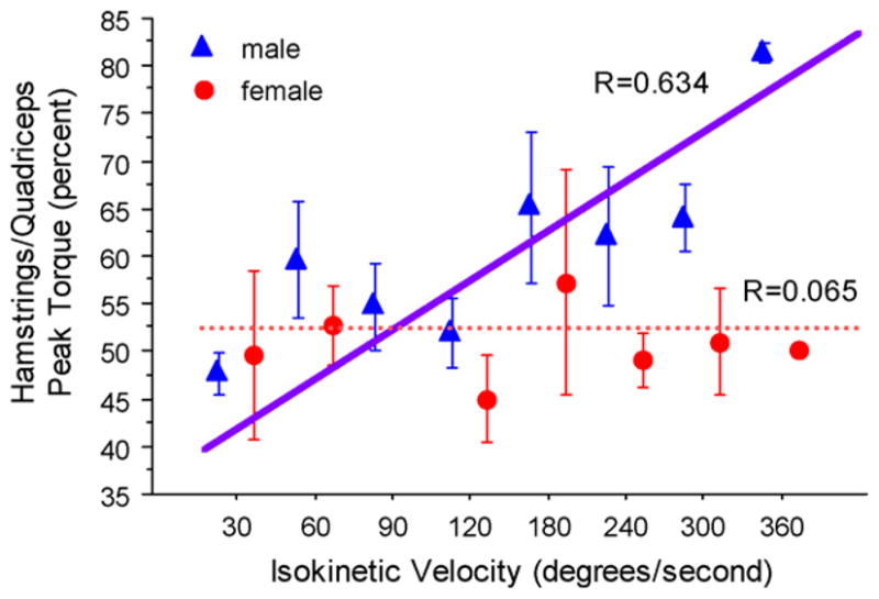
H/Q in female and male subjects diverges with increasing angular velocity.
Results
Twenty-two studies were included in this review and analysis based on the inclusionary and exclusionary criteria. These are presented in Table 1. The included studies contained a total of 1568 subjects (1145 male, 423 female). Isokinetic testing velocities ranged from 30°/s to 360°/s. The total mean H/Q ratios across all velocities were 51.9 ± 8.0% for females and 60.7 ± 9.5% for males (p ≤ 0.001).
The findings showed a significant correlation between H/Q ratio and isokinetic velocity in males (R = 0.634, p ≤ 0.0001, Fig. 1). The findings also demonstrated a significant increase in the H/Q ratio with increasing speed, from the lowest (mean 47.8 ± 2.2% at 30°/s) compared to the highest velocity (mean 81.4 ± 1.1% at 360°/s, p < 0.001, Fig. 1). Females did not demonstrate a correlation between H/Q ratio and testing velocity (R = 0.065, p = 0.77, Fig. 1). Females did not demonstrate a statistically significant difference in the H/Q ratio at the lowest versus the highest isokinetic angular velocity (mean 49.5 ± 8.8% at 30°/s versus 51.0 ± 5.7% at 360°/s, p = 0.84, Fig. 1).
Males demonstrated a significant increase in the H/Q ratio at increased velocity. ANOVA showed a significant difference in H/Q ratio in males between 30°/s and 60°/s, 180°/s, 240°/s, 300°/s and 360°/s isokinetic testing velocity (p < 0.05, Fig. 1). Females did not demonstrate any significant differences between H/Q ratios at different testing velocities. In addition, males demonstrated statistically significantly greater H/Q ratios than females not at 30°/s, but at 60°/s, 120°/s, 300°/s and 360°/s (p ≤ 0.05, Fig. 1; Table 1).
Discussion
The goal of this study was to review the effects of gender on H/Q ratio. An analysis of the published literature was employed to determine whether or not females and males respond to increased isokinetic angular velocity with increased hamstrings torque relative to their quadriceps torque. Review and analysis of the gravity-corrected isokinetic data published in the literature demonstrated significantly different changes in H/Q ratio with increased isokinetic velocity between males and females. At slower testing velocities, no gender differences in isokinetic H/Q ratio were observed. However, with increased knee flexion/extension angular velocities, approaching those that occur during sports activities, significantly greater H/Q ratios were observed in male than female athletes. Therefore, it does not appear that females, contrary to males, increase hamstrings to quadriceps peak torque ratios at functional knee motion velocities during seated open-chain isokinetic activity.
The preservation of “dynamic joint stability” depends on both the passive and active restraints. Passive joint stability is dependent on the ligaments associated with the direction of translation, such as the ACL, which restrains anterior tibial translation relative to the femur. The active muscular agonists and antagonists of the ACL are the hamstrings and the quadriceps, respectively. The relative strength and recruitment of these two muscle groups determine the physiologic range of motion and positioning of the bony articulations of the knee joint. As velocity of motion increases during seated, open-chain isokinetic activity, the forward momentum of the tibia increases to a point where increased hamstrings recruitment is required to limit both extension rotation and anterior translation of the joint. Therefore, as angular velocity increases, males increase their hamstrings to quadriceps peak torque output in order to stabilise the joint and protect the ACL.
Females with decreased H/Q peak torque ratios may be at increased risk of injury.2 Knapik et al. demonstrated that those female collegiate athletes with low H/Q ratios measured with high speed isokinetics had a higher incidence of ACL injury. The observed sex difference in the relationship between H/Q ratio and velocity may be related to females’ decreased ability to dynamically control the knee joint during sports activities.3,5 Aagaard et al.10 stated that eccentric hamstrings torque during deceleration minimises anterior shear forces at the proximal tibia and improves dynamic functionality of the joint.10 The hamstrings work synergistically with the ACL to resist quadriceps contraction during knee extension. Their relative activity is increased as the ligament is loaded by quadriceps contraction at knee flexion angles below 45° via the spinal level reflex arc between the ACL and the hamstrings.29 The absence of increased hamstrings torque relative to quadriceps torque may decrease the ability to control coronal and sagittal plane knee motion in female athletes, may increase strain on the knee and may predispose females to a higher rate of injury than males.5,30
Hamstrings and quadriceps may be 40–80% activated at the time that the foot touches the ground during cutting and landing.31 High rates of injury do occur during both cutting and landing.32–34 Both pivoting and landing are very common mechanisms of ACL injury, though they vary by sport (i.e., cutting injuries may occur more often in soccer and landing injuries may occur more often in basketball). Co-activation of the hamstrings and quadriceps protects the joint not only against anterior drawer but also against knee abduction and dynamic lower extremity valgus. If hamstrings are limited by weakness (peak torque output), quadriceps activation must be reduced, since a net external flexor moment is required to flex the knee. Hence, deficits in strength and activation of the hamstrings limit the potential for muscular co-contraction to protect ligaments. If hamstrings strength and recruitment is high, the quadriceps can be activated more while still producing a net external knee extensor moment (internal knee flexor or predominating hamstrings torque). Similar mechanisms apply to muscular protection against torsional loading, in which gender differences have been identified.35
Increased relative hamstrings recruitment and strength during dynamic tasks may reduce dangerous knee torques and decrease injury risk.5,12,36 Rudolph et al. demonstrated that, following ACL injury, subjects who did not function well with ACL deficiency (non-copers) had decreased co-contraction relative to subjects who did function well with ACL deficiency (copers), who had increased hamstrings activation. MacWilliams et al. stated that hamstrings strengthening following ACL injury may benefit ACL deficient and reconstructed knees by reducing the load in the ligament; however, they also imply that this comes at the expense of efficiency and higher patellofemoral and joint forces.
Muscular contraction can decrease the dynamic valgus motion of the knee, which potentially places the knee at increased risk of injury.37 Joint compression through muscular co-contraction allows more of the dynamic valgus load to be carried by articular contact forces, thus protecting the ligaments. It is likely that more equal distribution of forces transmitted across both the medial and lateral compartments of the knee joint would lead to decreased landing forces.12,38 In addition, a decreased dynamic valgus or varus moment would decrease the risk of femoral condylar lift-off from the tibial plateau. Biomechanical studies have established the relationship between femoral condylar lift-off and injury risk.37,39 The increased relative hamstrings recruitment demonstrated by the males at increased isokinetic velocity may provide a protective mechanism via co-contraction and decreased coronal plane motion during dynamic tasks.5,34,40
A thorough analysis of the published gravity corrected isokinetic literature demonstrated significant differences in the H/Q ratio between males and females. At slow testing speeds, gender differences are not significant. At knee flexion velocities near those of sports activities, gender differences are statistically significant. Females do not increase hamstrings torque relative to quadriceps torque at high velocities as do males at functional knee motion velocities. The absence of increased relative hamstrings to quadriceps peak torque may increase anterior shear on the tibia and valgus angulation of the lower extremity during high speed sporting activities, which may increase anterior drawer, valgus torque and tibial torsion and increase stress on the ACL and account for greater rates of certain knee injury in female athletes.5,12 The underlying mechanism to this divergence in H/Q ratio with increasing velocity may be due to growth and development differences in males and females.3 Decreased hamstrings strength and H/Q ratios of female athletes relative to males may be related to the development of neuromuscular imbalances associated with the onset on maturation.3 These neuromuscular imbalances may increase injury risk in pubertal and post pubertal female athletes.3–5
Limitations to this review and analysis included the inclusion of studies with inconsistent measures between studies; i.e., different types of dynamometers, different measurement protocols and differences in populations studied. In addition, additional databases could have been searched; however, we are confident that we located all applicable studies. Other limitations include the relatively basic statistical analysis and the limited generalisabilty of our review and analysis to open-chain isokinetic data, which have several of their own caveats and limitations, perhaps the most important of which is a potential lack of applicability to closed-chain sporting movements. However, strength of the current study is the inclusion of only gravity-corrected isokinetic studies. Other limitations include the variable age of subjects in the studies and the absence of complete methodologies in many studies in order that potential confounding variables could be better corrected for in the statistical analysis of the data. For example, the majority of the subjects in the current study were developmentally mature individuals which would not allow for a distribution of subjects to provide sufficiently powered populations for the inclusion of age as an independent variable in our statistical analysis. The advantages to the current study design are the power of the study results afforded by the large number of subjects and the generalisability of this large data set to multiple populations.
In summary, this study provides the largest comparison of isokinetic strength measures between genders reported in the literature. In addition, it provides a reference of normative female values, to be used for further exploration into the possible relationship of gender differences in strength and peak torque ratios to injury risk and athletic performance. The current analysis demonstrates that females do not increase hamstrings to quadriceps peak torque ratio at functional knee velocities. The underlying mechanism to this divergence in H/Q ratio with increasing velocity is likely the result of growth and development differences in males and females3 which may place females at increased risk of injury.5 Preseason screening programs that monitor relative hamstrings to quadriceps strength may be warranted to identify female athletes with potential deficits. Targeted neuromuscular interventions that increase relative hamstrings muscle strength and recruitment may decrease injury risk and potentially enhance performance in this population. These data suggest that future prospective investigations are warranted to determine the relationship between H/Q deficits and injury incidence in female athletes.
Acknowledgments
The authors would like to acknowledge funding support from National Institutes of Health Grant R01-AR049735 (TEH). In addition, we would like to acknowledge Paul Succop, Ph.D., for assistance with the statistical analysis. We would also like to acknowledge the assistance of Tiffany Evans for assistance with preparation and Thomas Guidone, PT, for critical reading of the manuscript.
References
- 1.Myer GD, Ford KR, Hewett TE. Rationale and clinical techniques for anterior cruciate ligament injury prevention in female athletes. J Athl Train. 2004;39(4):352–64. [PMC free article] [PubMed] [Google Scholar]
- 2.Knapik JJ, Bauman CL, Jones BH, et al. Preseason strength and flexibility imbalances associated with athletic injuries in female collegiate athletes. Am J Sports Med. 1991;19(1):76–81. doi: 10.1177/036354659101900113. [DOI] [PubMed] [Google Scholar]
- 3.Hewett TE, Myer GD, Ford KR. Decrease in neuromuscular control about the knee with maturation in female athletes. J Bone Joint Surg Am. 2004;86-A(8):1601–8. doi: 10.2106/00004623-200408000-00001. [DOI] [PubMed] [Google Scholar]
- 4.Shea KG, Pfeiffer R, Wang JH, et al. Anterior cruciate ligament injury in pediatric and adolescent soccer players: an analysis of insurance data. J Pediatr Orthop. 2004;24(6):623–8. doi: 10.1097/00004694-200411000-00005. [DOI] [PubMed] [Google Scholar]
- 5.Hewett TE, Myer GD, Ford KR, et al. Biomechanical measures of neuromuscular control and valgus loading of the knee predict anterior cruciate ligament injury risk in female athletes: a prospective study. Am J Sports Med. 2005;33(4):492–501. doi: 10.1177/0363546504269591. [DOI] [PubMed] [Google Scholar]
- 6.Wilk KE, Romaniello WT, Soscia SM, et al. The relationship between subjective knee scores, isokinetic testing, and functional testing in the ACL-reconstructed knee. J Orthop Sports Phys Ther. 1994;20(2):60–73. doi: 10.2519/jospt.1994.20.2.60. [DOI] [PubMed] [Google Scholar]
- 7.Draganich LF, Vahey JW. An in vitro study of anterior cruciate ligament strain induced by quadriceps and hamstrings forces. J Orthop Res. 1990;8(1):57–63. doi: 10.1002/jor.1100080107. [DOI] [PubMed] [Google Scholar]
- 8.Fillyaw M, Bevins T, Fernandez L. Importance of correcting isokinetic peak torque for the effect of gravity when calculating knee flexor to extensor muscle ratios. Phys Ther. 1986;66(1):23–31. doi: 10.1093/ptj/66.1.23. [DOI] [PubMed] [Google Scholar]
- 9.Westing SH, Seger JY. Eccentric and concentric torque-velocity characteristics, torque output comparisons, and gravity effect torque corrections for the quadriceps and hamstring muscles in females. Int J Sports Med. 1989;10(3):175–80. doi: 10.1055/s-2007-1024896. [DOI] [PubMed] [Google Scholar]
- 10.Aagaard P, Simonsen EB, Trolle M, et al. Isokinetic hamstring/quadriceps strength ratio: influence from joint angular velocity, gravity correction and contraction mode. Acta Physiol Scand. 1995;154(4):421–7. doi: 10.1111/j.1748-1716.1995.tb09927.x. [DOI] [PubMed] [Google Scholar]
- 11.Croce RV, Pitetti KH, Horvat M, et al. Peak torque, average power, and hamstrings/quadriceps ratios in nondisabled adults and adults with mental retardation. Arch Phys Med Rehabil. 1996;77(4):369–72. doi: 10.1016/s0003-9993(96)90086-6. [DOI] [PubMed] [Google Scholar]
- 12.Hewett TE, Stroupe AL, Nance TA, et al. Plyometric training in female athletes. Decreased impact forces and increased hamstring torques. Am J Sports Med. 1996;24(6):765–73. doi: 10.1177/036354659602400611. [DOI] [PubMed] [Google Scholar]
- 13.Huston LJ, Wojtys EM. Neuromuscular performance characteristics in elite female athletes. Am J Sports Med. 1996;24(4):427–36. doi: 10.1177/036354659602400405. [DOI] [PubMed] [Google Scholar]
- 14.Bennell K, Wajswelner H, Lew P, et al. Isokinetic strength testing does not predict hamstring injury in Australian Rules footballers. Br J Sports Med. 1998;32(4):309–14. doi: 10.1136/bjsm.32.4.309. [DOI] [PMC free article] [PubMed] [Google Scholar]
- 15.Pincivero DM, Coelho AJ, Campy RM. Perceived exertion and maximal quadriceps femoris muscle strength during dynamic knee extension exercise in young adult males and females. Eur J Appl Physiol. 2003;89(2):150–6. doi: 10.1007/s00421-002-0768-0. [DOI] [PubMed] [Google Scholar]
- 16.Magalhaes J, Oliveira J, Ascensao A, et al. Concentric quadriceps and hamstrings isokinetic strength in volleyball and soccer players. J Sports Med Phys Fitness. 2004;44(2):119–25. [PubMed] [Google Scholar]
- 17.Russell KW, Quinney HA, Hazlett CB, et al. Knee muscle strength in elite male gymnasts. J Orthop Sports Phys Ther. 1995;22(1):10–7. doi: 10.2519/jospt.1995.22.1.10. [DOI] [PubMed] [Google Scholar]
- 18.Tan J, Balci N, Sepici V, et al. Isokinetic and isometric strength in osteoarthrosis of the knee. A comparative study with healthy women. Am J Phys Med Rehabil. 1995;74(5):364–9. [PubMed] [Google Scholar]
- 19.Aagaard P, Simonsen EB, Beyer N, et al. Isokinetic muscle strength and capacity for muscular knee joint stabilization in elite sailors. Int J Sports Med. 1997;18(7):521–5. doi: 10.1055/s-2007-972675. [DOI] [PubMed] [Google Scholar]
- 20.Gerodimos V, Mandou V, Zafeiridis A, et al. Isokinetic peak torque and hamstring/quadriceps ratios in young basketball players. Effects of age, velocity, and contraction mode. J Sports Med Phys Fitness. 2003;43(4):444–52. [PubMed] [Google Scholar]
- 21.Schlinkman B. Norms for high school football players derived from Cybex data reduction computer. J Orthop Sports Phys Ther. 1984;5:243–5. doi: 10.2519/jospt.1984.5.5.243. [DOI] [PubMed] [Google Scholar]
- 22.Ozcakar L, Kunduracyoolu B, Cetin A, et al. Comprehensive isokinetic knee measurements and quadriceps tendon evaluations in footballers for assessing functional performance. Br J Sports Med. 2003;37(6):507–10. doi: 10.1136/bjsm.37.6.507. [DOI] [PMC free article] [PubMed] [Google Scholar]
- 23.Ergun M, Islegen C, Taskiran E. A cross-sectional analysis of sagittal knee laxity and isokinetic muscle strength in soccer players. Int J Sports Med. 2004;25(8):594–8. doi: 10.1055/s-2004-821116. [DOI] [PubMed] [Google Scholar]
- 24.Richards DP. Dynamic strength characteristics during isokinetic knee movements in healthy women. Physiother Canada. 1981;33:141–9. [Google Scholar]
- 25.Appen L, Duncan PW. Strength relationship of the knee nusculature: effects of gravity and sport. J Orthop Sports Phys Ther. 1986;7:232–5. [PubMed] [Google Scholar]
- 26.Ghena DR. Torque characteristics of the quadriceps and hamstring muscles during concentric and eccentric loading. J Orthop Sports Phys Ther. 1991;14:149–54. doi: 10.2519/jospt.1991.14.4.149. [DOI] [PubMed] [Google Scholar]
- 27.Perrin DH, Haskvitz EM, Weltman A. Effect of gravity correction on isokinetic average force of the quadriceps and hamstring muscle group in women runners. Isokinet Exerc Sci. 1991;1:9–102. [Google Scholar]
- 28.Griffin JW, Tooms RE, vander Zwaag R, et al. Eccentric muscle performance of elbow and knee muscle groups in untrained men and women. Med Sci Sports Exerc. 1993;25(8):936–44. [PubMed] [Google Scholar]
- 29.Solomonow M, Baratta R, Zhou BH, et al. The synergistic action of the anterior cruciate ligament and thigh muscles in maintaining joint stability. Am J Sports Med. 1987;15(3):207–13. doi: 10.1177/036354658701500302. [DOI] [PubMed] [Google Scholar]
- 30.Markolf KL, Burchfield DM, Shapiro MM, et al. Combined knee loading states that generate high anterior cruciate ligament forces. J Orthop Res. 1995;13(6):930–5. doi: 10.1002/jor.1100130618. [DOI] [PubMed] [Google Scholar]
- 31.Neptune RR, Wright IC, van den Bogert AJ. Muscle coordination and function during cutting movements. Med Sci Sports Exerc. 1999;31(2):29–302. doi: 10.1097/00005768-199902000-00014. [DOI] [PubMed] [Google Scholar]
- 32.Olsen OE, Myklebust G, Engebretsen L, et al. Injury mechanisms for anterior cruciate ligament injuries in team handball: a systematic video analysis. Am J Sports Med. 2004;32(4):1002–12. doi: 10.1177/0363546503261724. [DOI] [PubMed] [Google Scholar]
- 33.Boden BP, Dean GS, Feagin JA, et al. Mechanisms of anterior cruciate ligament injury. Orthopedics. 2000;23(6):573–8. doi: 10.3928/0147-7447-20000601-15. [DOI] [PubMed] [Google Scholar]
- 34.Ford KR, Myer GD, Toms HE, et al. Gender differences in the kinematics of unanticipated cutting in young athletes. Med Sci Sports. 2005;37(1):12–9. [PubMed] [Google Scholar]
- 35.Wojtys EM, Huston LJ, Schock HJ, et al. Gender differences in muscular protection of the knee in torsion in size-matched athletes. J Bone Joint Surg Am. 2003;85-A(5):782–9. doi: 10.2106/00004623-200305000-00002. [DOI] [PubMed] [Google Scholar]
- 36.Hewett TE, Lindenfeld TN, Riccobene JV, et al. The effect of neuromuscular training on the incidence of knee injury in female athletes. A prospective study. Am J Sports Med. 1999;27(6):699–706. doi: 10.1177/03635465990270060301. [DOI] [PubMed] [Google Scholar]
- 37.Markolf KL, Graff-Redford A, Amstutz HC. In vivo knee stability: a quantitative assessment using an instrumented clinical testing apparatus. J Bone Joint Surg. 1978;60A(5):664–74. [PubMed] [Google Scholar]
- 38.Crenshaw SJ, Pollo FE, Calton EF. Effects of lateralwedged insoles on kinetics at the knee. Clin Orthop. 2000;375:18–92. doi: 10.1097/00003086-200006000-00022. [DOI] [PubMed] [Google Scholar]
- 39.Tibone JE, Antich TJ, Fanton GS, et al. Functional analysis of anterior cruciate ligament instability. Am J Sport Med. 1986;14(4):276–84. doi: 10.1177/036354658601400406. [DOI] [PubMed] [Google Scholar]
- 40.Ford KR, Myer GD, Hewett TE. Valgus knee motion during landing in high school female and male basketball players. Med Sci Sports Exerc. 2003;35(10):1745–5170. doi: 10.1249/01.MSS.0000089346.85744.D9. [DOI] [PubMed] [Google Scholar]