The state of CENP-E–dependent BubR1 autophosphorylation in response to spindle microtubule capture regulates kinetochore function and accurate chromosome segregation.
Abstract
How the state of spindle microtubule capture at the kinetochore is translated into mitotic checkpoint signaling remains largely unknown. In this paper, we demonstrate that the kinetochore-associated mitotic kinase BubR1 phosphorylates itself in human cells and that this autophosphorylation is dependent on its binding partner, the kinetochore motor CENP-E. This CENP-E–dependent BubR1 autophosphorylation at unattached kinetochores is important for a full-strength mitotic checkpoint to prevent single chromosome loss. Replacing endogenous BubR1 with a nonphosphorylatable BubR1 mutant, as well as depletion of CENP-E, the BubR1 kinase activator, results in metaphase chromosome misalignment and a decrease of Aurora B–mediated Ndc80 phosphorylation at kinetochores. Furthermore, expressing a phosphomimetic BubR1 mutant substantially reduces the incidence of polar chromosomes in CENP-E–depleted cells. Thus, the state of CENP-E–dependent BubR1 autophosphorylation in response to spindle microtubule capture by CENP-E is important for kinetochore function in achieving accurate chromosome segregation.
Introduction
During mitosis, the kinetochore, the protein complex assembled at each centromere on each chromosome, serves as the attachment site for spindle microtubules and powers chromosome movement along the mitotic spindle (Cleveland et al., 2003; Santaguida and Musacchio, 2009; Joglekar et al., 2010). Unattached kinetochores generate the “waiting signal” for the mitotic checkpoint (also known as the spindle assembly checkpoint), which delays anaphase onset before successful attachment of every chromosome to microtubules of the spindle (Cleveland et al., 2003; Musacchio, 2011). Errors in this process cause aneuploidy, which early in development leads to lethal development defects and later is the hallmark of human tumor progression (Hartwell and Kastan, 1994).
BubR1, an essential mitotic checkpoint kinase (Chan et al., 1999; Chen, 2002), also plays an important role in kinetochore–microtubule attachment and metaphase chromosome alignment (Ditchfield et al., 2003; Lampson and Kapoor, 2005; Zhang et al., 2007). BubR1 has been shown to be phosphorylated by several other mitotic kinases, and these phosphorylations are important for BubR1 functions in kinetochore–microtubule attachment as well as the mitotic checkpoint (Elowe et al., 2007, 2010; Matsumura et al., 2007; Huang et al., 2008). However, how BubR1’s own kinase activity is involved in its kinetochore functions is largely unknown. Although BubR1 kinase activity is below a detectable level in vitro with purified components (Mao et al., 2003; Wong and Fang, 2007), its autophosphorylation activity is significantly increased upon either prephosphorylation by Cdk1 and Plx1 (Wong and Fang, 2007) or addition of CENP-E (Mao et al., 2003), a kinetochore-associated microtubule motor protein (Yen et al., 1992) and a BubR1 binding partner (Chan et al., 1998; Yao et al., 2000). Furthermore, microtubule capture by CENP-E can silence BubR1 kinase activity in a ternary complex of BubR1–CENP-E–microtubules (Mao et al., 2005). The BubR1 kinase activity has been shown to be important for the mitotic checkpoint in Xenopus laevis egg extracts (Mao et al., 2003) and human cells (Kops et al., 2004). Replacing endogenous BubR1 with a kinase-inactive (kinase dead [KD]) BubR1 in Xenopus egg extracts (Zhang et al., 2007), Drosophila melanogaster (Rahmani et al., 2009), and human cells (Matsumura et al., 2007) all results in metaphase spindles with misaligned chromosomes, indicating that BubR1 kinase activity also directly modulates microtubule capture at the kinetochore.
CENP-E, the activator of BubR1 kinase, is a kinetochore-associated kinesin motor protein. Interference with CENP-E function using anti–CENP-E antibody injection (McEwen et al., 2001) or CENP-E depletion by antisense oligonucleotides (Yao et al., 2000) or small interfering RNAs (Martin-Lluesma et al., 2002) results in an obvious but incomplete metaphase plate with variable numbers of polar chromosomes. Individual cells from cultured CENP-E–null embryos also show one or more misaligned chromosomes (Putkey et al., 2002). Furthermore, the mitotic checkpoint cannot be activated or maintained in Xenopus egg extracts depleted of CENP-E, probably because of the loss of Mad1–Mad2 from unattached kinetochores (Abrieu et al., 2000). Cells without CENP-E in vitro and in vivo also have reduced levels of Mad1–Mad2 associated with unattached kinetochores and produce premature anaphase onset with one or a few polar chromosomes, resulting in an increase of aneuploidy (Putkey et al., 2002; Weaver et al., 2003, 2007). These results indicate that the mitotic checkpoint cannot be maintained in the absence of CENP-E when there are only one or a few unattached kinetochores. Upon identifying a CENP-E–dependent BubR1 autophosphorylation site using purified components, we now show that BubR1 kinase activity and its autophosphorylation are important for kinetochore function in achieving accurate chromosome segregation to prevent single chromosome loss in human cells.
Results
BubR1 is a kinase and phosphorylates itself in human cells in a CENP-E–dependent manner
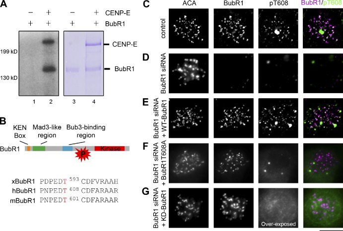
As we showed before (Mao et al., 2003, 2005), purified recombinant Xenopus BubR1 was able to phosphorylate itself in vitro but only in the presence of its binding partner, CENP-E (Fig. 1 A, lane 2). By mass spectrometry (MS), we subsequently identified a single CENP-E–dependent BubR1 autophosphorylation site, Thr593 (Thr608 in human BubR1), which lies just adjacent to the kinase domain and is conserved through frog to human (Fig. 1 B).
Figure 1.
BubR1 phosphorylates itself. (A) BubR1 kinase activity, as well as BubR1 autophosphorylation, is directly stimulated by CENP-E in vitro. Equal amounts of purified recombinant Xenopus BubR1 with (lanes 2 and 4) or without (lanes 1 and 3) purified recombinant Xenopus CENP-E were analyzed for BubR1 autokinase activity. (left) Autoradiography of SDS-PAGE gel of purified recombinant BubR1 autophosphorylated, as well as purified recombinant CENP-E phosphorylated, in the presence of γ-[32P]ATP. (right) Coomassie blue staining of purified recombinant BubR1 and CENP-E proteins. (B) BubR1 protein structure showing the relative positions of KEN box, the N-terminal Mad3-like domain, the Bub3-binding domain, the kinase domain, and the autophosphorylation site identified by LC-MS/MS, which is conserved in frog (xBubR1), human (hBubR1), and mouse (mBubR1). The autophosphorylation site is shown in red in the DNA sequences. P, phosphorylated. (C–G) Immunofluorescence images acquired using the indicated antibodies against BubR1, the autophosphorylation site (pT608), and anticentromere antigen (ACA; a centromere/kinetochore marker) in nocodazole-treated human T98G cells with endogenous BubR1 (C), with endogenous BubR1 depleted (D), replaced with siRNA-resistant mCherry-tagged wild-type (WT) BubR1 (E), the nonphosphorylatable BubR1T608A mutant (F), or kinase-inactive (KD) BubR1 (G). Bar, 10 µm.
We generated a rabbit polyclonal antibody specific for the phosphorylated Thr608 (pAb-T608) against human BubR1. Immunoblotting of cell lysates with this phosphoantibody revealed a clear increase in phosphorylation of this site during mitosis with a band corresponding to the size of human BubR1 (Fig. S1 A, compare lanes 1 and 2). Immunoreactivity with the pAb-T608 was eliminated either by treating the lysates with λ protein phosphatase (Fig. S1 A, compare lanes 3 and 4) or by adsorption of the antibody with the phosphopeptide but not with the nonphosphopeptide (Fig. S1 B). More importantly, indirect immunofluorescence analysis in human T98G cells revealed that the kinetochore staining of pAb-T608 was substantially reduced in cells transfected with BubR1 siRNA (Fig. 1, compare C and D). Expressing an siRNA-resistant mCherry-tagged wild-type BubR1, but not a nonphosphorylatable BubR1T608A mutant, restored pAb-T608 signals at the kinetochore (Fig. 1, compare E and F). Furthermore, replacing endogenous BubR1 with a kinase-inactive (KD) point mutant (K795A) did not display kinetochore-associated pAb-T608 signals even with an overexposed time compared with cells with wild-type BubR1 (Fig. 1 G). Collectively, these results strongly demonstrate that BubR1 is a kinase in human cells and phosphorylates itself at the kinetochore.
We further evaluated the BubR1 autophosphorylation at kinetochores during mitotic progression using the BubR1 pAb-T608 phosphoantibody. Cells in prometaphase showed BubR1 autophosphorylation signals at the kinetochores, whereas cells in metaphase displayed low levels of pAb-T608 signals at all aligned kinetochores (Fig. S1 C). Thus, BubR1 autophosphorylation is sensitive to kinetochore–microtubule attachment. Treatment with nocodazole to depolymerize microtubules indeed resulted in a relatively uniformly high level of BubR1 autophosphorylation on all unattached kinetochores (Fig. S1 C).
BubR1 kinase activity and its autophosphorylation were substantially increased in the presence of its binding partner, the kinetochore motor CENP-E, in vitro (Fig. 1 A, lane 2). Decreased levels of CENP-E after siRNA to CENP-E also reduced levels of BubR1 autophosphorylation on unattached kinetochores (Fig. S2, A–D). In contrast, the antibody that detects BubR1 regardless of its autophosphorylation state did not display substantial change in signal levels at kinetochores in CENP-E–depleted cells (Fig. S2, E–H). These results suggest that BubR1 kinase activity and its autophosphorylation, but not BubR1 itself, at the kinetochore in human cells depend on its interaction with CENP-E.
BubR1 kinase activity and its autophosphorylation are essential for accurate chromosome segregation
To determine whether the autophosphorylation is important for BubR1 functions in mitosis, we used site-directed mutagenesis to convert the autophosphorylatable residue to an amino acid mimicking constitutive phosphorylation (glutamic acid [E]) and to a nonphosphorylatable amino acid (alanine [A]). The endogenous BubR1 expression level in T98G cells can be reduced by an siRNA duplex targeted to a region of human BubR1, and the BubR1 expression can be restored with a cotransfected BubR1 gene mutated in three silent base positions within the site of action of the siRNA (Huang et al., 2008). Using this approach, we replaced endogenous BubR1 with mCherry-tagged siRNA-resistant wild-type BubR1 or BubR1 phosphorylation mutants to a similar expression level in human cells (Fig. 2 A).
Figure 2.
BubR1 autophosphorylation is essential for accurate chromosome segregation. (A) Immunoblotting of mitotic lysates of T98G cells 48 h after transfection with the indicated siRNAs and siRNA-resistant mCherry-BubR1 expression vectors as indicated. endo., endogenous; TA, T608A; TE, T608E. (B) Time spent in prometaphase and metaphase (or pseudometaphase) of transiently transfected HeLa cells (stably transfected with EYFP-H2B) with BubR1 siRNA and indicated plasmids encoding siRNA-resistant mCherry-tagged wild-type (WT) BubR1 or the BubR1 phosphorylation mutants. Each vertical bar represents a single cell. The transfected cells were identified by mCherry before live-cell imaging. Pseudometaphase is designated as when the majority of chromosomes are aligned with an obvious metaphase plate, but a few chromosomes are at the poles. (C) A summary of median time spent in prometaphase and metaphase (or pseudometaphase) as well as the percentage of cells with polar chromosomes (pseudometaphase) and chromosome missegregation during anaphase onset from data presented in B. (D and E) Stills of live-cell imaging showing a cell in which endogenous BubR1 was replaced with the nonphosphorylatable BubR1T608A mutant (D) or the phosphomimetic BubR1T608E mutant (E) during unperturbed mitoses. Bar, 10 µm.
Cells with BubR1 replaced by either type of mutants at the autophosphorylation site were assessed by live-cell imaging in unperturbed mitoses; each yielded overall time spent in mitosis was comparable to that of cells expressing wild-type BubR1 (Fig. 2, B and C). Although a majority of the cells expressing wild-type BubR1 (84%) progressed through normal mitosis with accurate chromosome segregation (Fig. 2, B and C), >75% of the cells expressing the nonphosphorylatable BubR1T608A mutant had misaligned (polar) chromosomes, which subsequently missegregated during anaphase (Fig. 2, B–D). Notably, cells that had many misaligned chromosomes spent a much longer time in mitosis. The majority of the cells expressing the phosphomimetic BubR1T608E mutant (86%) had full congression without any obvious misaligned chromosomes; however, 40% of them exhibited unequal chromosome segregation with chromosome bridges during anaphase (Fig. 2, B, C, and E). These results demonstrate that CENP-E–dependent BubR1 autophosphorylation is essential for both metaphase chromosome alignment and a full-strength mitotic checkpoint to prevent single chromosome loss.
To better understand the role of BubR1 kinase activity in chromosome segregation, we replaced endogenous BubR1 with a BubR1T608E-K795A double mutant, which is constitutively phosphorylated at the autophosphorylation site but lacks the kinase activity. Live-cell imaging analysis revealed that cells expressing this BubR1 double mutant exited mitosis with unaligned chromosomes more frequently than those expressing the BubR1T608E single mutant do (Fig. S3). Furthermore, in contrast to cells expressing the nonphosphorylatable or phosphomimetic BubR1 single mutant, cells expressing the BubR1T608E-K795A double mutant spent less time in mitosis in comparison to cells expressing wild-type BubR1 (Fig. S3 B). These results indicate that BubR1 kinase has other substrates that are important for achieving accurate chromosome segregation, further supporting a role of BubR1 kinase in mitosis.
The phosphorylated form of BubR1 is required for full levels of Mad1 and Mad2 association with unattached kinetochores
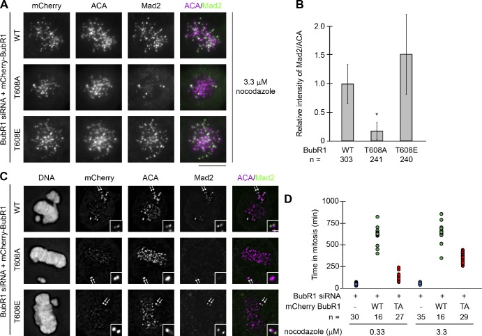
The basic plan of the mitotic checkpoint signaling cascade is established. Before spindle microtubule capture, the Mad1–Mad2 complex is targeted to unattached kinetochores (Chen et al., 1998), in which additional Mad2 is recruited (Luo et al., 2004; De Antoni et al., 2005) and converted into a rapidly released, inhibitory form (Shah et al., 2004; Vink et al., 2006), including possible assembly of complexes with other checkpoint proteins (Sudakin et al., 2001; Fang, 2002; Kulukian et al., 2009). To test whether BubR1 autophosphorylation at unattached kinetochores affects recruitment of Mad1 and Mad2, quantitative immunofluorescence was used to determine the levels of kinetochore-bound Mad1 and Mad2 upon treatment with nocodazole, which induces spindle microtubule disassembly. Relative to cells expressing wild-type BubR1 or the phosphomimetic BubR1 mutant, cells expressing the nonphosphorylatable BubR1T608A mutant had diminished levels of Mad1 on unattached kinetochores (58.4%; Fig. S4). Furthermore, cells expressing the nonphosphorylatable BubR1 mutant recruited even lower levels of Mad2 (17.3%) on unattached kinetochores compared with cells expressing wild-type BubR1 (Fig. 3, A and B). Thus, the nonphosphorylated form of BubR1 reduces Mad1–Mad2 recruitment at the kinetochore.
Figure 3.
BubR1 autophosphorylation is required for efficient kinetochore targeting of Mad2 and a prolonged mitosis induced by nocodazole. (A and C) Kinetochore localization of mCherry-BubR1 (mCherry; by an anti-mCherry antibody), ACA, and Mad2 in cells expressing BubR1 siRNA and siRNA-resistant mCherry-tagged wild-type (WT) BubR1 or BubR1 phosphorylation mutants as indicated and treated with 3.3 µM nocodazole (A) or not treated (C). In C, a pair of sister kinetochores on unaligned chromosomes indicated by arrows is shown in the insets. Bars: (main images) 10 µm; (insets) 1 µm. (B) Quantitation of the normalized integrated intensity of Mad2 signals against ACA signals at kinetochores in cells treated with 3.3 µM nocodazole. The experiments were repeated twice, and the numbers of kinetochores from ≥10 different cells quantified for each condition were shown. Error bars represent standard error (*, P < 0.05). (D) Time spent in mitosis (measured as the time of a cell’s rounding up by time-lapse imaging) at the indicated nocodazole concentrations for H2B-EYFP cells transfected with BubR1 siRNA along with or without an mCherry expression vector fused with wild-type BubR1 (WT) or the nonphosphorylatable BubR1 mutant (TA) as indicated. Each dot represents a single cell, and the numbers of cells filmed are presented. We should note that a majority of the cells expressing wild-type BubR1 were still in mitosis when the filming was ended; therefore, the time in mitosis for those cells showed here is underrepresented.
Kinetochore levels of Mad2 were also determined in the absence of the microtubule inhibitor nocodazole. Mad2 signals at metaphase kinetochores were largely undetectable in cells expressing wild-type BubR1 as well as the nonphosphorylatable BubR1T608A and the phosphomimetic BubR1T608E mutants (Fig. 3 C). However, kinetochores of unaligned chromosomes in cells expressing wild-type BubR1 or the phosphomimetic BubR1T608E mutant had strong Mad2 signals, whereas the levels of kinetochore-associated Mad2 signals on polar chromosomes were substantially diminished in cells expressing the nonphosphorylatable BubR1T608A mutant (Fig. 3 C).
BubR1 autophosphorylation inhibition weakens the mitotic checkpoint response in the absence of microtubules
Cells expressing the nonphosphorylatable BubR1T608A mutant enter anaphase with a few unaligned chromosomes and have reduced levels of Mad1–Mad2 on unattached kinetochores, suggesting a weakened mitotic checkpoint. We therefore compared the duration of the mitotic arrest in cells expressing either wild-type BubR1 or the BubR1T608A mutant by treating them with 0.33 and 3.3 µM nocodazole. As expected, we observed a prolonged mitotic arrest (>10 h) in cells expressing wild-type BubR1 at both concentrations (Fig. 3 D). In contrast, the duration of the mitotic arrest (359 ± 46 min) for cells expressing the nonphosphorylatable BubR1T608A mutant in the presence of high concentrations of nocodazole (3.3 µM) decreased significantly in comparison to cells expressing wild-type BubR1 (Fig. 3 D). Furthermore, cells expressing the BubR1T608A mutant left mitosis more rapidly (125 ± 36 min) at low concentrations of nocodazole (0.33 µM) than at high nocodazole concentrations (3.3 µM), though the duration of mitosis in those cells was still longer than cells without BubR1 (41 ± 10 min; Fig. 3 D). Collectively, these results support the notion that inhibition of BubR1 autophosphorylation results in a weakened mitotic checkpoint.
BubR1 autophosphorylation is essential for metaphase chromosome alignment
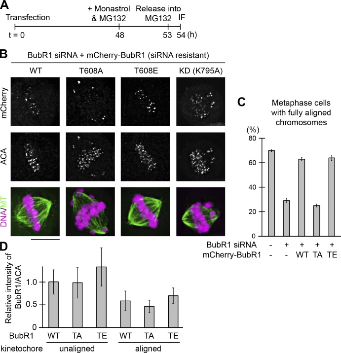
Though it is clear that BubR1 plays an important role in metaphase chromosome alignment (Lampson and Kapoor, 2005; Zhang et al., 2007), the molecular mechanism remains largely unknown. To specifically test for effects of BubR1 autophosphorylation on chromosome alignment, we treated cells with monastrol and MG132 for 4 h to accumulate mitotic cells and then released them into MG132 alone for 1 h (to prevent premature anaphase onset; Fig. 4 A). Consistent with an earlier study (Lampson and Kapoor, 2005), BubR1 siRNA resulted in only 30% of cells with fully aligned chromosomes, a decrease from 67% for cells with mismatch siRNA (Fig. 4, B and C). The siRNA-resistant wild-type BubR1 was able to effectively rescue the metaphase chromosome alignment defect (Fig. 4, B and C). Although 64% of cells expressing the phosphomimetic BubR1T608E mutant had all chromosomes fully aligned, only 25% of cells with the nonphosphorylatable BubR1T608A mutant achieved metaphase chromosome alignment (Fig. 4, B and C). Furthermore, replacing endogenous BubR1 with a kinase-inactive (KD) BubR1 mutant phenocopied the nonphosphorylatable BubR1 mutant, which produced bipolar spindles with misaligned chromosomes (Fig. 4, B and C). These results are consistent with a role of BubR1 kinase activity and its autophosphorylation in metaphase chromosome alignment.
Figure 4.
BubR1 autophosphorylation is necessary for metaphase chromosome alignment. (A) Schematic representation of BubR1 siRNA, rescue transfection, and chemical inhibitor treatment protocol. IF, immunofluorescence. (B) Replacing endogenous BubR1 with a nonphosphorylatable mutant (T608A) or a kinase-inactive (KD) mutant results in severe chromosome misalignment. Cells were imaged for mCherry-BubR1 (mCherry), ACA, microtubules (MT; by an antitubulin antibody), and DNA (visualized by DAPI). Bar, 10 µm. (C) The percentage of mitotic cells with fully aligned chromosomes was analyzed in cells transfected with BubR1 siRNA and the siRNA-resistant mCherry-BubR1 constructs as indicated. For each condition, >200 mitotic cells from three repeated experiments were counted to quantify the percentage of cells. Error bars represent standard error; n = 3. (D) Quantitation of the normalized integrated intensity of mCherry-BubR1 signals against ACA signals at unattached and attached kinetochores from ≥10 mitotic cells each from two experimental repeats. Error bars represent standard error. WT, wild type; TA, T608A; TE, T608E.
In comparison to wild-type BubR1, the relative levels of the nonphosphorylatable or phosphomimetic mutants on unaligned (polar) kinetochores and aligned kinetochores were not significantly altered, though, as expected, the levels of all three BubR1 proteins on aligned kinetochores were reduced (Fig. 4 D). Thus, the autophosphorylation state of BubR1 at kinetochores is important in regulating metaphase chromosome alignment.
The nonphosphorylated form of BubR1 reduces levels of Aurora B–mediated Ndc80 phosphorylation at kinetochores
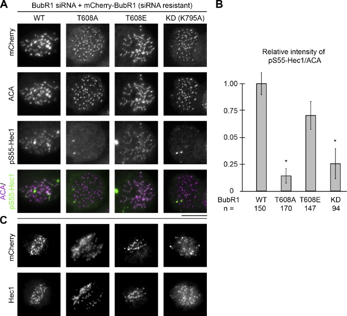
The high frequency of syntelic kinetochore–microtubule attachments even after cold treatment in cells expressing the nonphosphorylatable BubR1T608A mutant (Fig. S5 A) prompted us to investigate the Aurora B–mediated Ndc80 phosphorylation in these cells. Aurora B has been shown to function in destabilizing the improperly attached kinetochore microtubules by phosphorylating kinetochore-associated microtubule interactors, such as the KMN network (e.g., Ndc80; Cheeseman et al., 2006; DeLuca et al., 2006; Welburn et al., 2009) and the formin mDia3 (Cheng et al., 2011), to reduce their affinities to microtubules in the absence of tension (Lampson and Cheeseman, 2011; Mao, 2011). We first examined the levels of phosphorylated Hec1 (pS55-Hec1), a component of the Ndc80 complex and a major Aurora B substrate in attachment error correction (DeLuca et al., 2006), in nocodazole-treated cells. This revealed low levels of kinetochore-associated pS55-Hec1 signals in cells in which endogenous BubR1 was replaced by the nonphosphorylatable BubR1T608A mutant or the kinase-inactive (KD) mutant but not by the wild-type BubR1 or the phosphomimetic BubR1T608E mutant (Fig. 5, A and B). The kinetochore localization of Hec1 itself was obviously not affected in those cells (Fig. 5 C). Next, we examined the levels of phosphorylated pS55-Hec1 signals in untreated cells with both aligned and unaligned chromosomes. In agreement with the results obtained in cells treated with nocodazole, the levels of pS55-Hec1 signals on unattached kinetochores in cells expressing the nonphosphorylatable BubR1T608A mutant were also diminished in comparison to those in cells expressing wild-type BubR1 or the phosphomimetic mutant (Fig. S5 B). In addition, the levels of Aurora B phosphorylation of Hec1 at the S55 site on aligned kinetochores were diminished in all cells expressing any of the three BubR1 proteins (Fig. S5 B). Thus, our results support the hypothesis that the nonphosphorylated form of BubR1 is able to reduce Aurora B–mediated Ndc80 phosphorylation at kinetochores.
Figure 5.
The nonphosphorylated form of BubR1 reduces Aurora B–mediated Ndc80 phosphorylation at kinetochores. (A) Immunolocalization of mCherry-BubR1, ACA, and pS55-Hec1 in cells expressing wild-type (WT) BubR1 or BubR1 mutants as indicated. Bar, 10 µm. (B) Quantification of the normalized integrated intensities of the kinetochore signals of pS55-Hec1/ACA (±SD; the number of kinetochores was analyzed from ≥10 different cells as shown with three repeated experiments; *, P < 0.05). (C) Hec1 kinetochore localization is not affected in cells expressing BubR1 phosphorylation mutants. Immunolocalization of mCherry-BubR1 and Hec1 as indicated.
The effects of the nonphosphorylatable BubR1T608A mutant on the Hec1 phosphorylation by Aurora B at the kinetochore are surprising, given that BubR1 depletion has been shown to increase Aurora B–mediated CENP-A phosphorylation at kinetochores (Lampson and Kapoor, 2005). Thus, we tested the effects of BubR1 depletion on Hec1 phosphorylation at the S55 site by Aurora B. In contrast to the reduced kinetochore signals of pS55-Hec1 in cells expressing the nonphosphorylatable BubR1 mutant, the levels of pS55-Hec1 signals at kinetochores in BubR1-depleted cells increased or at least remained similar (Fig. S5 C). Therefore, the state of BubR1 autophosphorylation, not the mere presence of BubR1, is important for the regulation of Aurora B–mediated Ndc80 phosphorylation at the kinetochore.
BubR1 autophosphorylation depends on CENP-E, the kinetochore-associated motor and the activator of BubR1 kinase, in vitro (Fig. 1 A) and at the kinetochores in human cells (Fig. S2). Depletion of CENP-E resulted in metaphase chromosome misalignment with polar chromosomes (Martin-Lluesma et al., 2002; Putkey et al., 2002; Weaver et al., 2003), which phenocopies the expression of the BubR1 nonphosphorylatable mutant as well as the kinase-inactive (KD) BubR1 mutant (Fig. 4). Therefore, we next tested whether a decreased level of CENP-E expression upon treatment of CENP-E siRNA can also cause reduced levels of Ndc80 phosphorylation by Aurora B at kinetochores in human cells. We indeed observed substantially decreased levels of kinetochore-associated pS55-Hec1 signals in CENP-E–depleted cells (Fig. S5 D), which is consistent with our hypothesis that the Aurora B–mediated Ndc80 phosphorylation is reduced at kinetochores with a nonphosphorylated form of BubR1.
The expression of the BubR1 phosphomimetic mutant reduces the incidence of polar chromosomes in cells lacking CENP-E
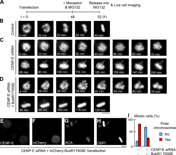
Expressing the nonphosphorylated BubR1 mutant or depletion of CENP-E results in reduced levels of Aurora B–mediated Ndc80 phosphorylation at kinetochores, leading to attachment errors. We next tested whether expressing the constitutively phosphorylated BubR1T608E mutant could correct chromosome misattachments in the absence of CENP-E. Cells expressing H2B-EYFP were treated with monastrol (and MG132 to prevent premature anaphase onset) to produce large amounts of syntelic and monotelic attachments. Upon release into MG132 medium alone, cells were followed by time-lapse microscopic analysis (Fig. 6 A). Control cells with wild-type CENP-E were able to position all chromosomes at the metaphase plate (Fig. 6 B), whereas CENP-E–depleted cells (11 out of 11) had persistent polar chromosomes (Fig. 6 C). In contrast, although it took a much longer time, a majority of the CENP-E–depleted cells (9 out of 10) expressing the phosphomimetic BubR1T608E mutant were able to align all chromosomes at the metaphase plate in the absence of CENP-E (Fig. 6 D).
Figure 6.
The polar chromosome phenotype in CENP-E–depleted cells can be rescued by expression of a phosphomimetic BubR1 mutant. (A) Schematic representation of transfection, chemical inhibitor treatment, and live-cell imaging protocol. (B–D) Representative still frames of live-cell microscopy of H2B-EYFP cells transfected without (B) or with CENP-E siRNA and mCherry (C) or mCherry-BubR1T608E (D) expression vectors. Arrows point to polar chromosome outside the metaphase plate. The transfected cells were identified by mCherry before live-cell imaging. In C, all 11 filmed cells had polar chromosomes by the end of the filming (300 min). In D, 9 out of 10 filmed cells had all chromosomes aligned at the metaphase plate. Bar, 10 µm. (E–H) Expression of the phosphomimetic BubR1T608E mutant rescues polar chromosome phenotype in CENP-E–depleted cells. Indirect immunofluorescence analysis of CENP-E (E), mCherry-BubR1T608E (F), ACA (G), and DNA (H). Arrows point to polar chromosomes outside the metaphase plate. (I) The percentage of mitotic cells with or without polar chromosomes analyzed in cells transfected with CENP-E siRNA and with or without the mCherry-BubR1T608 construct as indicated (± SD; ≥50 cells each from two repeated experiments were counted). Bars, 10 µm.
To further confirm that attachment errors are able to be corrected in CENP-E–depleted cells expressing the phosphomimetic BubR1 mutant, we quantified our results in fixed cells upon indirect immunofluorescence. Compared with control cells, depletion of CENP-E produced polar chromosomes in 82% of cells. In contrast, 70% of CENP-E–depleted cells expressing the BubR1T608E mutant did not show any obvious polar chromosomes and had a complete metaphase plate (Fig. 6, E–I). These results clearly suggest that CENP-E–dependent BubR1 autophosphorylation regulates Aurora B–mediated Ndc80 phosphorylation at kinetochores in response to spindle microtubule capture.
Discussion
BubR1 is a kinase, and its kinase activity, as well as its autophosphorylation, is essential for a full-strength mitotic checkpoint to prevent single chromosome loss
Although the yeast homologue of BubR1, Mad3, does not harbor a kinase domain, several lines of evidence in Drosophila (Rahmani et al., 2009), Xenopus egg extracts (Mao et al., 2003; Zhang et al., 2007), and human cells (Kops et al., 2004; Matsumura et al., 2007) support a role of BubR1 kinase activity in metaphase chromosome alignment and/or the mitotic checkpoint. However, this role has been challenged by reports that replacing endogenous BubR1 with a kinase-inactive BubR1 mutant (Elowe et al., 2007) or a BubR1 mutant without the kinase domain (Suijkerbuijk et al., 2010) is still able to achieve accurate chromosome segregation in unperturbed mitoses. Furthermore, even a cytosolic murine BubR1 fragment (amino acids 1–386) without the Bub3-binding domain (essential for BubR1 kinetochore localization) and the kinase domain has been shown to be able to support metaphase chromosome alignment, the mitotic checkpoint, and cell survival (Malureanu et al., 2009). Adding to these is the undetectable kinase activity of purified recombinant BubR1 alone in vitro (Mao et al., 2003; Wong and Fang, 2007) as well as the lack of substrates. The disparity of those results could be attributed to inefficient knockdown or depletion of endogenous BubR1 (Chen, 2002) or different experimental approaches, as well as different systems, to examine subtle phenotypic differences.
In this study, we have identified a CENP-E–dependent BubR1 autophosphorylation site using purified components and MS. This site has also been identified in human cells using systematic phosphorylation analysis of mitotic protein complexes (Hegemann et al., 2011). Using a specific phosphoantibody against this site, we show that kinetochore-associated signals are eliminated by BubR1 depletion (siRNA) in mitotic cells. More importantly, the kinetochore-associated signals are restored only by expressing the siRNA-resistant wild-type BubR1 but not a BubR1 kinase-inactive (KD) point mutant (Fig. 1). To our knowledge, this is the first direct evidence to support the kinase activity of BubR1 in human cells. Furthermore, the kinetochore-associated BubR1 autophosphorylation signals in cells also depend on the presence of CENP-E (Fig. S2), just as in the previous results that CENP-E can stimulate BubR1 kinase activity in vitro (Mao et al., 2003; Weaver et al., 2003) and in Xenopus egg extracts (Mao et al., 2003).
Live-cell imaging analysis has revealed that BubR1 autophosphorylation is not only important for metaphase chromosome alignment (for details see next Discussion section) but also a full-strength mitotic checkpoint. Cells harboring the nonphosphorylatable BubR1 mutant spend a similar time in mitosis to that of cells having wild-type BubR1, suggesting that BubR1 autophosphorylation is not required for mitotic timing and the basic mitotic checkpoint strength when there are lots of unattached kinetochores in a short time frame. However, a majority of those cells with the nonphosphorylatable BubR1 mutant initiate anaphase onset with polar chromosomes (Fig. 2), phenocopying what has been shown in cells without CENP-E function (Putkey et al., 2002; Weaver et al., 2003, 2007). Cells expressing the nonphosphorylatable BubR1 mutant also have a shorter duration of mitotic arrest induced by nocodazole in comparison with cells having wild-type BubR1 (Fig. 3 D). Furthermore, similar to what has been shown in the absence of CENP-E (Weaver et al., 2003), unattached kinetochores in cells expressing the nonphosphorylatable BubR1 mutant have reduced levels of checkpoint components Mad1 and Mad2 (Fig. 3, A–C; and Fig. S4). Therefore, the CENP-E–dependent BubR1 kinase activity and BubR1 autophosphorylation are essential for a full-strength mitotic checkpoint to prevent chromosome missegregation when there are only one or a few unattached kinetochores (Fig. 7 A). A study using BubR1 conditional knockout cells and BubR1 domain mutants has suggested that a prolonged mitotic checkpoint signaling requires BubR1 kinase activity at kinetochores (Malureanu et al., 2009). The CENP-E–dependent BubR1 kinase activity is also important for the mitotic checkpoint in Xenopus egg extracts (Mao et al., 2003), a system known to produce a weak mitotic checkpoint signal to the extent that >9,000 sperm head nuclei (haploid without sister kinetochores)/microliter are needed to activate the checkpoint (Murray, 1991).
Figure 7.
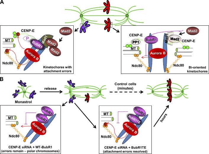
A model for the role of CENP-E–dependent BubR1 autophosphorylation at the kinetochore. (A) Incorrect kinetochore–microtubule attachments (purple chromosomes) trigger Aurora B–mediated phosphorylation of the KMN network (represented by the Ndc80 complex in the cartoon) and CENP-E, resulting in destabilization of kinetochore microtubules. At unattached kinetochores, CENP-E stimulates BubR1 kinase activity and its autophosphorylation. Mad1 and Mad2 are also recruited onto unattached kinetochore to generate mitotic checkpoint signaling. At attached bioriented kinetochores (red chromosomes), spindle microtubule capture by CENP-E silences BubR1 kinase activity and recruits Phosphatase 1 (PP1) to outer kinetochores. The nonphosphorylated form of BubR1 reduces Aurora B–mediated Ndc80 phosphorylation and Mad1–Mad2 association at kinetochores. These coordinated events result in dephosphorylation of key components at the kinetochore–microtubule interface (e.g., Ndc80), leading to stable attachment upon biorientation and separation of the inner centromeric Aurora B from outer kinetochore substrates, as well as silencing the mitotic checkpoint signaling. (B) During the formation of the bipolar spindle upon release from monastrol treatment, a large number of attachment errors needs to be corrected. In the absence of CENP-E, the nonphosphorylated form of endogenous BubR1 resides on incorrectly attached kinetochores, which reduces Aurora B–mediated Ndc80 phosphorylation, leading to the persistent existence of polar chromosomes. Expressing a phosphomimetic BubR1 mutant to replace some of the nonphosphorylated endogenous BubR1 at kinetochores helps to correct misattachments, although it takes a much longer time for those cells to achieve metaphase chromosome alignment. MT, microtubule; P, phosphorylated.
Although the crystal structure of BubR1 kinase has not been resolved, superposition of a 3D model structure of BubR1 kinase generated by comparative modeling using the known Bub1 kinase as a template has suggested that these two kinases have very similar structures (Bolanos-Garcia and Blundell, 2011). One of the particularly intriguing features is the interaction of the N-terminal extension with the kinase domain, similar to what has been shown for the activation of Cdks by cyclins (Kang et al., 2008). The interaction between BubR1 and CENP-E and the subsequent BubR1 autophosphorylation could cause a structural change of BubR1 and, therefore, influence the functional roles of BubR1 at the kinetochore.
The nonphosphorylated form of BubR1 can reduce levels of Aurora B–mediated Ndc80 phosphorylation at attached kinetochores upon spindle microtubule capture by CENP-E
The current spatial separation model suggests that centromere tension produces spatial separation of Aurora B from the kinetochore substrates, subsequently reduces their phosphorylation, and therefore, stabilizes kinetochore–microtubule interactions (Lampson and Cheeseman, 2011). KNL1- and CENP-E–dependent PP1 recruitment at the outer kinetochore has also been suggested to reverse Aurora B–mediated phosphorylation of the core microtubule-binding proteins (e.g., the Ndc80 complex) for stable microtubule capture by chromosomes (Kim et al., 2010; Liu et al., 2010; Rosenberg et al., 2011). Here, our findings support another mechanism involved in reducing Aurora B–mediated Ndc80 phosphorylation at attached kinetochores to stabilize microtubule attachment (Fig. 7 A). CENP-E–dependent activation of BubR1 is sensitive to the kinetochore attachment state, occurring only on unattached kinetochores (Mao et al., 2003, 2005). The capture of spindle microtubules by kinetochore-associated motor CENP-E silences BubR1 kinase activity (Mao et al., 2005). Therefore, the nonphosphorylated form of BubR1 residing on aligned kinetochores is able to reduce Aurora B–mediated Ndc80 phosphorylation through an unknown mechanism, leading to subsequent stable attachment between kinetochores and dynamic spindle microtubule ends.
Cells without CENP-E, the BubR1 kinase activator, cannot correct attachment errors upon release from monastrol with large amounts of syntelic attachments caused by the reduced levels of Aurora B–mediated Ndc80 phosphorylation in the absence of CENP-E–dependent BubR1 autophosphorylation, whereas expression of a phosphomimetic BubR1 mutant is able to largely rescue the phenotypes (Fig. 6 and Fig. 7 B). However, it takes a much longer time for CENP-E–depleted cells expressing the phosphomimetic BubR1 mutant to align all chromosomes at the metaphase plate, and a quarter of the cells remain to have polar chromosomes. This could be attributed to two possibilities. First, the levels of Aurora B–mediated Ndc80 phosphorylation at the kinetochores are influenced by the balance of the phosphomimetic BubR1 mutant and the endogenous nonphosphorylated form of BubR1 in the absence of CENP-E to achieve correct kinetochore–microtubule attachment. And second, CENP-E could play an important role in achieving metaphase chromosome alignment. CENP-E has been proposed to stabilize microtubule capture at kinetochores because most CENP-E–free aligned kinetochores bound only half the normal number of microtubules, and polar chromosomes have no obvious attached microtubules in primary mouse fibroblasts (Putkey et al., 2002). It has also been proposed that congression of polar chromosomes to the metaphase plate can be powered by the processive, plus end–directed kinetochore motor CENP-E (Kim et al., 2008; Yardimci et al., 2008) alongside kinetochore fibers attached to other already bioriented chromosomes (Kapoor et al., 2006).
Materials and methods
In vitro kinase assay and MS
Recombinant full-length Xenopus BubR1 was expressed in Escherichia coli with a His tag at the N terminus and purified over an Ni–nitrilotriacetic acid column (QIAGEN) following the manufacturer’s protocols. Recombinant Xenopus CENP-E protein was produced as previously described (Abrieu et al., 2000). In summary, the full-length CENP-E was expressed in Hi5 insect cells using the expression system (Bac-to-Bac; Gibco/Life Technologies). The recombinant bacmids were produced as directed by the manufacturer. The CENP-E protein was then purified from whole-cell extracts upon being frozen in liquid nitrogen and thawed, through a two-step ion-exchange chromatography, a HiTrap SP Sepharose ion-exchange column, and a Source 15Q column. The protein was eluted using a linear gradient from 100 mM to 1 M KCl in ion-exchange chromatography buffer (10 mM Pipes, pH 6.8, 0.5 mM EGTA, 2 mM MgCl2, and 50 µM ATP).
Purified BubR1 were incubated with or without CENP-E at room temperature for 30 min with 25 mM Hepes, pH 7.5, 10 mM MgCl2, and 200 µM ATP. BubR1 from both reactions were recovered from the SDS-PAGE. The gel bands were subjected to liquid chromatography (LC)–MS/MS analysis.
Tissue culture, transfection, and drug treatment
T98G cells were cultured in DME with 10% FBS at 37°C in 5% CO2. BubR1 siRNA were purchased from QIAGEN. Transfection was performed using HiPerFect (QIAGEN) according to the manufacturer’s instruction. In all chromosome alignment analyses, 48 h after transfection, cells were treated with monastrol to accumulate mitotic cells for 4 h and released into MG132 medium for 1 h. Cells were then fixed and subjected to immunofluorescence analysis. Nocodazole, monastrol, and MG132 (Sigma-Aldrich) were added to a final concentration of 100 ng/ml, 100 µM, and 10 µM, respectively, or as indicated in figures and/or figure legends.
Immunofluorescence microscopy and live-cell imaging
A synthesized phosphorylated peptide, KPNPED-pT-CDFARAAR-amide, was used to raise BubR1 pT608 antibodies in rabbits, and the sera were affinity purified (YenZym Antibodies, LLC). The pS55-Hec1 and Mad2 antibodies are provided by J. DeLuca (Colorado State University, Fort Collins, CO) and G. Kops (The University Medical Center Utrecht, Utrecht, Netherlands), respectively. Other antibodies were obtained from Abcam.
For indirect immunofluorescence, cells grown on poly-l-lysine–coated coverslips were washed once with microtubule-stabilizing buffer (MTSB; 100 mM Pipes, 1 mM EGTA, 1 mM MgSO4, and 30% of glycerol), extracted with 0.5% Triton X-100 in MTSB for 1 min, fixed with 4% paraformaldehyde in MTSB or cold methanol (−20°C) for 10 min, and blocked in TBS containing 0.1% Tween 20 and 4% BSA (Sigma-Aldrich) for 1 h. Coverslips were subjected to primary antibodies diluted in blocking buffer for 1 h and then to FITC or Rhodamine secondary antibodies (Jackson ImmunoResearch Laboratories, Inc.). Cover glasses were mounted with antifade reagent with DAPI (ProLong Gold; Molecular Probes). Image acquisition and data analysis were performed at room temperature using an inverted microscope (IX81; Olympus) with a 60×, NA 1.42 Plan Apochromat oil immersion objective lens (Olympus), a monochrome charge-coupled device camera (Sensicam QE; Cooke Corporation), and the SlideBook software package (Olympus). All images in each experiment were collected on the same day using identical exposure times and were deconvolved and presented as stacked images. Quantitative analysis of the immunofluorescence was performed with the SlideBook software. For quantification of kinetochore intensities, a mask was created with a circular region covering each kinetochore, and the mean pixel intensities were obtained within the mask. For live-cell imaging, HeLa cells stably expressing H2B-EYFP were plated onto 35-mm glass-bottom dishes (MatTek Corporation). Images were acquired using a 40×, NA 0.6 dry objective lens every 3 or 5 min with a 37°C, 5% CO2 chamber and processed using SlideBook software. All statistical significance of quantification experiments was verified by Student’s t test using Excel software (Microsoft).
Online supplemental material
Fig. S1 shows that BubR1 autophosphorylation at the kinetochores is sensitive to spindle microtubule capture. Fig. S2 demonstrates that the kinetochore-associated BubR1 autophosphorylation signals are dependent on CENP-E, the BubR1 kinase activator. Fig. S3 indicates that BubR1 kinase has substrates other than the BubR1T608 autophosphorylation site that are important for accurate chromosome segregation. Fig. S4 shows the reduction of the signal levels of kinetochore-associated Mad1 in nocodazole-treated cells in which the endogenous BubR1 is replaced by a nonphosphorylatable BubR1 mutant. Fig. S5 demonstrates that CENP-E depletion, but not BubR1 depletion, causes a decrease of Aurora B–mediated Ndc80 phosphorylation at kinetochores. Online supplemental material is available at http://www.jcb.org/cgi/content/full/jcb.201202152/DC1.
Acknowledgments
We thank Dr. Mary Ann Gawinowicz at the Protein Core Facility, Columbia University College of Physicians and Surgeons, for help in identifying the BubR1 autophosphorylation site using LC-MS/MS. We also thank Catherine Dai for technical help and data analysis and Hilary Going for critical reading of the manuscript. We are especially grateful to Jennifer DeLuca and Geert Kops for valuable reagents.
This work has been supported by a Research Scholar Grant (RSG-09-027-01-CCG) from the American Cancer Society and a grant (P09-000694) from New York Community Trust Program in Blood Disease to Y. Mao. Y. Mao is a recipient of the Irma T. Hirschl/Monique Weill-Caulier Trusts Research Award.
Footnotes
Abbreviations used in this paper:
- ACA
- anticentromere antigen
- KD
- kinase dead
- LC
- liquid chromatography
- MS
- mass spectrometry
- MTSB
- microtubule-stabilizing buffer
References
- Abrieu A., Kahana J.A., Wood K.W., Cleveland D.W. 2000. CENP-E as an essential component of the mitotic checkpoint in vitro. Cell. 102:817–826 10.1016/S0092-8674(00)00070-2 [DOI] [PubMed] [Google Scholar]
- Bolanos-Garcia V.M., Blundell T.L. 2011. BUB1 and BUBR1: multifaceted kinases of the cell cycle. Trends Biochem. Sci. 36:141–150 10.1016/j.tibs.2010.08.004 [DOI] [PMC free article] [PubMed] [Google Scholar]
- Chan G.K., Schaar B.T., Yen T.J. 1998. Characterization of the kinetochore binding domain of CENP-E reveals interactions with the kinetochore proteins CENP-F and hBUBR1. J. Cell Biol. 143:49–63 10.1083/jcb.143.1.49 [DOI] [PMC free article] [PubMed] [Google Scholar]
- Chan G.K., Jablonski S.A., Sudakin V., Hittle J.C., Yen T.J. 1999. Human BUBR1 is a mitotic checkpoint kinase that monitors CENP-E functions at kinetochores and binds the cyclosome/APC. J. Cell Biol. 146:941–954 10.1083/jcb.146.5.941 [DOI] [PMC free article] [PubMed] [Google Scholar]
- Cheeseman I.M., Chappie J.S., Wilson-Kubalek E.M., Desai A. 2006. The conserved KMN network constitutes the core microtubule-binding site of the kinetochore. Cell. 127:983–997 10.1016/j.cell.2006.09.039 [DOI] [PubMed] [Google Scholar]
- Chen R.-H. 2002. BubR1 is essential for kinetochore localization of other spindle checkpoint proteins and its phosphorylation requires Mad1. J. Cell Biol. 158:487–496 10.1083/jcb.200204048 [DOI] [PMC free article] [PubMed] [Google Scholar]
- Chen R.-H., Shevchenko A., Mann M., Murray A.W. 1998. Spindle checkpoint protein Xmad1 recruits Xmad2 to unattached kinetochores. J. Cell Biol. 143:283–295 10.1083/jcb.143.2.283 [DOI] [PMC free article] [PubMed] [Google Scholar]
- Cheng L., Zhang J., Ahmad S., Rozier L., Yu H., Deng H., Mao Y. 2011. Aurora B regulates formin mDia3 in achieving metaphase chromosome alignment. Dev. Cell. 20:342–352 10.1016/j.devcel.2011.01.008 [DOI] [PMC free article] [PubMed] [Google Scholar]
- Cleveland D.W., Mao Y., Sullivan K.F. 2003. Centromeres and kinetochores: from epigenetics to mitotic checkpoint signaling. Cell. 112:407–421 10.1016/S0092-8674(03)00115-6 [DOI] [PubMed] [Google Scholar]
- De Antoni A., Pearson C.G., Cimini D., Canman J.C., Sala V., Nezi L., Mapelli M., Sironi L., Faretta M., Salmon E.D., Musacchio A. 2005. The Mad1/Mad2 complex as a template for Mad2 activation in the spindle assembly checkpoint. Curr. Biol. 15:214–225 10.1016/j.cub.2005.01.038 [DOI] [PubMed] [Google Scholar]
- DeLuca J.G., Gall W.E., Ciferri C., Cimini D., Musacchio A., Salmon E.D. 2006. Kinetochore microtubule dynamics and attachment stability are regulated by Hec1. Cell. 127:969–982 10.1016/j.cell.2006.09.047 [DOI] [PubMed] [Google Scholar]
- Ditchfield C., Johnson V.L., Tighe A., Ellston R., Haworth C., Johnson T., Mortlock A., Keen N., Taylor S.S. 2003. Aurora B couples chromosome alignment with anaphase by targeting BubR1, Mad2, and Cenp-E to kinetochores. J. Cell Biol. 161:267–280 10.1083/jcb.200208091 [DOI] [PMC free article] [PubMed] [Google Scholar]
- Elowe S., Hümmer S., Uldschmid A., Li X., Nigg E.A. 2007. Tension-sensitive Plk1 phosphorylation on BubR1 regulates the stability of kinetochore microtubule interactions. Genes Dev. 21:2205–2219 10.1101/gad.436007 [DOI] [PMC free article] [PubMed] [Google Scholar]
- Elowe S., Dulla K., Uldschmid A., Li X., Dou Z., Nigg E.A. 2010. Uncoupling of the spindle-checkpoint and chromosome-congression functions of BubR1. J. Cell Sci. 123:84–94 10.1242/jcs.056507 [DOI] [PubMed] [Google Scholar]
- Fang G. 2002. Checkpoint protein BubR1 acts synergistically with Mad2 to inhibit anaphase-promoting complex. Mol. Biol. Cell. 13:755–766 10.1091/mbc.01-09-0437 [DOI] [PMC free article] [PubMed] [Google Scholar]
- Hartwell L.H., Kastan M.B. 1994. Cell cycle control and cancer. Science. 266:1821–1828 10.1126/science.7997877 [DOI] [PubMed] [Google Scholar]
- Hegemann B., Hutchins J.R., Hudecz O., Novatchkova M., Rameseder J., Sykora M.M., Liu S., Mazanek M., Lénárt P., Hériché J.K., et al. 2011. Systematic phosphorylation analysis of human mitotic protein complexes. Sci. Signal. 4:rs12 10.1126/scisignal.2001993 [DOI] [PMC free article] [PubMed] [Google Scholar]
- Huang H., Hittle J., Zappacosta F., Annan R.S., Hershko A., Yen T.J. 2008. Phosphorylation sites in BubR1 that regulate kinetochore attachment, tension, and mitotic exit. J. Cell Biol. 183:667–680 10.1083/jcb.200805163 [DOI] [PMC free article] [PubMed] [Google Scholar]
- Joglekar A.P., Bloom K.S., Salmon E.D. 2010. Mechanisms of force generation by end-on kinetochore-microtubule attachments. Curr. Opin. Cell Biol. 22:57–67 10.1016/j.ceb.2009.12.010 [DOI] [PMC free article] [PubMed] [Google Scholar]
- Kang J., Yang M., Li B., Qi W., Zhang C., Shokat K.M., Tomchick D.R., Machius M., Yu H. 2008. Structure and substrate recruitment of the human spindle checkpoint kinase Bub1. Mol. Cell. 32:394–405 10.1016/j.molcel.2008.09.017 [DOI] [PMC free article] [PubMed] [Google Scholar]
- Kapoor T.M., Lampson M.A., Hergert P., Cameron L., Cimini D., Salmon E.D., McEwen B.F., Khodjakov A. 2006. Chromosomes can congress to the metaphase plate before biorientation. Science. 311:388–391 10.1126/science.1122142 [DOI] [PMC free article] [PubMed] [Google Scholar]
- Kim Y., Heuser J.E., Waterman C.M., Cleveland D.W. 2008. CENP-E combines a slow, processive motor and a flexible coiled coil to produce an essential motile kinetochore tether. J. Cell Biol. 181:411–419 10.1083/jcb.200802189 [DOI] [PMC free article] [PubMed] [Google Scholar]
- Kim Y., Holland A.J., Lan W., Cleveland D.W. 2010. Aurora kinases and protein phosphatase 1 mediate chromosome congression through regulation of CENP-E. Cell. 142:444–455 10.1016/j.cell.2010.06.039 [DOI] [PMC free article] [PubMed] [Google Scholar]
- Kops G.J., Foltz D.R., Cleveland D.W. 2004. Lethality to human cancer cells through massive chromosome loss by inhibition of the mitotic checkpoint. Proc. Natl. Acad. Sci. USA. 101:8699–8704 10.1073/pnas.0401142101 [DOI] [PMC free article] [PubMed] [Google Scholar]
- Kulukian A., Han J.S., Cleveland D.W. 2009. Unattached kinetochores catalyze production of an anaphase inhibitor that requires a Mad2 template to prime Cdc20 for BubR1 binding. Dev. Cell. 16:105–117 10.1016/j.devcel.2008.11.005 [DOI] [PMC free article] [PubMed] [Google Scholar]
- Lampson M.A., Cheeseman I.M. 2011. Sensing centromere tension: Aurora B and the regulation of kinetochore function. Trends Cell Biol. 21:133–140 10.1016/j.tcb.2010.10.007 [DOI] [PMC free article] [PubMed] [Google Scholar]
- Lampson M.A., Kapoor T.M. 2005. The human mitotic checkpoint protein BubR1 regulates chromosome-spindle attachments. Nat. Cell Biol. 7:93–98 10.1038/ncb1208 [DOI] [PubMed] [Google Scholar]
- Liu D., Vleugel M., Backer C.B., Hori T., Fukagawa T., Cheeseman I.M., Lampson M.A. 2010. Regulated targeting of protein phosphatase 1 to the outer kinetochore by KNL1 opposes Aurora B kinase. J. Cell Biol. 188:809–820 10.1083/jcb.201001006 [DOI] [PMC free article] [PubMed] [Google Scholar]
- Luo X., Tang Z., Xia G., Wassmann K., Matsumoto T., Rizo J., Yu H. 2004. The Mad2 spindle checkpoint protein has two distinct natively folded states. Nat. Struct. Mol. Biol. 11:338–345 10.1038/nsmb748 [DOI] [PubMed] [Google Scholar]
- Malureanu L.A., Jeganathan K.B., Hamada M., Wasilewski L., Davenport J., van Deursen J.M. 2009. BubR1 N terminus acts as a soluble inhibitor of cyclin B degradation by APC/C(Cdc20) in interphase. Dev. Cell. 16:118–131 10.1016/j.devcel.2008.11.004 [DOI] [PMC free article] [PubMed] [Google Scholar]
- Mao Y. 2011. FORMIN a link between kinetochores and microtubule ends. Trends Cell Biol. 21:625–629 10.1016/j.tcb.2011.08.005 [DOI] [PMC free article] [PubMed] [Google Scholar]
- Mao Y., Abrieu A., Cleveland D.W. 2003. Activating and silencing the mitotic checkpoint through CENP-E-dependent activation/inactivation of BubR1. Cell. 114:87–98 10.1016/S0092-8674(03)00475-6 [DOI] [PubMed] [Google Scholar]
- Mao Y., Desai A., Cleveland D.W. 2005. Microtubule capture by CENP-E silences BubR1-dependent mitotic checkpoint signaling. J. Cell Biol. 170:873–880 10.1083/jcb.200505040 [DOI] [PMC free article] [PubMed] [Google Scholar]
- Martin-Lluesma S., Stucke V.M., Nigg E.A. 2002. Role of Hec1 in spindle checkpoint signaling and kinetochore recruitment of Mad1/Mad2. Science. 297:2267–2270 10.1126/science.1075596 [DOI] [PubMed] [Google Scholar]
- Matsumura S., Toyoshima F., Nishida E. 2007. Polo-like kinase 1 facilitates chromosome alignment during prometaphase through BubR1. J. Biol. Chem. 282:15217–15227 10.1074/jbc.M611053200 [DOI] [PubMed] [Google Scholar]
- McEwen B.F., Chan G.K., Zubrowski B., Savoian M.S., Sauer M.T., Yen T.J. 2001. CENP-E is essential for reliable bioriented spindle attachment, but chromosome alignment can be achieved via redundant mechanisms in mammalian cells. Mol. Biol. Cell. 12:2776–2789 [DOI] [PMC free article] [PubMed] [Google Scholar]
- Murray A.W. 1991. Cell cycle extracts. Methods Cell Biol. 36:581–605 10.1016/S0091-679X(08)60298-8 [DOI] [PubMed] [Google Scholar]
- Musacchio A. 2011. Spindle assembly checkpoint: the third decade. Philos. Trans. R. Soc. Lond. B Biol. Sci. 366:3595–3604 10.1098/rstb.2011.0072 [DOI] [PMC free article] [PubMed] [Google Scholar]
- Putkey F.R., Cramer T., Morphew M.K., Silk A.D., Johnson R.S., McIntosh J.R., Cleveland D.W. 2002. Unstable kinetochore-microtubule capture and chromosomal instability following deletion of CENP-E. Dev. Cell. 3:351–365 10.1016/S1534-5807(02)00255-1 [DOI] [PubMed] [Google Scholar]
- Rahmani Z., Gagou M.E., Lefebvre C., Emre D., Karess R.E. 2009. Separating the spindle, checkpoint, and timer functions of BubR1. J. Cell Biol. 187:597–605 10.1083/jcb.200905026 [DOI] [PMC free article] [PubMed] [Google Scholar]
- Rosenberg J.S., Cross F.R., Funabiki H. 2011. KNL1/Spc105 recruits PP1 to silence the spindle assembly checkpoint. Curr. Biol. 21:942–947 10.1016/j.cub.2011.04.011 [DOI] [PMC free article] [PubMed] [Google Scholar]
- Santaguida S., Musacchio A. 2009. The life and miracles of kinetochores. EMBO J. 28:2511–2531 10.1038/emboj.2009.173 [DOI] [PMC free article] [PubMed] [Google Scholar]
- Shah J.V., Botvinick E., Bonday Z., Furnari F., Berns M., Cleveland D.W. 2004. Dynamics of centromere and kinetochore proteins; implications for checkpoint signaling and silencing. Curr. Biol. 14:942–952 [DOI] [PubMed] [Google Scholar]
- Sudakin V., Chan G.K., Yen T.J. 2001. Checkpoint inhibition of the APC/C in HeLa cells is mediated by a complex of BUBR1, BUB3, CDC20, and MAD2. J. Cell Biol. 154:925–936 10.1083/jcb.200102093 [DOI] [PMC free article] [PubMed] [Google Scholar]
- Suijkerbuijk S.J., van Osch M.H., Bos F.L., Hanks S., Rahman N., Kops G.J. 2010. Molecular causes for BUBR1 dysfunction in the human cancer predisposition syndrome mosaic variegated aneuploidy. Cancer Res. 70:4891–4900 10.1158/0008-5472.CAN-09-4319 [DOI] [PMC free article] [PubMed] [Google Scholar]
- Vink M., Simonetta M., Transidico P., Ferrari K., Mapelli M., De Antoni A., Massimiliano L., Ciliberto A., Faretta M., Salmon E.D., Musacchio A. 2006. In vitro FRAP identifies the minimal requirements for Mad2 kinetochore dynamics. Curr. Biol. 16:755–766 10.1016/j.cub.2006.03.057 [DOI] [PubMed] [Google Scholar]
- Weaver B.A., Bonday Z.Q., Putkey F.R., Kops G.J., Silk A.D., Cleveland D.W. 2003. Centromere-associated protein-E is essential for the mammalian mitotic checkpoint to prevent aneuploidy due to single chromosome loss. J. Cell Biol. 162:551–563 10.1083/jcb.200303167 [DOI] [PMC free article] [PubMed] [Google Scholar]
- Weaver B.A., Silk A.D., Montagna C., Verdier-Pinard P., Cleveland D.W. 2007. Aneuploidy acts both oncogenically and as a tumor suppressor. Cancer Cell. 11:25–36 10.1016/j.ccr.2006.12.003 [DOI] [PubMed] [Google Scholar]
- Welburn J.P., Grishchuk E.L., Backer C.B., Wilson-Kubalek E.M., Yates J.R., III, Cheeseman I.M. 2009. The human kinetochore Ska1 complex facilitates microtubule depolymerization-coupled motility. Dev. Cell. 16:374–385 10.1016/j.devcel.2009.01.011 [DOI] [PMC free article] [PubMed] [Google Scholar]
- Wong O.K., Fang G. 2007. Cdk1 phosphorylation of BubR1 controls spindle checkpoint arrest and Plk1-mediated formation of the 3F3/2 epitope. J. Cell Biol. 179:611–617 10.1083/jcb.200708044 [DOI] [PMC free article] [PubMed] [Google Scholar]
- Yao X., Abrieu A., Zheng Y., Sullivan K.F., Cleveland D.W. 2000. CENP-E forms a link between attachment of spindle microtubules to kinetochores and the mitotic checkpoint. Nat. Cell Biol. 2:484–491 10.1038/35019518 [DOI] [PubMed] [Google Scholar]
- Yardimci H., van Duffelen M., Mao Y., Rosenfeld S.S., Selvin P.R. 2008. The mitotic kinesin CENP-E is a processive transport motor. Proc. Natl. Acad. Sci. USA. 105:6016–6021 10.1073/pnas.0711314105 [DOI] [PMC free article] [PubMed] [Google Scholar]
- Yen T.J., Li G., Schaar B.T., Szilak I., Cleveland D.W. 1992. CENP-E is a putative kinetochore motor that accumulates just before mitosis. Nature. 359:536–539 10.1038/359536a0 [DOI] [PubMed] [Google Scholar]
- Zhang J., Ahmad S., Mao Y. 2007. BubR1 and APC/EB1 cooperate to maintain metaphase chromosome alignment. J. Cell Biol. 178:773–784 10.1083/jcb.200702138 [DOI] [PMC free article] [PubMed] [Google Scholar]


