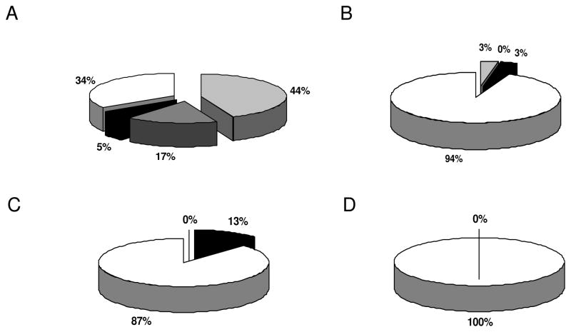
Figure 7.
Immunohistochemical analysis for annexin A3 expression by tissue micro arrays technology. RCC tissue micro arrays were stained with the annexin A3 specific antibody as described in Material and methods section. Panel A shows the staining pattern of RCC of clear cell subtype, panel B that of chromophilic subtype, panel C that of chromophobic subtype and panel D renal cell adenomas of oncocytic type. Light grey segments indicate strong positive, dark grey segments intermediate, black segments weak staining and blank segments indicate no staining. The relative staining frequencies are indicated by providing the respective percentage for each segment.

