Fig. 5.
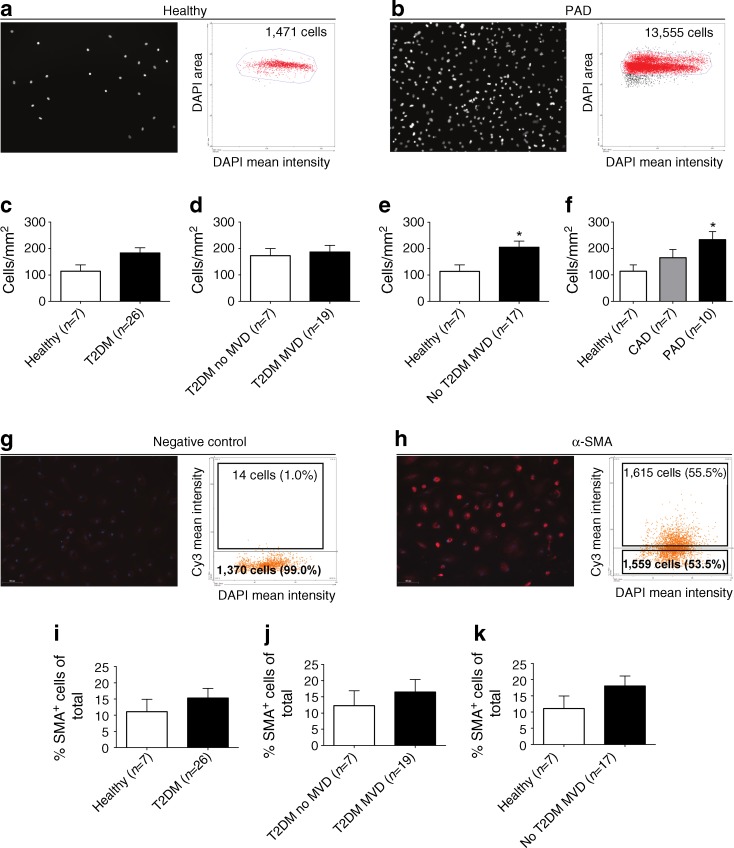
SMPC outgrowth is increased in non-type 2 diabetic patients with PAD. (a,b) Representative images and corresponding scatterplots of DAPI-stained SMPC nuclei quantified with the TissueFAXS system. The scatterplots show the total number of nuclei that were quantified in a fixed region in a representative healthy control (a) and a non-type 2 diabetic patient with PAD (b). (c) There was no significant difference in SMPC numbers between type 2 diabetic patients and healthy controls. (d) Within type 2 diabetic patients, similar SMPC numbers were detected in patients with and without MVD. (e) Within non-diabetic individuals, patients with MVD had a 1.8-fold increase in the number of outgrowth SMPCs compared with healthy controls. (f) Within non-diabetic patients, SMPC levels were increased 2.0-fold in PAD patients compared with healthy controls. (g,h) The presence of α-SMA was assessed using TissueFAXS analysis: (g) negative control staining; and (h) α-SMA staining. There was a tendency towards increased percentages of α-SMA+ outgrowth SMPCs in: (i) type 2 diabetic patients with and without MVD compared with healthy controls; (j) type 2 diabetic patients with MVD, compared with those without MVD; and (k) non-type 2 diabetic patients with MVD compared with healthy controls. Data are expressed as mean values ± SEM; *p < 0.05. T2DM, type 2 diabetes mellitus

