Abstract
The use of the lipid lowering agent niacin is hampered by a frequent flush response which is largely mediated by prostaglandin (PG) D2. Therefore, concomitant administration of the D-type prostanoid (DP) receptor antagonist laropiprant has been proposed to be a useful approach in preventing niacin-induced flush. However, antagonizing PGD2, which is a potent inhibitor of platelet aggregation, might pose the risk of atherothrombotic events in cardiovascular disease. In fact, we found that in vitro treatment of platelets with laropiprant prevented the inhibitory effects of PGD2 on platelet function, i.e. platelet aggregation, Ca2+ flux, P-selectin expression, activation of glycoprotein IIb/IIIa and thrombus formation. In contrast, laropiprant did not prevent the inhibitory effects of acetylsalicylic acid or niacin on thrombus formation. At higher concentrations, laropiprant by itself attenuated platelet activation induced by thromboxane (TP) and E-type prostanoid (EP)-3 receptor stimulation, as demonstrated in assays of platelet aggregation, Ca2+ flux, P-selectin expression, and activation of glycoprotein IIb/IIIa. Inhibition of platelet function exerted by EP4 or I-type prostanoid (IP) receptors was not affected by laropiprant. These in vitro data suggest that niacin/laropiprant for the treatment of dyslipidemias might have a beneficial profile with respect to platelet function and thrombotic events in vascular disease.
Introduction
Prostanoids are important regulators of platelet function and are involved in hemostasis by differentially influencing platelet aggregation [1]. The endothelium and, under inflammatory conditions, infiltrating leukocytes, vascular smooth muscle cells, and activated platelets release prostaglandins (PG) such as thromboxane (TX) A2, PGI2, PGE2 and PGD2 [2]–[7]. Platelets express corresponding receptors for these prostaglandins [1].
While TXA2 by activating the TP receptor is a very potent inducer and amplifier of platelet aggregation, PGI2 and PGD2 are clearly anti-aggregatory in their action. In contrast, PGE2 evokes a biphasic response, at nanomolar concentrations facilitating, while at micromolar concentrations inhibiting platelet aggregation [8]–[11]. The pro-aggregatory effect of PGE2 has been ascribed to the activation of the E-type prostanoid (EP) 3 receptor [12], [13]. An EP3 antagonist has been proposed to be useful for antithrombotic therapy [12]. Our group and others have simultaneously shown that the anti-aggregatory action of PGE2 in human platelets is mediated by EP4 receptors and a selective EP4 agonist potently inhibits platelet aggregation, Ca2+ mobilization, upregulation of P-selectin, and the activation of glycoprotein (GP) IIb/IIIa [14]–[17]. We could demonstrate that these inhibitory effects of EP4 receptors on platelet activation translate to potent antithrombotic activity as shown by an in vitro thrombus formation assay using whole blood [14].
Niacin has been shown to improve all lipoprotein abnormalities, by lowering cholesterol, triglycerides, low density lipoproteins (LDL), and apolipoprotein(a), while increasing high density lipoproteins (HDL) [18], alone or in combination with statins [19]. However, a frequent adverse effect in patients receiving niacin (1–2 g/day) is the development of significant cutaneous warmth and facial vasodilation. Although flushing is transient following intake of niacin, 5–6% percent of patients discontinue niacin because of that side effect [20]. Recent studies have elucidated the molecular mechanism that mediates niacin-induced flushing: Niacin acting through the G protein-coupled receptor GPR109A stimulates the production of several prostaglandins, including PGE2 and PGD2, in mast cells, keratinocytes and monocytes/macrophages [21], [22]. Particularly, PGD2 acting through the DP receptor has been alleged to cause the niacin-induced flush [23]. Consequently, a combination of the DP receptor antagonist laropiprant with niacin (Tredaptive®) is currently marketed for treatment of dyslipidemias for Europe [24]. In contrast, the U.S. Federal Drug Administration rejected the drug in 2008; although the reasons for the decision have not been published safety concerns are likely to have played a role. Although niacin/laropiprant has been reported to be effective and well tolerated [25]–[28], its effect on thrombotic cardiovascular events, such as myocardial infarction and stroke has not been revealed yet. Since prostaglandins are important regulators of platelet function, laropiprant, by interfering with the anti-aggregatory action of PGD2 [10], might confer additional cardiovascular risks to patients, thus outweighing the beneficial effects of niacin on lipid metabolism. However, laropiprant has also been purported to block the thromboxane receptor (TP) at high concentration, but the therapeutic relevance of this finding has not been followed up yet [25].
Prompted by these open issues we investigated the effects of laropiprant and niacin on in vitro thrombus formation in flowing human whole blood and found that both compounds have anti-platelet properties. While the inhibitory effect of niacin does not involve prostanoids such as PGD2, laropiprant at higher concentrations inhibits platelet function by blocking TP- and EP3-mediated platelet activation. Our results suggest that niacin/laropiprant might have beneficial effects on platelet function.
Methods
Ethics statement
The study was approved by the Institutional Review Board (Ethics committee of the Medical University Graz). Blood was drawn from healthy volunteers after they signed an informed consent form.
Material
All laboratory reagents were from Sigma (Vienna, Austria), unless specified. Assay buffer as used in Ca2+ flux and flow cytometric immunostaining was Dulbecco's modified phosphate-buffered saline (PBS; with or without 0.9 mM Ca2+ and 0.5 mM Mg2+; Invitrogen, Vienna, Austria). Laropiprant, sulprostone, PGD2, BW245c, BWA868c, U46619, CAY10598 and iloprost were purchased from Cayman (Ann Arbor, MI, USA). Fixative solution was prepared by adding 9 ml distilled water and 30 mL FACS-Flow to 1 mL CellFix. ADP and equine fibrillar collagen were obtained from Probe&Go (Osburg, Germany). Agonists/antagonists were dissolved in water, ethanol or dimethyl sulfoxide (DMSO) and further diluted in assay buffer to give a final concentration of the solvents <0.1%. CD62P-FITC and PAC-1-FITC antibodies were obtained from Becton Dickenson (Vienna, Austria), CD41 antibody from Invitrogen and VASP (pSer 157)-FITC from Acris Antibodies (Herford, Germany). D, L-lysin acetylsalicylic acid was obtained from Bayer (Munich, Germany).
Platelet aggregation
Platelet-rich and platelet-poor plasma were prepared from citrated whole blood by centrifugation and aggregation was performed by addition of ADP, collagen or U46619 in an Aggrecorder II (KDK Corp, Kyoto, Japan) [14], [29]–[31]. CaCl2 at a final concentration of 1 mmol/L was added 2 min before ADP or collagen [32]. In some experiments, acetylsalicylic acid (1 mmol/L as lysine acetylsalicylic acid) was added to platelet-rich plasma 30 minutes before the proaggregatory stimulus. To measure inhibition or enhancement of aggregation, the agonists were added 5 min before the Ca2+ stimulus. Antagonists were added 10 min before the agonists. Data were expressed as percentage of maximum light transmission, with nonstimulated platelet-rich plasma being 0% and platelet-poor plasma 100%. The used concentrations of laropiprant were within the peak concentrations obtained by oral administration of the clinically used dose (40 mg/day) or within the peak concentration of a well-tolerated high dose (200 mg/day) in patients [25].
Ca2+ flux
Intracellular Ca2+ levels were analyzed by flow cytometry [33]. Platelet rich plasma was loaded with 5 µmol/L of the acetoxymethyl ester of Fluo-3 in the presence of 2.5 mmol/L probenecid at 37°C for 30 minutes and were then resuspended in Tyrode's buffer (134 mM NaCl, 1 mM CaCl2, 12 mM NaHCO3, 2.9 mM KCl, 0.34 mM Na2HPO4, 1 mM MgCl2 and 0.055 mM glucose, supplemented with 10 mM HEPES). Changes in intracellular free Ca2+ levels in response to ADP (30 to 1000 nmol/L) were detected as increase in fluorescence intensity of the Ca2+-sensitive dye Fluo-3 in the FL-1 channel. Data were expressed as percent change of baseline.
Detection of vasodilator stimulatory phosphoprotein (VASP) phosphorylation
Platelet rich plasma was suspended in PBS containing Ca2+ and Mg2+. Platelets were pre-treated with the antagonist/vehicle for 5 min followed by agonist/vehicle treatment for 5 min before stimulation with 3 μM ADP for another 5 min. The samples were fixed by adding twice the volume of 2% formaldehyde for 10 min. PBS without Ca2+ and Mg2+ was added to the reaction mixture and then centrifuged at 400 g for 7 min. Thereafter, platelets were permeabilized by adding 0.5% triton X-100 and incubated for 10 min washed with PBS without Ca2+ and Mg2+ and stained using the VASP pSer 137 FITC-conjugated antibody (1 μg/ml in antibody diluent) for 30 min at room temperature in the dark [34]. After washing and fixation (with fixation solution as described in materials) the samples were analyzed by flow cytometry. Data were expressed in fluorescence units.
Flow cytometric immunofluorescence staining of P-selectin and GPIIb/IIIa
Platelet rich plasma was suspended in PBS containing Ca2+ and Mg2+ for immunofluorescence staining. Incubation of the platelets with prostanoids/agonists was done for 5 min. Pretreatment with antagonists started 10 min, and with acetylsalicylic acid 30 min, before the prostanoid/agonist treatment. For P-selectin staining platelets were activated by ADP (3 μM) and cytochalasin B (5 μg/ml) for 15 min at 37°C in the presence of the anti-CD62P-FITC onjugated antibody [14], [35]. The samples were washed and fixed, and P-selectin upregulation was detected by flow cytometry.
Activation of the fibrinogen receptor GPIIb/IIIa was assessed using the PAC-1-FITC conjugated antibody that recognizes a conformation-dependent determinant on the GPIIb/IIIa complex [36]. Total receptor expression was determined with an anti-CD41 FITC-conjugated antibody directed against GPIIb alone. For staining after the pre-treatment with antagonist/agonist, stimulation with ADP (3 μM) was carried out at 37°C for 5 min in the presence of the antibody. Samples were washed and fixed, and then analyzed by flow cytometry [14], [36]. Data were expressed as percent of control (vehicle) response.
In Vitro thrombogenesis
Vena8Fluoro+ Biochips (Cellix, Dublin, Ireland) were coated with collagen (200 µg/mL) at 4°C overnight and thereafter blocked with bovine serum albumin (10 µg/mL) for 30 minutes at room temperature followed by washing steps. Whole blood collected in sodium citrate was incubated with 3, 3-dihexyloxacarbocyanine iodide (1 µmol/L) in the dark for 10 minutes [14]. PGD2 (30 nmol/L), BW245c (3 nmol/L) were added 10 min before the start of perfusion, and the DP antagonist BWA868c or laropiprant (1 µmol/L) were added 10 min before the agonists. In another set of experiments whole blood was treated with niacin (3 mmol/L), acetylsalicylic acid (1 mmol/L) or laropiprant (1 µmol/L and 10 µmol/L) for 30 min. CaCl2 at a final concentration of 1 mmol/L was added 2 minutes before the perfusion over the collagen-coated chip. Perfusion was carried out at a shear rate of 30 dynes cm2. Thrombus formation was recorded by a Zeiss Axiovert 40 CFL microscope, Zeiss A-Plan 10X/0.25 Ph1 lens using Hamamatsu ORCA-03G digital camera and Cellix VenaFlux software. Computerized image analysis was performed by DucoCell analysis software (Cellix, Dublin), where the area covered by the thrombus was calculated. Data were expressed as percent of area covered in a control sample.
Statistical analysis
Data are shown as mean+SEM for n observations. Repeated measurements were performed using blood from the same healthy individual. Comparisons of groups were performed using Wilcoxon signed rank test or one-way ANOVA for repeated measurements with two sided – Dunnett's post test or Bonferroni's test. Probability values of P<0.05 were considered as statistically significant.
Results
Laropiprant, an antagonist for DP receptor-mediated inhibition of platelet function
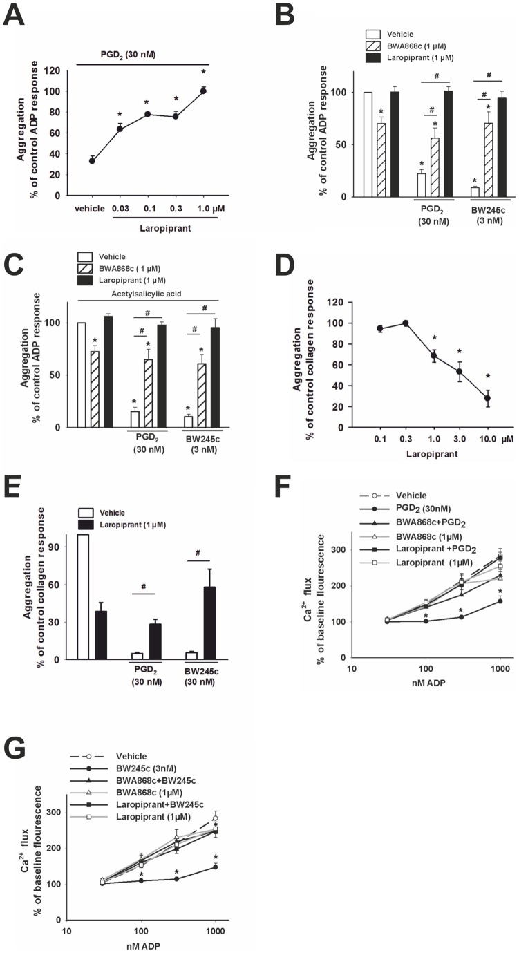
PGD2 is known to inhibit platelet aggregation via activation of the DP receptor. First we investigated whether laropiprant counteracted the effects of PGD2 and the DP agonist BW245c, on platelet aggregation. Platelet rich plasma was treated with PGD2 (30 nmol/L, a concentration which corresponds with the EC50 of the prostanoid [30]) or the DP agonist BW245c (3 nmol/L [37]) for 7 min. Platelet aggregation was induced with ADP (1.5–10 µmol/L) to give a submaximal effect (50– 60% aggregation). PGD2 (30 nmol/L) caused a significant inhibition of aggregation that was concentration dependently counteracted by laropiprant (with a maximal inhibition seen at 1 µmol/L Figure 1 A, B) and the selective DP antagonist BWA868c (1 µmol/L, Figure 1 B). Also the significant inhibition of aggregation caused by BW245, was abrogated by BWA868c (1 µmol/L) as well as by laropiprant (1 µmol/L; Figure 1 B). While laropiprant on its own showed no effect, BWA868c caused a slight inhibition of ADP- induced aggregation (Figure 1 B) which is consistent with the known intrinsic activity of BWA868c. To exclude the possibility that the inhibitory effect of PGD2 or BW245c was due to modulation of prostanoid release from platelets, samples were pretreated with acetylsalicylic acid (1 mmol/L). Acetylsalicylic acid treatment caused a slight reduction of the ADP-induced aggregation (−20.3±7.9%, as compared to non- treated samples; n = 4), but the effects of PGD2, BW245c, as well as the inhibition by BW868c or laropiprant were unaffected (Figure 1 C). When platelet aggregation was induced by collagen at a concentration that resulted in a 50–60% aggregation (1.25 to 10 mg/L), we found that laropiprant already at a concentration of 1 µmol/L caused a significant inhibition of the aggregation (Figure 1 D, E) but still counteracted the pronounced inhibition caused by PGD2 (30 nmol/L) and BW245c (3 nmol/L) (Figure 1 E).
Figure 1. DP receptor activation inhibits platelet aggregation and intracellular Ca2+ mobilization, effects that are counteracted by laropiprant.
(A–E) Aggregation was induced using ADP or collagen at concentrations which were adjusted to give submaximal aggregation. Data were expressed as percent of the control response. (A) ADP-induced aggregation was inhibited by PGD2 (30 nmol/L) and this effect was concentration dependently counteracted by laropiprant (n = 6). (B) The ADP- induced aggregation was slightly reduced by the DP receptor antagonist BWA868c (1 μmol/L), but not by laropiprant (1 μmol/L) alone. PGD2 (30 nmol/L) and the DP agonist, BW245c (3 nmol/L) caused pronounced inhibition of platelet aggregation and these effects were reversed by BWA868c and laropiprant (n = 4). (C) Pre-treatment of platelets with acetylsalicylic acid (1 mmol/L) did not affect the inhibitory effects of PGD2 (30 nmol/L) and BW245c (3 nmol/L) and the reversal of these effects by BWA868c and laropiprant at 1 μmol/L (n = 4). (D) Laropiprant caused a concentration-dependent inhibition of collagen-induced aggregation (n = 6). (E) Laropiprant (1 μmol/L) counteracted the inhibition of aggregation by PGD2 (30 nmol/L) and BW245c (3 nmol/L) (n = 4–6). (F, G) Ca2+ responses were detected by flow cytometry as changes in fluorescence of the Ca2+-sensitive dye Fluo-3 by and are presented as percent of baseline fluorescence. Ca2+ flux induced by ADP (30–1000 nmol/L) was significantly reduced by pre-treatment of platelets with (E) PGD2 (30 nmol/L) and (F) BW245c (3 nmol/L). The inhibition of the Ca2+ flux by DP receptor activation was reversed by laropiprant (1 μmol/L) and the DP antagonist, BWA868c (1 μmol/L) (n = 6). Values are shown as mean+SEM. *P<0.05 as compared to vehicle and # P<0.05 as compared to agonist treatment.
Elevation of intracellular free Ca2+ is an important prerequisite for platelet activation subsequently leading to cytoskeletal changes and subserving platelet aggregation. We investigated whether laropiprant affects the DP receptor-mediated inhibition of Ca2+ responses using a flow cytometric Ca2+ flux assay. ADP caused a concentration dependent increase in Ca2+ flux that was inhibited by PGD2 (30 nmol/L) as well as by the DP agonist BW245c (3 nmol/L). These effects were completely reversed by pretreatment of platelets with the DP antagonist BWA868c as well as by laropiprant (each 1 µmol/L) (Figure 1 F, G).
Laropiprant blocks DP receptor-dependent increase in VASP phosphorylation, as well as inhibition of P-selectin expression, GPIIb/IIIa activation and in vitro thrombus formation
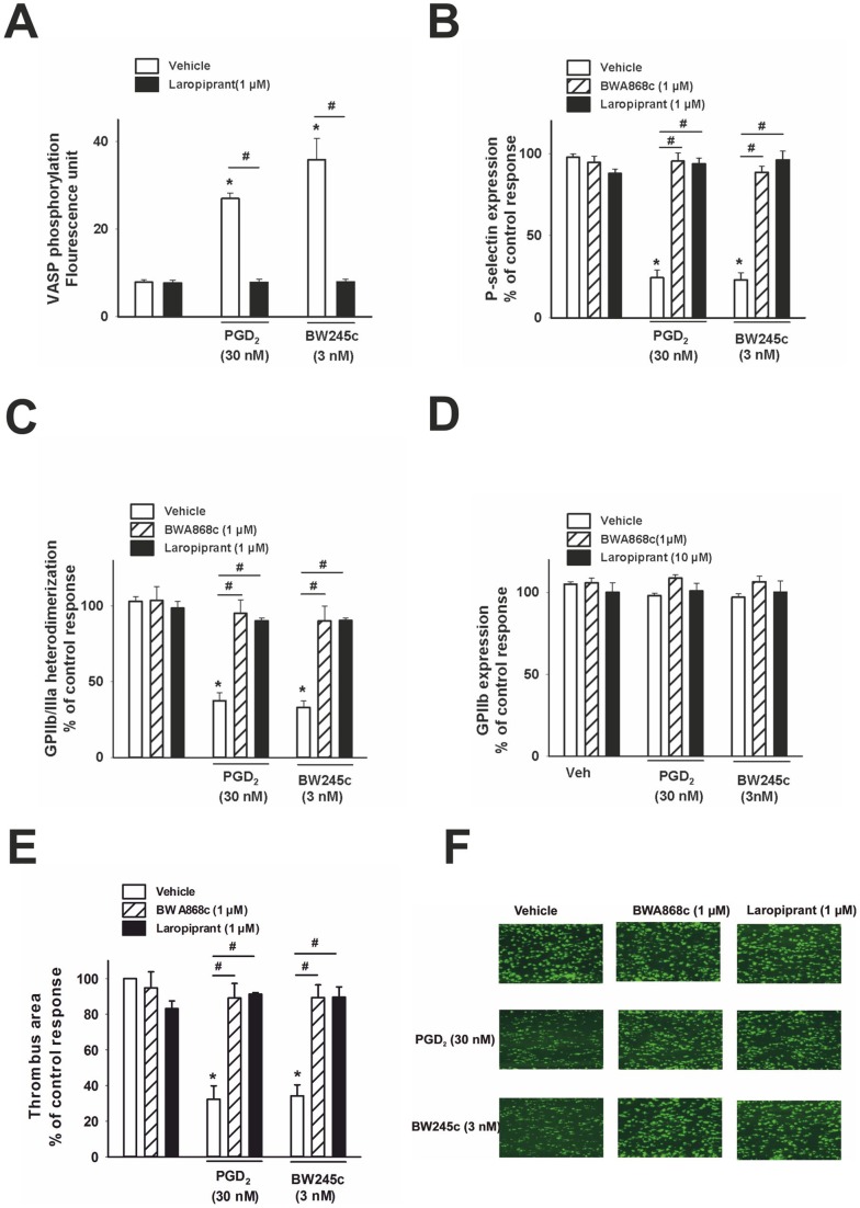
VASP is a key regulator for rapid dynamic changes in the cytoskeleton of platelets [38] and is phosphorylated on Ser157 in a cAMP-dependent manner. This phosphorylation leads to conformational changes of cell surface molecules and hence constitutes an important inhibitory pathway of platelet aggregation. It is known that DP receptor activation leads to Gs-mediated increases in intracellular cAMP [39]. Therefore, the effect of DP receptor activation on VASP phosphorylation was investigated. PGD2 (30 nM) or BW245c (3 nM) significantly enhanced VASP phosphorylation in platelets stimulated with ADP (3 µmol/L). The DP antagonists BWA868c (n = 4; data not shown) and laropiprant (1 µmol/L each) had no effect alone, but completely reversed the effect of DP receptor stimulation (Figure 2 A).
Figure 2. DP receptor activation increases VASP phosphorylation, and inhibits P-selectin expression, GPIIb/IIIa activation and in vitro thrombus formation. (A).
VASP phosphorylation was visualized using an anti-VASP p-Ser 157 antibody in flow cytometry and data were expressed in fluorescence units. VASP phosphorylation in platelets stimulated with ADP (3 µmol/L) was significantly enhanced by PGD2 (30 nmol/L) as well as the DP agonist BW245c (3 nmol/L), which was completely prevented by pre-treatment of platelets with laropiprant (1 μmol/L) (n = 4). (B) ADP (3 μmol/L) increased the surface expression of P-selectin, detected using a CD62P antibody, in platelets primed with cytochalasin B (5 μg/mL). PGD2 (30 nmol/L) and BW245c (3 nmol/L) caused a significant inhibition of P-selectin expression on the platelet surface and this effect was revoked by pre-treatment with DP antagonists, BWA868c (1 μmol/L) and laropiprant (1 μmol/L; n = 6). (C) The ADP (3 μmol/L) induced activation of GPIIb/IIIa, detected using a conformation dependent antibody PAC-1, was attenuated by PGD2 (30 nmol/L) and the DP agonist BW245c (3 nmol/L). These effects were reversed by BWA868c and laropiprant at 1 μmol/L (n = 6). (D) GPIIb expression was determined using an anti-CD41 antibody by flow cytometry. None of the treatments affected the GPIIb expression. Data were expressed as percent of control ADP response. (E) Whole blood incubated with the fluorescent dye 3,3′-dihexyloxacarbocyanine iodide was perfused over collagen-coated channels and thrombus formation was recorded by fluorescence microscopy. Thrombus-covered area was calculated by computerized image analysis and is expressed as percent of control (vehicle) response. Vehicle-treated samples showed pronounced thrombogenesis over collagen. Both PGD2 (30 nmol/L) and BW245c (3 nmol/L) markedly decreased the formation of thrombi. The DP antagonist BWA868c and laropiprant (1 µmol/L each) had no effect on thrombus formation by themselves but reversed the inhibition of thrombogenesis by DP receptor stimulation (n = 4). (F) Original fluorescence images. Values are shown as mean+SEM. *P<0.05 as compared to vehicle and # P<0.05 as compared to agonist treatment.
Since elevation of intracellular free Ca2+ is essential for the upregulation of P-selectin and activation of GPIIb/IIIa, we further investigated the effects of DP receptor activation on these responses. Platelets were treated with ADP (3 µmol/L) and cytochalasin (5 µg/mL) to stimulate P-selectin expression on the surface of platelets as determined by an anti-CD62P antibody. The ADP-induced activation of GPIIb/IIIa was detected by staining with the conformation-dependent antibody PAC-1. Both PGD2 (30 nmol/L) and BW245c (3 nmol/L) effectively prevented P-selectin expression as well as GPIIb/IIIa activation (Figure 2 B, C). The DP antagonist BWA868c and laropiprant alone (each 1 µmol/L) were without an effect but completely prevented the effect of DP receptor stimulation. The total amount GPIIb/IIIa was not changed by any of these treatments as determined using an anti-CD41 antibody (Figure 2 D).
Platelet adhesion is the first step in thrombus formation and this is governed by (i) platelet-endothelial-leukocyte interactions mediated by P-selectin and (ii) by platelet-platelet interactions mediated by activated GPIIb/IIIa which act as fibrin receptors. Since PGD2 via DP receptor activation inhibited both processes, we next assessed whether DP receptor activation also prevented thrombogenesis in vitro. Whole blood was perfused through collagen-coated channels at a shear rate of 30 dynes/cm2, and thrombus formation was recorded by fluorescence microscopy. PGD2 (30 nmol/L) as well as BW245c (3 nmol/L) effectively inhibited thrombogenesis. This inhibition was counteracted by BWA868c and by laropiprant (both 1 µmol/L), at concentrations at which none of them showed significant effects on thrombus formation by themselves (Figure 2 E, F).
These data showed that activation of the DP receptor attenuates platelet aggregation by inhibiting Ca2+ mobilization, increasing VASP phosphorylation and inhibition of P-selectin expression and GPIIb/IIIa activation, translating to inhibition of thrombogenesis. These effects were prevented by laropiprant as a DP receptor antagonist.
Laropiprant prevents TP and EP3 receptor-mediated platelet responses
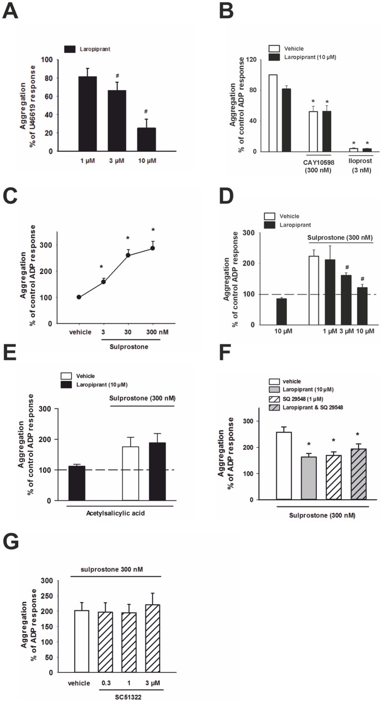
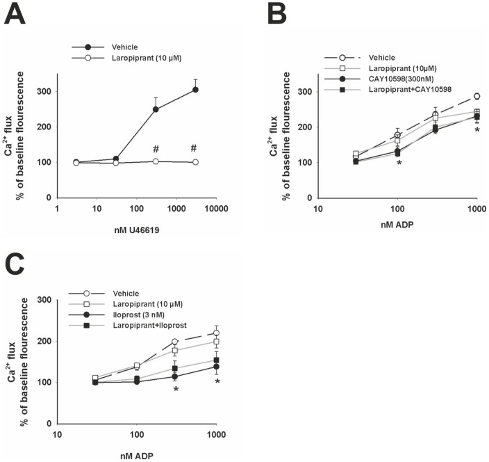
In addition to its antagonistic effect on the DP receptor, laropiprant purportedly binds to the TP receptor, albeit with a lower affinity [25]. However, it is not known whether laropiprant acts also on other prostanoid receptors. First we investigated the effect of laropiprant on platelet aggregation induced by the TP agonist, U46619. At 300 nmol/L, U46619 caused a pronounced increase in aggregation, which was concentration-dependently inhibited by laropiprant (Figure 3 A). In further experiments, we addressed the possibility that laropiprant in addition to its effect on DP and TP receptors also affects EP3, EP4 and IP receptor-mediated responses. The EP4 agonist CAY10598 (300 nmol/L) significantly reduced, and the EP1/IP agonist iloprost (3 nmol/L) almost completely prevented, the ADP-induced platelet aggregation. These effects were not influenced by laropiprant up to 10 µmol/L, (Figure 3 B). The EP3 agonist sulprostone caused a concentration-dependent increase of ADP-induced platelet aggregation (Figure 3 C) and the response to 300 nmol/L of sulprostone was concentration-dependently inhibited by laropiprant (Figure 3 D). Pretreatment of platelets with acetylsalicylic acid diminished the proaggregatory effect of sulprostone by 70%, and rendered laropiprant ineffective in preventing the remaining sulprostone response (Figure 3 E). Additional experiments showed that the enhanced aggregation induced by sulprostone was also inhibited by the TP receptor antagonist SQ29548 (1 µmol/L) and the combination of laropiprant and SQ29548 caused no further attenuation (Figure 3 F). Since sulprostone can also activate the EP1 receptor in addition to its effect on the EP3 receptor, we employed the EP1 antagonist SC51322 which, however, showed no inhibitory effect towards sulprostone (Figure 3 G). To further elucidate the role of laropiprant we investigated the TP receptor-induced Ca2+ flux. U46619 caused a concentration-dependent increase in Ca2+ flux which was prevented by pretreating platelets with laropiprant at 10 µmol/L (Figure 4 A). In contrast, laropiprant at 10 µmol/L did not reverse the inhibition of ADP-induced Ca2+ flux by the EP4 agonist CAY10598 (300 nmol/L) or the IP agonist iloprost (3 nmol/L) (Figure 4 B, C).
Figure 3. Laropiprant antagonizes the increased platelet aggregation by TP and EP3 receptor activation.
In (A), aggregation was induced by U46619 (300 nmol/L) which was concentration-dependently inhibited by laropiprant (n = 4). In B–G, ADP concentrations (1.25–10 μmol/L) were adjusted to give 30–50% of maximal aggregation. (B) The inhibiton of ADP-induced aggregation by the EP4 agonist CAY10598 (300 nmol/L) and the IP agonist iloprost (3 nmol/L) was not affected by laropiprant (10 nmol/L, (n = 7)). (C) The EP3 agonist sulprostone concentration dependently amplified ADP-induced aggregation (n = 4–6). (D) The effect of sulprostone (300 nmol/L) was concentration dependently inhibited by laropiprant (n = 4). (E) Pretreatment with acetylsalicylic acid (1 mmol/L) markedly attenuated the pro-aggregatory effect of sulprostone, and in this case, laropiprant was unable to reverse the stimulatory effect of the EP3 agonist. Data were expressed as percent of control response. (F) The TP receptor antagonist SQ29578 (1 µmol/L) inhibited the sulprostone-induced increase in platelet aggregation to the same extend as laropiprant (10 µmol/L). The combination of SQ29578 and laropiprant did not cause further inhibition as compared to laropiprant or SQ29578 alone (n = 4–6) (G) The pro-aggregatory effect of sulprostone was not inhibited by the EP1 receptor antagonist SC51322 (n = 4). Data were expressed as percent of control ADP response and are shown as mean+SEM. *P<0.05 as compared to vehicle and #P<0.05 as compared to the respective agonist treatment.
Figure 4. Laropiprant antagonizes Ca2+ mobilization induced by TP receptor activation.
Ca2+ responses were detected by flow cytometry as changes in fluorescence of the Ca2+ -sensitive dye Fluo-3 by flow cytometry and are presented as percent of baseline fluorescence. (A) The TP agonist, U46619 (3–3000 nmol/L), induced Ca2+flux in a concentration-dependent manner and this effect was completely inhibited by laropiprant at 10 μmol/L (n = 6). (B) The EP4 receptor agonist CAY10598 (300 nmol/L) and (C) the IP receptor agonist iloprost (3 nmol/L) caused a significant inhibition of the ADP-induced Ca2+ flux (n = 4). Laropiprant (10 µmol/L) did not antagonize these effects (n = 4). Values are shown as mean+SEM. *P<0.05 as compared to vehicle and #P<0.05 as compared to the respective agonist treatment treatment.
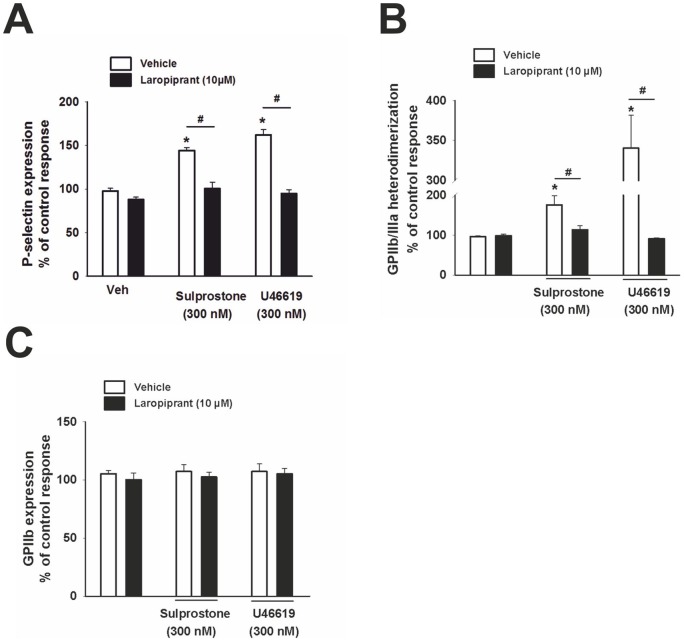
Activation of the EP3 receptor by sulprostone (300 nmol/L) and of the TP receptor by U46619 (300 nmol/L) enhanced the ADP- induced P-selectin surface expression and GPIIb/IIIa activation. Importantly, both effects were totally prevented by laropiprant (10 µmol/L; Figure 5 A, B). The total amount of GPIIb was not changed by any of these treatments as determined using an anti-CD41 antibody (Figure 5 C). Thus in total, laropiprant reversed TP- and EP3-mediated platelet responses, such as platelet aggregation, P-selectin expression, GPIIb/IIIa activation and Ca2+ flux.
Figure 5. Laropiprant antagonizes the P-selectin expression and GPIIb/IIIa activation induced by TP and EP3 receptor activation. (A).
ADP (3 μmol/L) increased the surface expression of P-selectin in platelets primed with cytochalasin B (5 μg/mL), detected using a CD62P antibody. The EP3 agonist sulprostone (300 nmol/L) and U46619 (300 nmol/L) elevated P-selectin expression on the surface of platelets. These effects were counteracted by laropiprant at 10 μmol/L (n = 5). (B) The ADP (3 μmol/L) induced activation of GPIIb/IIIa, detected using a conformation-dependent antibody, PAC-1, was increased by sulprostone (300 nmol/L) and U46619 (300 nmol/L). Laropiprant at 10 µmol/L antagonized these effects (n = 5). (C) GPIIb expression was determined using an anti-CD41 antibody by flow cytometry. None of the treatments affected the GPIIb expression (n = 5). Data were expressed as percentage of ADP control response and are shown as mean+SEM. *P<0.05 as compared to vehicle and # P<0.05 as compared to the respective agonist treatment.
Effect of laropiprant and niacin on in vitro thrombus formation
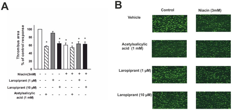
Based on the results that laropiprant inhibited the DP, TP and EP3 mediated effects, we investigated how this translates to thrombus formation in whole blood. While laropiprant at 1 µmol/L had no effect on thrombus formation, it caused a pronounced inhibition at 10 µmol/L, comparable to the inhibition of thrombogenesis by acetylsalicylic acid (1 mmol/L) (Figure 6). It has been shown that niacin and its metabolite niceritrol inhibit platelet aggregation in vitro [40], [41] and cause the release of TXA2, PGD2 and PGE2 at a clinically relevant concentration of 3 mmol/L [41]. Therefore, we investigated the effect of niacin on thrombus formation. Incubation of whole blood with niacin (3 mmol/L for 30 min) caused a pronounced inhibition of thrombus formation, an effect that was not influenced by pretreatment of blood with acetylsalicylic acid (1 mmol/L) or laropiprant (10 µmol/L). This suggests that – in contrast to the flushing response – formation of prostaglandins, e.g. PGD2, are not involved in the inhibitory effect of niacin on platelet aggregation.
Figure 6. Laropiprant at 10 µmol/L and niacin inhibit in vitro thrombus formation.
Whole blood was incubated with the fluorescent dye 3,3′-dihexyloxacarbocyanine iodide, perfused over collagen-coated channels, and thrombus formation was recorded by fluorescence microscopy. The images were taken 3 minutes after the start of the perfusion and are representative of 4 different experiments. (A) Vehicle-treated samples showed pronounced and rapid thrombogenesis over collagen. Treatment of whole blood with acetylsalicylic acid (1 mmol/L), laropiprant 10 µmol/L and niacin 3 mmol/L for 30 min, caused a marked reduction of thrombus formation, while laropiprant 1 µmol/L had no effect. The antithrombotic effect of niacin was not influenced by pretreatment of blood with acetylsalicylic acid or laropiprant. Thrombus-covered area was calculated by computerized image analysis and is expressed as percent of control (vehicle) response. (B) Original fluorescence images. Values are shown as mean+SEM. *P<0.05 versus vehicle.
Discussion
In this study we investigated the effects of niacin and laropiprant, the two active substances in the lipid-lowering drug Tredaptive®, on platelet function in vitro, in order to address concerns that the drug may harbor the risk of atherothrombotic events. In addition to mediating the “flush” response to niacin, PGD2 is a potent anti-aggregatory agent in platelets via activation of the DP receptor [42]. Lipocalin-type PGD2 synthase, one of the key enzymes responsible for the biosynthesis of PGD2 has been shown to be expressed in vascular endothelial cells [43] in serum of atherosclerotic patients and to be accumulated in the atherosclerotic plaque of coronary arteries with severe stenosis [44], [45]. Therefore, it can be assumed that PGD2 is formed in atherosclerotic lesions and may prevent the adhesion and subsequent aggregation of platelets. Moreover, studies have shown that niacin intake increases the plasma concentrations of PGD2, PGI2 and TXA2 [46]–[49] and that the increase in PGD2 is by far the most pronounced. Accordingly, laropiprant might prevent the anti-thrombotic effect of PGD2 thereby fostering vascular events such as stroke or myocardial infarction.
PGD2, apart from being released from mast cells [50], [51], dendritic cells, eosinophils, and Th2 cells [52]–[55], is also synthesized in the vasculature, i.e. by endothelial cells, macrophages and platelets [6], [56]–[59]. PGD2 exerts it biological effects via two G protein-coupled receptors, the DP receptor and chemoattractant receptor homologous molecule expressed on Th2 cells (CRTH2). Activation of the DP receptor [30], [60] causes an increase in cAMP production [61] and inhibits Ca2+ mobilization in platelets [62]. In this study we further elucidated the cellular mechanisms involved in PGD2 inhibition of platelet aggregation and confirmed the potent antagonist effect of laropiprant on the DP receptor. We found that activation of the DP receptor inhibited ADP- and collagen induced platelet aggregation, without involving other inhibitory prostanoid receptors or modulating TXA2 release, since identical results were obtained in acetylsalicylic acid-treated platelets. Moreover, the ADP-induced increase in Ca2+ mobilization was abrogated by DP receptor activation, which is in line with previously published results [62]. VASP phosphorylation, which is an indicator of inhibition of platelet aggregation by preventing shape change in platelets [63], [64], was increased by DP receptor activation. Since VASP phosphorylation was assessed at Ser 157, this can be attributed to a cAMP protein kinase-mediated effect [64] which is consistent with the known increase of cAMP by DP receptor stimulation [39].
Surface expression of P-selectin is upregulated in activated platelets and plays an important role in platelet-leukocyte-endothelial interaction. Conformational change of GPIIb/IIIa to a high affinity state for fibrinogen enables thrombus formation. Activation of the DP receptor abrogated the agonist-induced P-selectin expression and GPIIb/IIIa activation. All effects elicited by activation of the DP receptor were counteracted by the DP receptor antagonist BWA868c and by laropiprant at 1 µmol/L. These findings translated to the inhibitory effects of DP receptor activation on thrombogenesis under flow condition. Laropirant at 1 µmol/L had no effect on its own on thrombus formation, while counteracting the inhibition induced by DP receptor stimulation, which suggests that laropiprant has potential pro-thrombotic effects in vitro. However, residual activity of laropiprant on pro-aggregatory TP receptors has been purported [25] which might compensate for the loss of anti-aggregatory DP activity. We have further advanced this notion by showing that platelet aggregation, Ca2+ mobilization, P-selectin expression and GPIIb/IIIa activation induced by the TP receptor agonist U46619 were completely blocked by laropiprant at a concentration of 10 µmol/L. Interestingly, laropiprant already at a concentration of 1 µmol/L markedly attenuated collagen-induced platelet aggregation (Figure 1 D). Lai et al [25], [65], described an inhibitory effect on collagen-induced platelet aggregation ex vivo when plasma levels of laropiprant were about 5 µmol/L. Collagen has been found to induce platelet aggregation partially through TXA2 formation leading to TP receptor activation [66], and we have observed that the TP antagonist SQ29548 (1 µmol/L) exhibited similar inhibitory effect on collagen-induced aggregation as laropiprant (n = 4, data not shown). These data suggest that laropiprant attenuates collagen-induced platelet aggregation by blocking TP receptors. In contrast, no effects on the IP or EP4 receptors were observed in assays of platelet aggregation and Ca2+ flux, showing that the promiscuity of laropiprant does not extend to these inhibitory platelet receptors.
An interesting novel finding of the current study is that laropiprant also attenuates EP3-mediated responses. PGE2 has a biphasic, concentration-dependent effect on platelet aggregation. While at high concentrations it counteracts aggregation and thrombus formation of human platelets via activation of the EP4 receptor [14]–[17], at lower concentrations it aggravates agonist-induced aggregation by activation of the EP3 receptor, by eliciting Ca2+ mobilization, decreased VASP phosphorylation and enhanced P-selectin expression [12]. An EP3 antagonist has been proposed to be useful for antithrombotic therapy [12]. We observed that laropiprant antagonized the EP3-mediated enhancement of ADP-induced platelet aggregation, P-selectin expression and GPIIb/IIIa activation. Interestingly, inhibition of prostaglandin synthesis using acetylsalicylic acid attenuated the sulprostone-induced facilitation of platelet aggregation and abolished the inhibitory effect of laropiprant. Sulprostone acts as an EP3/EP1 agonist, however, the EP1 antagonist SC51322 did not abrogate the stimulatory effect of sulprostone. In addition, we found that the TP antagonist SQ29548 reduced the effect of sulprostone and did not further enhance the effect of laropiprant. Therefore, in agreement with previous reports [12] we hypothesize that EP3 activation triggers TXA2 release which in turn activates TP receptors to promote platelet aggregation, and TP blockade by laropiprant disrupts this signaling cascade. Similar observations were made with regard to regulation of vascular tone by the EP3 receptor: vasoconstriction induced by EP3 receptor activation is mediated through increasing TP-mediated signaling in guinea-pig aorta [67], and a TP antagonist attenuates vasoconstrictor responses to EP3 agonist in rat mesenteric artery [68]. Therefore, laropiprant might be capable of opposing EP3/TP-mediated vasospasm in vascular inflammation in patients with atherosclerosis.
Together, our results suggest that laropiprant can interfere with platelet aggregation by inhibiting pro-aggregatory targets, TP and EP3 receptors. This was further substantiated by our results of in vitro thrombogenesis of whole blood under flow condition. At the concentration of 10 µmol/L, laropiprant inhibited thrombogenesis to an extent comparable to acetylsalicylic acid (1 mmol/L). Therefore, it can be assumed that, at this concentration, laropiprant acts as a TP antagonist and hence inhibits thrombogenesis. Even though the dose given in combination with niacin is 40 mg/day, which results in peak concentrations of 1.5–2.5 µmol/L [25], [69], laropiprant up to a dose of 600 mg per day is well tolerated in healthy subjects and a multiple oral dose of 200 mg/day for 10 days results in a peak plasma concentration of 11.4 µmol/L [25]. Our in vitro results demonstrate the potential of laropiprant to antagonize DP, TP and EP3 mediated effects; however, whether this is clinical relevant depends on the endogenous PGD2, thromboxane and EP3 agonist activity.
It has been shown that niacin and its metabolite niceritrol inhibit platelet aggregation in vitro [40], [41] and incubation of whole blood with niacin at a clinically relevant concentration increases the concentrations of PGD2, PGE2 and TXA2 [41]. Our results show that niacin causes a profound inhibition of thrombus formation in vitro; however, prostaglandins such as PGD2 do not seem to be involved in this process, since pre-treatment of blood with acetylsalicylic acid as well as laropiprant did not counteract this effect. Therefore, the mechanisms involved in niacin-induced inhibition of platelet aggregation remain to be investigated.
In conclusion our data show that laropiprant at a low concentration antagonizes the effects of DP receptor activation on platelets while it does not reverse the anti-aggregatory effect of niacin. However, at a concentration that has been shown to be well tolerated [25], laropiprant attenuates TP and EP3 receptor function and effectively inhibits thrombus formation. Although further in vivo studies with oral application of the drugs are needed, our in vitro findings suggest that higher concentrations of laropiprant may have beneficial effects with respect to platelet function and thrombotic events in vascular disease.
Acknowledgments
The authors are indebted to Martina Ofner and Geri Parzmair for their excellent technical work.
Funding Statement
Dr. Philipose was funded by the PhD Program in Molecular Medicine of the Medical University of Graz. This study was supported by the Jubiläumsfonds of the Austrian National Bank (OeNB, grants 13487 and 14263) and the Austrian Science Fund (FWF; grants P22521-B18, P19473-B05, P21004-B02 and P22976-B18. The funders had no role in study design, data collection and analysis, decision to publish, or preparation of the manuscript.
References
- 1. Armstrong RA (1996) Platelet prostanoid receptors. Pharmacol Ther 72: 171–91. [DOI] [PubMed] [Google Scholar]
- 2. Svensson J, Hamberg M, Samuelsson B (1976) On the formation and effects of thromboxane A2 in human platelets. Acta Physiol Scand 98: 285–94. [DOI] [PubMed] [Google Scholar]
- 3. Vane JR, Botting RM (1995) Pharmacodynamic profile of prostacyclin. Am J Cardiol 75: 3A–10A. [DOI] [PubMed] [Google Scholar]
- 4. Bishop-Bailey D, Pepper JR, Haddad EB, Newton R, Larkin SW, et al. (1997) Induction of cyclooxygenase-2 in human saphenous vein and internal mammary artery. Arterioscler Thromb Vasc Biol 17: 1644–8. [DOI] [PubMed] [Google Scholar]
- 5. Bishop-Bailey D, Pepper JR, Larkin SW, Mitchell JA (1998) Differential induction of cyclooxygenase-2 in human arterial and venous smooth muscle: role of endogenous prostanoids. Arterioscler Thromb Vasc Biol 18: 1655–61. [DOI] [PubMed] [Google Scholar]
- 6. Oelz O, Oelz R, Knapp HR, Sweetman BJ, Oates JA (1977) Biosynthesis of prostaglandin D2. 1. Formation of prostaglandin D2 by human platelets. Prostaglandins 13: 225–34. [DOI] [PubMed] [Google Scholar]
- 7. Hata AN, Breyer RM (2004) Pharmacology and signaling of prostaglandin receptors: multiple roles in inflammation and immune modulation. Pharmacol Ther 103: 147–66. [DOI] [PubMed] [Google Scholar]
- 8. Shio H, Ramwell PW, Jessup SJ (1972) Prostaglandin E2: effects on aggregation, shape change and cyclic AMP of rat platelets. Prostaglandins 1: 29–36. [DOI] [PubMed] [Google Scholar]
- 9. Vezza R, Roberti R, Nenci GG, Gresele P (1993) Prostaglandin E2 potentiates platelet aggregation by priming protein kinase C. Blood. 82: 2704–13. [PubMed] [Google Scholar]
- 10. Feinstein MB, Egan JJ, Sha'afi RI, White J (1983) The cytoplasmic concentration of free calcium in platelets is controlled by stimulators of cyclic AMP production (PGD2, PGE1, forskolin). Biochem Biophys Res Commun 113: 598–604. [DOI] [PubMed] [Google Scholar]
- 11. Gresele P, Blockmans D, Deckmyn H, Vermylen J (1988) Adenylate cyclase activation determines the effect of thromboxane synthase inhibitors on platelet aggregation in vitro. Comparison of platelets from responders and nonresponders. J Pharmacol Exp Ther 246: 301–7. [PubMed] [Google Scholar]
- 12. Heptinstall S, Espinosa DI, Manolopoulos P, Glenn JR, White AE, et al. (2008) DG-041 inhibits the EP3 prostanoid receptor–a new target for inhibition of platelet function in atherothrombotic disease. Platelets 19: 605–13. [DOI] [PubMed] [Google Scholar]
- 13. Gross S, Tilly P, Hentsch D, Vonesch JL, Fabre JE (2007) Vascular wall-produced prostaglandin E2 exacerbates arterial thrombosis and atherothrombosis through platelet EP3 receptors. J Exp Med 204: 311–20. [DOI] [PMC free article] [PubMed] [Google Scholar]
- 14. Philipose S, Konya V, Sreckovic I, Marsche G, Lippe IT, et al. (2010) The prostaglandin E2 receptor EP4 is expressed by human platelets and potently inhibits platelet aggregation and thrombus formation. Arterioscler Thromb Vasc Biol 30: 2416–23. [DOI] [PubMed] [Google Scholar]
- 15. Smith JP, Haddad EV, Downey JD, Breyer RM, Boutaud O (2010) PGE2 decreases reactivity of human platelets by activating EP2 and EP4. Thromb Res 126: e23–9. [DOI] [PMC free article] [PubMed] [Google Scholar]
- 16. Iyu D, Glenn JR, White AE, Johnson AJ, Fox SC, et al. (2010) The role of prostanoid receptors in mediating the effects of PGE(2) on human platelet function. Platelets 21: 329–42. [DOI] [PubMed] [Google Scholar]
- 17. Kuriyama S, Kashiwagi H, Yuhki K, Kojima F, Yamada T, et al. (2010) Selective activation of the prostaglandin E2 receptor subtype EP2 or EP4 leads to inhibition of platelet aggregation. Thromb Haemost 104: 796–803. [DOI] [PubMed] [Google Scholar]
- 18. Bodor ET, Offermanns S (2008) Nicotinic acid: an old drug with a promising future. Br J Pharmacol 153 Suppl 1S68–75. [DOI] [PMC free article] [PubMed] [Google Scholar]
- 19. Yiu KH, Cheung BM, Tse HF (2010) A new paradigm for managing dyslipidemia with combination therapy: laropiprant + niacin + simvastatin. Expert Opin Investig Drugs 19: 437–49. [DOI] [PubMed] [Google Scholar]
- 20. Brinton EA, Kashyap ML, Vo AN, Thakkar RB, Jiang P, et al. (2011) Niacin extended-release therapy in phase III clinical trials is associated with relatively low rates of drug discontinuation due to flushing and treatment-related adverse events: a pooled analysis. Am J Cardiovasc Drugs 11: 179–87. [DOI] [PubMed] [Google Scholar]
- 21. Hanson J, Gille A, Zwykiel S, Lukasova M, Clausen BE, et al. (2010) Nicotinic acid- and monomethyl fumarate-induced flushing involves GPR109A expressed by keratinocytes and COX-2-dependent prostanoid formation in mice. J Clin Invest 120: 2910–9. [DOI] [PMC free article] [PubMed] [Google Scholar]
- 22. Kamanna VS, Ganji SH, Kashyap ML (2009) The mechanism and mitigation of niacin-induced flushing. Int J Clin Pract 63: 1369–77. [DOI] [PMC free article] [PubMed] [Google Scholar]
- 23. Papaliodis D, Boucher W, Kempuraj D, Michaelian M, Wolfberg A, et al. (2008) Niacin-induced “flush” involves release of prostaglandin D2 from mast cells and serotonin from platelets: evidence from human cells in vitro and an animal model. J Pharmacol Exp Ther 327: 665–72. [DOI] [PubMed] [Google Scholar]
- 24. Sanyal S, Kuvin JT, Karas RH (2010) Niacin and laropiprant. Drugs Today (Barc) 46: 371–8. [DOI] [PubMed] [Google Scholar]
- 25. Lai E, Wenning LA, Crumley TM, De Lepeleire I, Liu F, et al. (2008) Pharmacokinetics, pharmacodynamics, and safety of a prostaglandin D2 receptor antagonist. Clin Pharmacol Ther 83: 840–7. [DOI] [PubMed] [Google Scholar]
- 26. Bays HE, Shah A, Lin J, McCrary Sisk C, Paolini JF, et al. (2010) Efficacy and tolerability of extended-release niacin/laropiprant in dyslipidemic patients with metabolic syndrome. J Clin Lipidol 4: 515–21. [DOI] [PubMed] [Google Scholar]
- 27. McKenney J, Bays H, Koren M, Ballantyne CM, Paolini JF, et al. (2010) Safety of extended-release niacin/laropiprant in patients with dyslipidemia. J Clin Lipidol 4: 105–112.e1. [DOI] [PubMed] [Google Scholar]
- 28. Maccubbin D, Bays HE, Olsson AG, Elinoff V, Elis A, et al. (2008) Lipid-modifying efficacy and tolerability of extended-release niacin/laropiprant in patients with primary hypercholesterolaemia or mixed dyslipidaemia. Int J Clin Pract 62: 1959–70. [DOI] [PubMed] [Google Scholar]
- 29. Schuligoi R, Sedej M, Waldhoer M, Vukoja A, Sturm EM, et al. (2009) Prostaglandin H2 induces the migration of human eosinophils through the chemoattractant receptor homologous molecule of Th2 cells, CRTH2. J Leukoc Biol 85: 136–45. [DOI] [PubMed] [Google Scholar]
- 30. Schuligoi R, Schmidt R, Geisslinger G, Kollroser M, Peskar BA, et al. (2007) PGD2 metabolism in plasma: kinetics and relationship with bioactivity on DP1 and CRTH2 receptors. Biochem Pharmacol 74: 107–17. [DOI] [PubMed] [Google Scholar]
- 31. Bohm E, Sturm GJ, Weiglhofer I, Sandig H, Shichijo M, et al. (2004) 11-Dehydro-thromboxane B2, a stable thromboxane metabolite, is a full agonist of chemoattractant receptor-homologous molecule expressed on TH2 cells (CRTH2) in human eosinophils and basophils. J Biol Chem 279: 7663–70. [DOI] [PubMed] [Google Scholar]
- 32. Hu H, Forslund M, Li N (2005) Influence of extracellular calcium on single platelet activation as measured by whole blood flow cytometry. Thromb Res 116: 241–7. [DOI] [PubMed] [Google Scholar]
- 33. Heinemann A, Ofner M, Amann R, Peskar BA (2003) A novel assay to measure the calcium flux in human basophils: effects of chemokines and nerve growth factor. Pharmacology 67: 49–54. [DOI] [PubMed] [Google Scholar]
- 34. Laky M, Assinger A, Esfandeyari A, Bertl K, Haririan H, et al. (2011) Decreased phosphorylation of platelet vasodilator-stimulated phosphoprotein in periodontitis–a role of periodontal pathogens. Thromb Res 128: 155–60. [DOI] [PubMed] [Google Scholar]
- 35. Curvers J, de Wildt-Eggen J, Heeremans J, Scharenberg J, de Korte D, et al. (2008) Flow cytometric measurement of CD62P (P-selectin) expression on platelets: a multicenter optimization and standardization effort. Transfusion (Paris) 48: 1439–46. [DOI] [PubMed] [Google Scholar]
- 36. Rossi F, Rossi E, Pareti FI, Colli S, Tremoli E, et al. (2001) In vitro measurement of platelet glycoprotein IIb/IIIa receptor blockade by abciximab: interindividual variation and increased platelet secretion. Haematologica 86: 192–8. [PubMed] [Google Scholar]
- 37. Schratl P, Royer JF, Kostenis E, Ulven T, Sturm EM, et al. (2007) The role of the prostaglandin D2 receptor, DP, in eosinophil trafficking. J Immunol 179: 4792–9. [DOI] [PubMed] [Google Scholar]
- 38. Kohler D, Straub A, Weissmuller T, Faigle M, Bender S, et al. (2011) Phosphorylation of vasodilator-stimulated phosphoprotein prevents platelet-neutrophil complex formation and dampens myocardial ischemia-reperfusion injury. Circulation 123: 2579–90. [DOI] [PubMed] [Google Scholar]
- 39. Hirata M, Kakizuka A, Aizawa M, Ushikubi F, Narumiya S (1994) Molecular characterization of a mouse prostaglandin D receptor and functional expression of the cloned gene. Proc Natl Acad Sci U S A 91: 11192–6. [DOI] [PMC free article] [PubMed] [Google Scholar]
- 40. Nagakawa Y, Orimo H, Harasawa M (1985) The anti-platelet effect of niceritrol in patients with arteriosclerosis and the relationship of the lipid-lowering effect to the anti-platelet effect. Thromb Res 40: 543–53. [DOI] [PubMed] [Google Scholar]
- 41. Serebruany V, Malinin A, Aradi D, Kuliczkowski W, Norgard NB, et al. (2010) The in vitro effects of niacin on platelet biomarkers in human volunteers. Thromb Haemost 104: 311–7. [DOI] [PubMed] [Google Scholar]
- 42. Gray SJ, Giles H, Posner J (1992) The effect of a prostaglandin DP-receptor partial agonist (192C86) on platelet aggregation and the cardiovascular system in healthy volunteers. Br J Clin Pharmacol 34: 344–51. [DOI] [PMC free article] [PubMed] [Google Scholar]
- 43. Taba Y, Sasaguri T, Miyagi M, Abumiya T, Miwa Y, et al. (2000) Fluid shear stress induces lipocalin-type prostaglandin D(2) synthase expression in vascular endothelial cells. Circ Res 86: 967–73. [DOI] [PubMed] [Google Scholar]
- 44. Eguchi Y, Eguchi N, Oda H, Seiki K, Kijima Y, et al. (1997) Expression of lipocalin-type prostaglandin D synthase (beta-trace) in human heart and its accumulation in the coronary circulation of angina patients. Proc Natl Acad Sci U S A 94: 14689–94. [DOI] [PMC free article] [PubMed] [Google Scholar]
- 45. Inoue T, Eguchi Y, Matsumoto T, Kijima Y, Kato Y, et al. (2008) Lipocalin-type prostaglandin D synthase is a powerful biomarker for severity of stable coronary artery disease. Atherosclerosis 201: 385–91. [DOI] [PubMed] [Google Scholar]
- 46. Morrow JD, Parsons WG 3rd, Roberts LJ 2nd (1989) Release of markedly increased quantities of prostaglandin D2 in vivo in humans following the administration of nicotinic acid. Prostaglandins 38: 263–74. [DOI] [PubMed] [Google Scholar]
- 47. Olsson AG, Carlson LA, Anggard E, Ciabattoni G (1983) Prostacyclin production augmented in the short term by nicotinic acid. Lancet 2: 565–6. [DOI] [PubMed] [Google Scholar]
- 48. Nozaki S, Kihara S, Kubo M, Kameda K, Matsuzawa Y, et al. (1987) Increased compliance of niceritrol treatment by addition of aspirin: relationship between changes in prostaglandins and skin flushing. Int J Clin Pharmacol Ther Toxicol 25: 643–7. [PubMed] [Google Scholar]
- 49. Saareks V, Ylitalo P, Mucha I, Riutta A (2002) Opposite effects of nicotinic acid and pyridoxine on systemic prostacyclin, thromboxane and leukotriene production in man. Pharmacol Toxicol 90: 338–42. [DOI] [PubMed] [Google Scholar]
- 50. Schleimer RP, Fox CC, Naclerio RM, Plaut M, Creticos PS, et al. (1985) Role of human basophils and mast cells in the pathogenesis of allergic diseases. J Allergy Clin Immunol 76: 369–74. [DOI] [PubMed] [Google Scholar]
- 51. Anhut H, Peskar BA, Bernauer W (1978) Release of 15-keto-13, 14-dihydro-thromboxane B2 and prostaglandin D2 during anaphylaxis as measured by radioimmunoassay. Naunyn Schmiedebergs Arch Pharmacol 305: 247–52. [DOI] [PubMed] [Google Scholar]
- 52. Urade Y, Ujihara M, Horiguchi Y, Ikai K, Hayaishi O (1989) The major source of endogenous prostaglandin D2 production is likely antigen-presenting cells. Localization of glutathione-requiring prostaglandin D synthetase in histiocytes, dendritic, and Kupffer cells in various rat tissues. J Immunol 143: 2982–9. [PubMed] [Google Scholar]
- 53. Vinall SL, Townsend ER, Pettipher R (2007) A paracrine role for chemoattractant receptor-homologous molecule expressed on T helper type 2 cells (CRTH2) in mediating chemotactic activation of CRTH2+ CD4+ T helper type 2 lymphocytes. Immunology 121: 577–84. [DOI] [PMC free article] [PubMed] [Google Scholar]
- 54. Hyo S, Kawata R, Kadoyama K, Eguchi N, Kubota T, et al. (2007) Expression of prostaglandin D2 synthase in activated eosinophils in nasal polyps. Arch Otolaryngol Head Neck Surg 133: 693–700. [DOI] [PubMed] [Google Scholar]
- 55. Soler M, Camacho M, Escudero JR, Iniguez MA, Vila L (2000) Human vascular smooth muscle cells but not endothelial cells express prostaglandin E synthase. Circ Res 87: 504–7. [DOI] [PubMed] [Google Scholar]
- 56. Watanabe T, Narumiya S, Shimizu T, Hayaishi O (1982) Characterization of the biosynthetic pathway of prostaglandin D2 in human platelet-rich plasma. J Biol Chem 257: 14847–53. [PubMed] [Google Scholar]
- 57. Camacho M, Lopez-Belmonte J, Vila L (1998) Rate of vasoconstrictor prostanoids released by endothelial cells depends on cyclooxygenase-2 expression and prostaglandin I synthase activity. Circ Res 83: 353–65. [DOI] [PubMed] [Google Scholar]
- 58. Tajima T, Murata T, Aritake K, Urade Y, Hirai H, et al. (2008) Lipopolysaccharide induces macrophage migration via prostaglandin D(2) and prostaglandin E(2). J Pharmacol Exp Ther 326: 493–501. [DOI] [PubMed] [Google Scholar]
- 59. Ali M, Cerskus AL, Zamecnik J, McDonald JW (1977) Synthesis of prostaglandin D2 and thromboxane B2 by human platelets. Thromb Res 11: 485–96. [DOI] [PubMed] [Google Scholar]
- 60. Giles H, Leff P, Bolofo ML, Kelly MG, Robertson AD (1989) The classification of prostaglandin DP-receptors in platelets and vasculature using BW A868C, a novel, selective and potent competitive antagonist. Br J Pharmacol 96: 291–300. [DOI] [PMC free article] [PubMed] [Google Scholar]
- 61. Ito S, Narumiya S, Hayaishi O (1989) Prostaglandin D2: a biochemical perspective. Prostaglandins Leukot Essent Fatty Acids 37: 219–34. [DOI] [PubMed] [Google Scholar]
- 62. Smith JB, Dangelmaier C, Selak MA, Ashby B, Daniel J (1992) Cyclic AMP does not inhibit collagen-induced platelet signal transduction. Biochem J 283 (Pt 3): 889–92. [DOI] [PMC free article] [PubMed] [Google Scholar]
- 63. Bearer EL, Prakash JM, Manchester RD, Allen PG (2000) VASP protects actin filaments from gelsolin: an in vitro study with implications for platelet actin reorganizations. Cell Motil Cytoskeleton 47: 351–64. [DOI] [PMC free article] [PubMed] [Google Scholar]
- 64. Butt E, Abel K, Krieger M, Palm D, Hoppe V, et al. (1994) cAMP- and cGMP-dependent protein kinase phosphorylation sites of the focal adhesion vasodilator-stimulated phosphoprotein (VASP) in vitro and in intact human platelets. J Biol Chem 269: 14509–17. [PubMed] [Google Scholar]
- 65. Lai E, Schwartz JI, Dallob A, Jumes P, Liu F, et al. (2010) Effects of extended release niacin/laropiprant, laropiprant, extended release niacin and placebo on platelet aggregation and bleeding time in healthy subjects. Platelets 21: 191–8. [DOI] [PubMed] [Google Scholar]
- 66. Watts IS, Wharton KA, White BP, Lumley P (1991) Thromboxane (Tx) A2 receptor blockade and TxA2 synthase inhibition alone and in combination: comparison of anti-aggregatory efficacy in human platelets. Br J Pharmacol 102: 497–505. [DOI] [PMC free article] [PubMed] [Google Scholar]
- 67. Jones RL, Woodward DF (2011) Interaction of prostanoid EP and TP receptors in guinea-pig isolated aorta: contractile self-synergism of 11-deoxy-16,16-dimethyl PGE. Br J Pharmacol 162: 521–31. [DOI] [PMC free article] [PubMed] [Google Scholar]
- 68. Kobayashi K, Murata T, Hori M, Ozaki H (2011) Prostaglandin E2-prostanoid EP3 signal induces vascular contraction via nPKC and ROCK activation in rat mesenteric artery. Eur J Pharmacol 660: 375–80. [DOI] [PubMed] [Google Scholar]
- 69. Karanam B, Madeira M, Bradley S, Wenning L, Desai R, et al. (2007) Absorption, metabolism, and excretion of. Drug Metab Dispos 35: 1196–202. [DOI] [PubMed] [Google Scholar]


